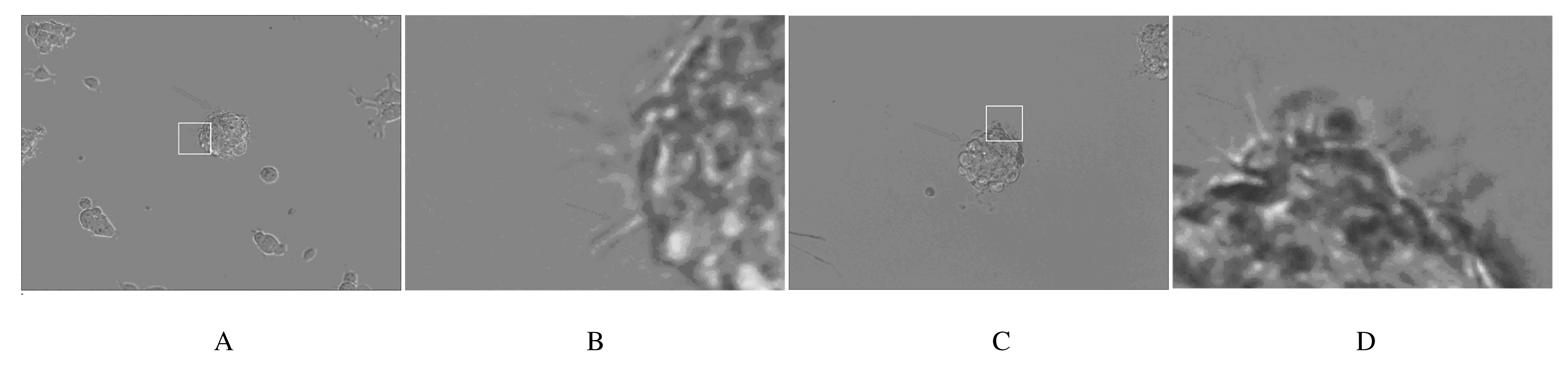
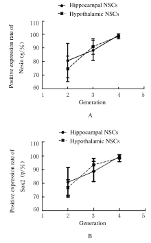
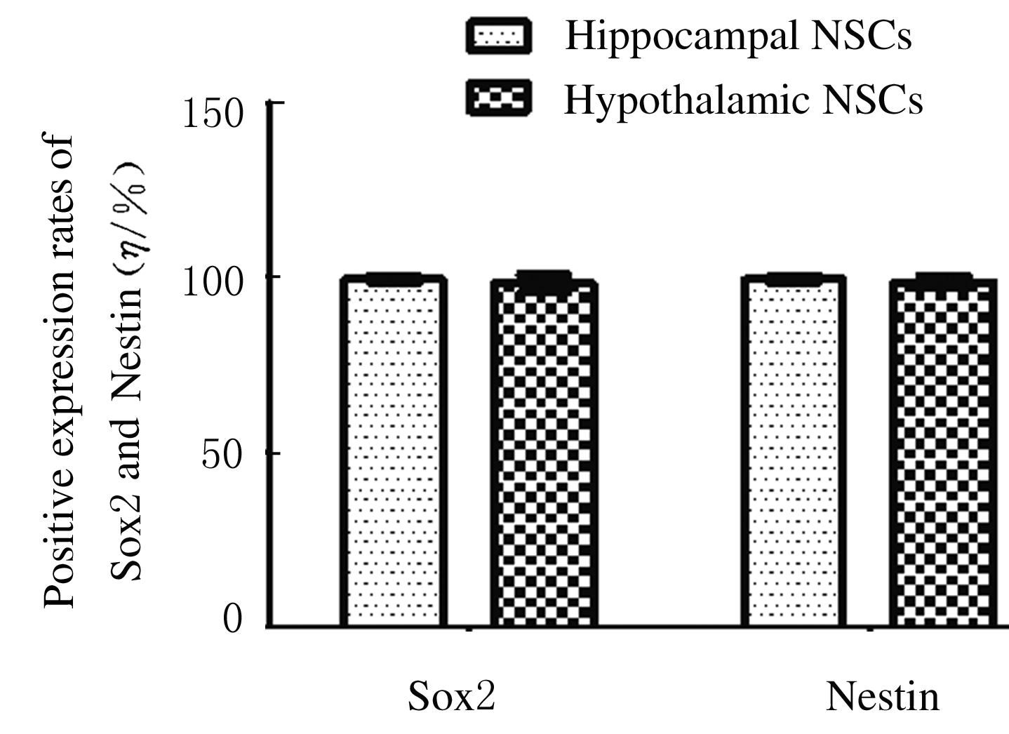
| 1 |
REYNOLDS B A, RIETZE R L. Neural stem cells and neurospheres: re-evaluating the relationship[J]. Nat Methods, 2005, 2(5): 333-336.
|
| 2 |
GROCHOWSKI C, RADZIKOWSKA E, MACIEJEWSKI R. Neural stem cell therapy-Brief review[J]. Clin Neurol Neurosurg, 2018, 173: 8-14.
|
| 3 |
ZHANG T, KE W, ZHOU X, et al. Human neural stem cells reinforce hippocampal synaptic network and rescue cognitive deficits in a mouse model of Alzheimer’s disease[J]. Stem Cell Reports, 2019, 13(6): 1022-1037.
|
| 4 |
ZHAO C M, DENG W, GAGE F H. Mechanisms and functional implications of adult neurogenesis[J]. Cell, 2008, 132(4): 645-660.
|
| 5 |
PELLEGRINO G, TRUBERT C, TERRIEN J, et al. A comparative study of the neural stem cell niche in the adult hypothalamus of human, mouse, rat and gray mouse lemur (Microcebus murinus)[J]. J Comp Neurol, 2018, 526(9): 1419-1443.
|
| 6 |
LEE D A, BEDONT J L, PAK T, et al. Tanycytes of the hypothalamic Median eminence form a diet-responsive neurogenic niche[J]. Nat Neurosci, 2012, 15(5): 700-702.
|
| 7 |
HVAN PRAAG, SCHINDER A F, CHRISTIE B R, et al. Functional neurogenesis in the adult hippocampus[J]. Nature, 2002, 415(6875): 1030-1034.
|
| 8 |
KOZAREVA D A, CRYAN J F, NOLAN Y M. Born this way: Hippocampal neurogenesis across the lifespan[J]. Aging Cell, 2019, 18(5): e13007.
|
| 9 |
LLEDO P M, SAGHATELYAN A. Integrating new neurons into the adult olfactory bulb: joining the network, life-death decisions, and the effects of sensory experience[J]. Trends Neurosci, 2005, 28(5): 248-254.
|
| 10 |
ALVAREZ-BUYLLA A, GARCı́A-VERDUGO J M. Neurogenesis in adult subventricular zone [J]. J Neurosci, 2002, 22(3): 629-634.
|
| 11 |
BLURTON-JONES M, KITAZAWA M, MARTINEZ-CORIA H, et al. Neural stem cells improve cognition via BDNF in a transgenic model of Alzheimer disease[J]. Proc Natl Acad Sci U S A, 2009, 106(32): 13594-13599.
|
| 12 |
GUO W, PATZLAFF N E, JOBE E M, et al. Isolation of multipotent neural stem or progenitor cells from both the dentate gyrus and subventricular zone of a single adult mouse[J]. Nat Protoc, 2012, 7(11): 2005-2012.
|
| 13 |
ZHANG R, LI Y, HU B, et al. Traceable nanoparticle delivery of small interfering RNA and retinoic acid with temporally release ability to control neural stem cell differentiation for Alzheimer's disease therapy[J]. Adv Mater, 2016, 28(30): 6345-6352.
|
| 14 |
SUKSUPHEW S, NOISA P. Neural stem cells could serve as a therapeutic material for age-related neurodegenerative diseases[J]. World J Stem Cells, 2015, 7(2): 502-511.
|
| 15 |
KIM K, CHOE H K. Role of hypothalamus in aging and its underlying cellular mechanisms[J]. Mech Ageing Dev, 2019, 177: 74-79.
|
| 16 |
SAPER C B, LOWELL B B. The hypothalamus [J]. Curr Biol, 2014, 24(23): R1111-R1116.
|
| 17 |
ZHANG Y L, KIM M S, JIA B S, et al. Hypothalamic stem cells control ageing speed partly through exosomal miRNAs[J]. Nature, 2017, 548(7665): 52-57.
|
| 18 |
STROJNIK T, RØSLAND G V, SAKARIASSEN P O, et al. Neural stem cell markers, nestin and musashi proteins, in the progression of human glioma: correlation of nestin with prognosis of patient survival[J]. Surg Neurol, 2007, 68(2): 133-143.
|
| 19 |
LIU D X, NATH N, CHELLAPPAN S P, et al. Regulation of neuron survival and death by p130 and associated chromatin modifiers[J]. Genes Dev, 2005, 19(6): 719-732.
|
| 20 |
HATSUZAWA K, HIROSE H, TANI K, et al. Syntaxin 18, a SNAP receptor that functions in the endoplasmic reticulum, intermediate compartment, and cis-Golgi vesicle trafficking[J]. J Biol Chem, 2000, 275(18): 13713-13720.
|
| 21 |
IINUMA T, AOKI T, ARASAKI K, et al. Role of syntaxin 18 in the organization of endoplasmic reticulum subdomains[J]. J Cell Sci, 2009, 122(pt 10): 1680-1690.
|
 ),Chun CUI2(