Journal of Jilin University(Medicine Edition) ›› 2026, Vol. 52 ›› Issue (1): 105-115.doi: 10.13481/j.1671-587X.20260111
• Research in basic medicine • Previous Articles Next Articles
Improvement effect of empagliflozin on ameliorating doxorubicin-induced myocardial injury rat model and its mechanism
Jiawei LI1, Adilijiang2,Li WU1,Yun JIANG1( )
)
- 1.Department of Oncology and Cardiology,Affiliated Cancer Hospital,Xinjiang Medical University,Urumqi 830000,China
2.Department of Respiratory Medicine,Karamay Central Hospital,Karamay 834000,China
-
Received:2025-03-31Accepted:2025-06-03Online:2026-01-28Published:2026-02-24 -
Contact:Yun JIANG E-mail:95379368@qq.com
CLC Number:
- R541
Cite this article
Jiawei LI, Adilijiang,Li WU,Yun JIANG. Improvement effect of empagliflozin on ameliorating doxorubicin-induced myocardial injury rat model and its mechanism[J].Journal of Jilin University(Medicine Edition), 2026, 52(1): 105-115.
share this article
Tab. 1
LVIDs, LVEF, and LVFS of rats iv various groups"
| Group | LVIDs | LVEF (η/%) | LVFS (η/%) |
|---|---|---|---|
| Control | 4.06±1.11 | 82.4±1.3 | 53.2±2.6 |
| HI | 5.82±0.83* | 43.1±1.7* | 22.6±1.4* |
| HI+EMPA | 4.24±1.77△ | 66.7±2.2△ | 51.4±2.1△ |
| F | 4.45 | 997.00 | 538.90 |
| P | <0.05 | <0.05 | <0.05 |
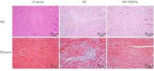
Fig. 1
Pathomorphology of myocardium tissue and deposition of collagen fibers in myocardium tissue of rats in various groups"
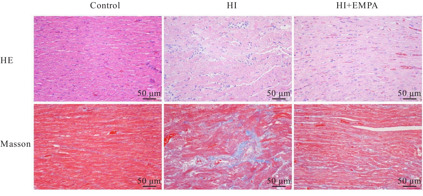

Fig. 2
Apoptosis of myocardium tissue cells (A) and number of TUNEL positive cells(B) of rats in various groups"
Tab.2
Levels of serum LDH and CK of rats in various groups [n=8,x±s, λB/(U·L-1)]"
| Group | Serum LDH | Serum CK |
|---|---|---|
| Control | 1 967±213 | 1 233±239 |
| HI | 3 056±261* | 4 136±312* |
| HI+EMPA | 1 745±188△ | 875±178△ |
| F | 79.39 | 412.40 |
| P | <0.001 | <0.001 |
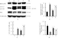
Fig. 3
Electrophoregram(A) and histograms(B-D) of expressions of BECLIN1, LC3-Ⅰ, LC3-Ⅱ, and P62 protein in myocardial H9c2 cells of rats in various groups"

Fig. 4
Electrophoregram(A) and histograms(B-D) of expressions of SIRT1, PGC-1α and Drp-1 proteins in myocardial H9c2 cells of rats in various groups"
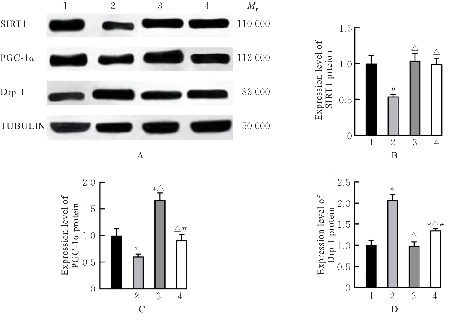
Fig. 5
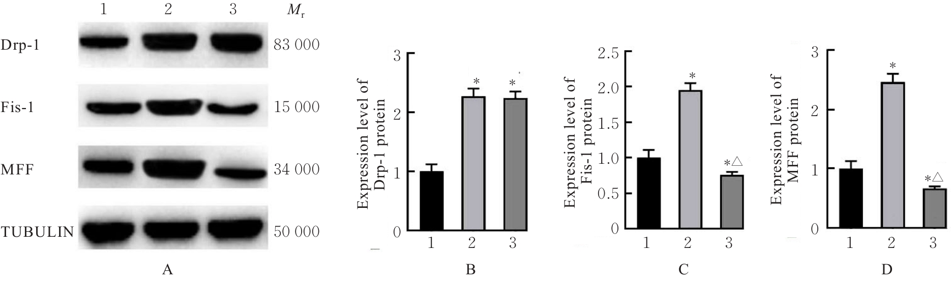
Electrophoregram(A) and histograms(B-D) of expressions of Drp-1, Fis-1 and MFF proteins in myocardial H9c2 cells of rats in various groups"
| [1] | MATTIOLI R, ILARI A, COLOTTI B, et al. Doxorubicin and other anthracyclines in cancers: activity, chemoresistance and its overcoming [J]. Mol Aspects Med, 2023, 93:101205. |
| [2] | CHRISTIDI E, BRUNHAM L R. Regulated cell death pathways in doxorubicin-induced cardiotoxicity [J]. Cell death Dis, 2021, 12(4):339. |
| [3] | WALLACE K B, SARDÃO V A, OLIVEIRA P J. Mitochondrial determinants of doxorubicin-induced cardiomyopathy [J]. Circ Res, 2020, 126(7):926-941. |
| [4] | SHEIBANI M, AZIZI Y, SHAYAN M, et al. Doxorubicin-induced cardiotoxicity: an overview on pre-clinical therapeutic approaches [J]. Cardiovasc Toxicol, 2022, 22(4):292-310. |
| [5] | 刘诗瑶,张梦晓,刘浩. 线粒体质量控制在阿霉素心肌损伤中的作用研究进展[J]. 中国药理学通报,2024,40(10):1814-1818. |
| [6] | SANO R, SHINOZAKI Y, OHTA T. Sodium-glucose cotransporters: functional properties and pharmaceutical potential[J]. J Diabetes Investig, 2020, 11(4): 770-782. |
| [7] | GARCÍA-ROPERO Á, VARGAS-DELGADO A P, SANTOS-GALLEGO C G, et al. Inhibition of sodium glucose cotransporters improves cardiac performance [J]. Int J Mol Sci, 2019, 20(13):3289. |
| [8] | LUCONI M, RAIMONDI L, DI FRANCO A, et al. Which is the main molecular target responsible for the cardiovascular benefits in the EMPA-REG OUTCOME trial? A journey through the kidney, the heart and other interesting places[J]. Nutr Metab Cardiovasc Dis, 2020, 26(12):1071-1078. |
| [9] | KAUR S, KHULLAR N, NAVIK U, et al. Multifaceted role of dynamin-related protein 1 in cardiovascular disease: from mitochondrial fission to therapeutic interventions [J]. Mitochondrion, 2024, 78:101904. |
| [10] | KANE A E, SINCLAIR D A. Sirtuins and NAD+ in the development and treatment of metabolic and cardiovascular diseases [J]. Circ Res, 2018, 123(7):868-885. |
| [11] | RAKSHE P S, DUTTA B J, CHIB S, et al. Unveiling the interplay of AMPK/SIRT1/PGC-1α axis in brain health: promising targets against aging and NDDs [J]. Ageing Res Rev, 2024, 96:102255. |
| [12] | ZHANG J, REN D, FEDOROVA J, et al. SIRT1/SIRT3 Modulates redox homeostasis during ischemia/reperfusion in the aging heart [J]. Antioxidants (Basel), 2020, 9(9):858. |
| [13] | 李登科,张伟,黄从新. SIRT1介导的信号通路在阿霉素诱导心脏毒性中的作用机制[J]. 心血管病学进展,2024,45(3):257-260. |
| [14] | HUANG P, BAI L, LIU L, et al. Redd1 knockdown prevents doxorubicin-induced cardiac senescence[J]. Aging (Albany NY), 2021, 13(10):13788-13806. |
| [15] | ABDULRAHMAN N, IBRAHIM M, JOSEPH J M, et al. Empagliflozin inhibits angiotensin Ⅱ-induced hypertrophy in H9c2 cardiomyoblasts through inhibition of NHE1 expression [J]. Mol Cell Biochem, 2022, 477(6):1865-1872. |
| [16] | GUAN S, XIN Y, DING Y, et al. Ginsenoside Rg1 protects against cardiac remodeling in heart failure via SIRT1/PINK1/parkin-mediated mitophagy [J]. Chem Biodivers, 2023, 20(2):e202200730. |
| [17] | SU Z D, LI C Q, WANG H W, et al. Inhibition of DRP1-dependent mitochondrial fission by Mdivi-1 alleviates atherosclerosis through the modulation of M1 polarization [J]. J Transl Med, 2023, 21(1):427. |
| [18] | LINDERS A N, DIAS I B, LÓPEZ FERNÁNDEZ T, et al. A review of the pathophysiological mechanisms of doxorubicin-induced cardiotoxicity and aging [J]. NPJ Aging, 2024, 10(1):9. |
| [19] | WU B B, LEUNG K T, POON E N. Mitochondrial-Targeted therapy for doxorubicin-induced cardiotoxicity[J]. Int J Mol Sci, 2022, 23(3):1912. |
| [20] | PATORNO E, PAWAR A, FRANKLIN J M, et al. Empagliflozin and the risk of heart failure hospitalization in routine clinical care[J]. Circulation, 2019, 139(25):2822-2830. |
| [21] | KOSIBOROD M N, ANGERMANN C E, COLLINS S P, et al. Effects of empagliflozin on symptoms, physical limitations, and quality of life in patients hospitalized for acute heart failure: results from the EMPULSE trial [J]. Circulation, 2022, 146(4):279-288. |
| [22] | LI Y, ZHANG Z, ZHANG Z, et al. Empagliflozin, a sodium-glucose cotransporter inhibitor enhancing mitochondrial action and cardioprotection in metabolic syndrome [J]. J Cell Physiol, 2024, 239(6):e31264. |
| [23] | CAI C, GUO Z, CHANG X, et al. Empagliflozin attenuates cardiac microvascular ischemia/reperfusion through activating the AMPKα1/ULK1/FUNDC1/mitophagy pathway [J]. Redox Biol, 2022, 52:102288. |
| [24] | ZOU R, SHI W, QIU J, et al. Empagliflozin attenuates cardiac microvascular ischemia/reperfusion injury through improving mitochondrial homeostasis[J]. Cardiovasc Diabetol, 2022, 21(1):106. |
| [25] | CAI C, WU F, ZHUANG B, et al. Empagliflozin activates Wnt/β-catenin to stimulate FUNDC1-dependent mitochondrial quality surveillance against type-3 cardiorenal syndrome [J]. Mol Metab, 2022, 64:101553. |
| [26] | KOIZUMI T, WATANABE M, YOKOTA T, et al. Empagliflozin suppresses mitochondrial reactive oxygen species generation and mitigates the inducibility of atrial fibrillation in diabetic rats [J]. Front Cardiovasc Med, 2023, 10:1005408. |
| [27] | YANG X, LIU Q, LI Y, et al. The diabetes medication canagliflozin promotes mitochondrial remodelling of adipocyte via the AMPK-SIRT1-PGC-1α signalling pathway [J]. Adipocyte, 2020, 9(1):484-494. |
| [28] | WU S K, WANG L, WANG F, et al. Resveratrol improved mitochondrial biogenesis by activating SIRT1/PGC-1α signal pathway in SAP [J]. Sci Rep, 2024, 14(1):26216. |
| [29] | YANG T T, ZHOU L H, GU L F, et al. CHK1 attenuates cardiac dysfunction via suppressing SIRT1-ubiquitination [J]. Metabolism, 2025, 162:156048. |
| [30] | HUANG Q, SU H, QI B, et al. A SIRT1 activator, ginsenoside Rc, promotes energy metabolism in cardiomyocytes and neurons [J]. J Am Chem Soc, 2021, 143(3):1416-1427. |
| [31] | AN X, MA X, LIU H, et al. Inhibition of PDGFRβ alleviates endothelial cell apoptotic injury caused by DRP-1 overexpression and mitochondria fusion failure after mitophagy [J]. Cell Death Dis, 2023, 14(11):756. |
| [32] | KELM N Q, BEARE J E, WEBER G J, et al. Thrombospondin-1 mediates DRP-1 signaling following ischemia reperfusion in the aging heart[J]. FASEB Bioadv, 2020, 2(5):304-314. |
| [1] | Fengmei XIONG,Yuxiang CAI,Zhuo LIU,Na SUN,Yang LI. Alleviatory effect of curcumin on cardiomyocyte toxicity induced by doxorubicin by regulating SIRT3/SOD2 signaling pathway [J]. Journal of Jilin University(Medicine Edition), 2024, 50(5): 1339-1347. |
| [2] | Xiaodong GAI,Ying ZHAO,Hefei WANG,Chengyuan HE,Xingxiang WANG,Chun LI. Regulatory effect of FOXP3 on chemosensitivity of non small-cell lung cancer A549 cells to doxorubicin and its mechnism [J]. Journal of Jilin University(Medicine Edition), 2023, 49(5): 1161-1167. |
| [3] | Yintao ZHAO, Yingying YANG, Xiangqin ZHANG, Lu ZHENG, Yawei XU, Haibo YANG, Yuan LIU. Improvement effect of follistatin-like 1 on doxorubicin-induced acute myocardial injury in mice and its mechanism [J]. Journal of Jilin University(Medicine Edition), 2023, 49(3): 565-572. |
| [4] | Xin LI,Xu DING,Xiaomeng LIU,Xinchan LIU,Zhou WU,Weixian YU. Effect of silencing information regulator 1 on kidney injury in chronic periodontitis model rats [J]. Journal of Jilin University(Medicine Edition), 2022, 48(5): 1200-1208. |
| [5] | Ying YANG, Wei ZHAO, Dan LYU. Effect of C19ORF12 on proliferation and chemo-sensitivity of gastric cancer MKN45 cells and its mechanism [J]. Journal of Jilin University(Medicine Edition), 2021, 47(3): 687-693. |
| [6] | YANG Zebin, WANG Mingyue, CHEN Li, LIU Ning, WANG Hao, CUI Meiying, FANG Kaiyi, XIA Wei, GUAN Xingang. Killing effect of doxorubicin-loaded cell membrane nanovesicles on melanoma B16F10 cells [J]. Journal of Jilin University(Medicine Edition), 2020, 46(05): 905-910. |
| [7] | YU Yang, XU Lu, LIU Shibing, LI Songyan, XU Ye. Induction effect of myricetin on apoptosis of human ovarian cancer SKOV3 cells by promoting DRP1-dependent mitochondrial fission [J]. Journal of Jilin University(Medicine Edition), 2018, 44(05): 903-907. |
| [8] | MA Hongyun, ZHUANG Xinming, XU Weiguo, LIU Yi. Inhibitory effects of hyaluronic acid nanoparticles loading doxorubicin and cisplatin on allograft breast cancer in mice [J]. Journal of Jilin University Medicine Edition, 2018, 44(02): 243-248. |
| [9] | ZHANG Fan, ZHENG Yi, YANG Xiaohong, YE Caifen, WANG Liangyu. Application of two dimension speckle tracking imaging in evaluating on cardiac dysfunction in patients with malignant lymphoma after anthracycline treatment [J]. Journal of Jilin University Medicine Edition, 2017, 43(06): 1193-1198. |
| [10] | YU Yang, LIU Shibing, LI Songyan, XU Lu, XU Ye. Induction effect of myricetin on autophagy in SKOV3 cells and promoting effect on mitochondrial fission [J]. Journal of Jilin University Medicine Edition, 2017, 43(04): 685-689. |
| [11] | JIN Xin1,SHEN Wei-zhang1,JIN Li-fang,JIA Jiao-yuan,LI Xiao-feng,WANG Xiu-li,DI Xin,ZHANG Hong-juan,LI Ping-ya. Protective effect of pseudo-ginsenoside GQ on doxorubicin-induced acute myocardial injury in rats [J]. Journal of Jilin University Medicine Edition, 2013, 39(6): 1164-1168. |
| [12] | ZHANG Hai-shan, ZHANG Xue-wen,WANG Da-min,LIU De-quan,ZHANG De-heng. Protective effect of heme oxygenase-1 induced by doxorubicin on hepatic ischemia-reperfusion in rats [J]. J4, 2007, 33(4): 686-689. |
| [13] | ZHANG Hai-shan,ZHANG Xue-wen, MA Shu-rong,ZHANG De-heng,QIU Dong-tao. Expression of heme oxygenase-1 induced by doxorubicin in liver,spleen, and kidney and effect of doxorubicin on liver function [J]. J4, 2004, 30(1): 82-84. |