吉林大学学报(医学版) ›› 2021, Vol. 47 ›› Issue (2): 284-291.doi: 10.13481/j.1671-587X.20210205

• 基础研究 • 上一篇    下一篇

线粒体分裂融合蛋白表达失衡在IgA肾病小鼠肾脏病理学变化中的作用

张旭1,2,刘乃萌3,马娇艳2,林楠2,孙珉丹4()   

  1. 1.长春医学高等专科学校临床医学部妇儿教研室,吉林 长春 130031
    2.吉林大学基础医学院 病理生理学教研室,吉林 长春 130021
    3.吉林大学第一医院乳腺外科,吉林 长春 130021
    4.吉林大学第一医院肾病内科,吉林 长春 130021
  • 收稿日期:2020-09-12 出版日期:2021-03-28 发布日期:2021-03-25
  • 通讯作者: 孙珉丹 E-mail:mindansun@sina.com
  • 作者简介:张 旭(1962-),女,吉林省长春市人,副教授,主要从事慢性肾病基础方面的研究。
  • 基金资助:
    吉林省科技厅科研基金项目(20191008012TC)

Effect of mitochondrial fission/fussion protein expression imbalance in pathological changes of kidney tissue in IgA nephropathy mice

Xu ZHANG1,2,Naimeng LIU3,Jiaoyan MA2,Nan LIN2,Mindan SUN4()   

  1. 1.Department of Gynecology and Pediatrics,Clinical Medicine Center,Changchun Medical College,Changchun 130031,China
    2.Department of Pathophysiology,School of Basic Medical Sciences,Jilin University,Changchun 130021,China
    3.Department of Breast Surgery,First Hospital,Jilin University,Changchun 130021,China
    4.Department of Nephropathy,First Hospital,Jilin University,Changchun 130021,China
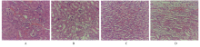
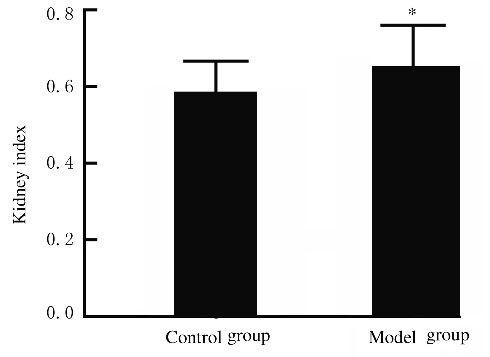
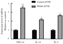
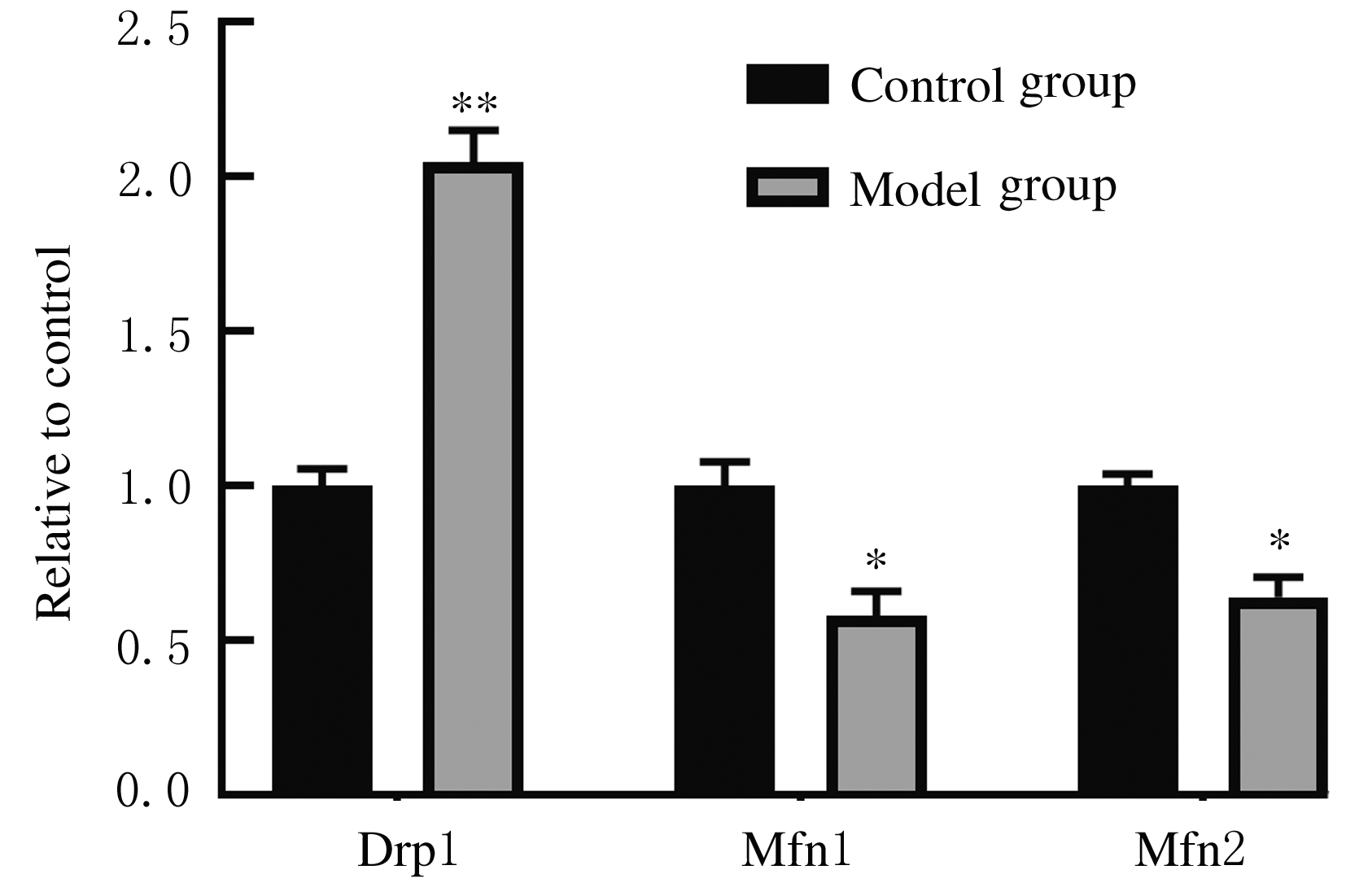
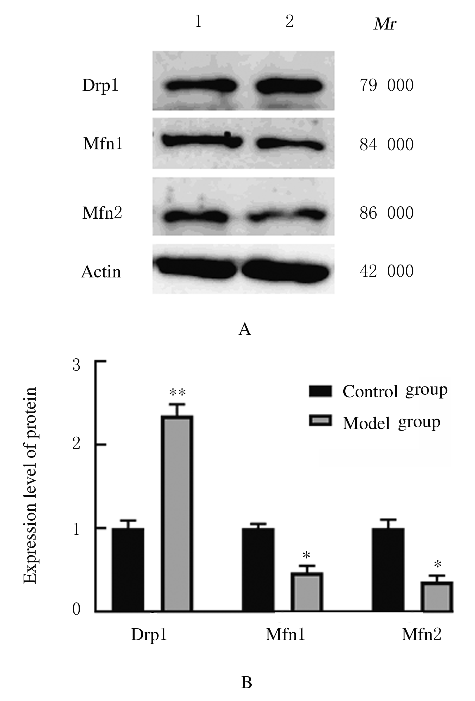
  • Received:2020-09-12 Online:2021-03-28 Published:2021-03-25
  • Contact: Mindan SUN E-mail:mindansun@sina.com

摘要: 目的

探讨线粒体分裂融合蛋白表达失衡在IgA肾病(IgAN)小鼠肾脏病理学变化中的作用,为IgAN的治疗提供一定的实验依据。

方法

20只健康SPF级雄性BALB/C小鼠, 6~8周龄,随机分为对照组和模型组,每组10只。采用牛血清白蛋白(BSA)、脂多糖(LPS)和四氯化碳(CCl4)联合给药的方式建立小鼠IgAN模型。采用免疫荧光技术检测肾小球中IgA沉积,苏木精-伊红(HE)染色观察小鼠肾组织病理形态表现,以评价模型是否构建成功。称量小鼠体质量和肾脏质量,计算各组小鼠肾脏指数;酶活性法检测小鼠血清中肌酐和尿素氮水平。采用酶联免疫吸附试验(ELISA)法检测各组小鼠血清中肿瘤坏死因子α(TNF-α)、白细胞介素1β(IL-1β)和白细胞介素6(IL-6)水平,实时荧光定量聚合酶链式反应(RT-qPCR)法检测小鼠肾组织中调控线粒体分裂融合的关键基因动力相关蛋白1(Drp1)、线粒体融合蛋白1(Mfn1)和线粒体融合蛋白2(Mfn2)mRNA表达水平,Western blotting法检测小鼠肾组织中Drp1、Mfn1和Mfn2蛋白表达水平。

结果

与对照组比较,模型组小鼠肾小球出现明显的IgA沉积,伴随肾小球萎缩,肾小球系膜细胞和基质增生导致系膜区增宽,肾小管部分细胞肿胀坏死,表明IgAN模型构建成功。与对照组比较,模型组小鼠肾脏指数、血清肌酐和尿素氮水平均明显升高(P<0.05),小鼠血清中TNF-α、IL-1β和IL-6水平明显升高(P<0.05或P<0.01),小鼠肾组织中Drp1 mRNA和蛋白表达水平明显升高(P<0.05),而小鼠肾组织中Mfn1和Mfn2 mRNA和蛋白表达水平明显降低(P<0.05)。

结论

线粒体分裂融合蛋白表达失衡可能是IgAN发生发展的机制之一,以分裂融合稳态为靶点的研究可能为IgAN的治疗提供新思路。

关键词: 免疫球蛋白A肾病, 线粒体, 动力相关蛋白1, 线粒体融合蛋白1, 线粒体融合蛋白2

Abstract:

Objective: To investigate the effect of imbalanced expression of mitochondrial fission/fusion proteins in the pathological changes of kidney tissue of the mice with IgA nephropathy (IgAN), and to provide an experimental data for the therapy of IgAN.

Methods

A total of 20 healthy SPF male BALB/C mice aged 6-8 weeks were randomly divided into control group and model group, with 10 mice in each group. The IgAN model was constructed by the combined administration of bovine serum albumin (BSA), lipopolysaccharide (LPS), and carbon tetrachloride (CCl4).IgA deposits in glomeruli were detected by immunofluorescence method, and hematoxylin-eosin (HE) staining was used to observe the pathomorphology of kidney tissue of the mice to evaluate whether the model was successfully constructed.The body weights and kidney weights of the mice were weighed, and the kidney index was caculated.The levels of serum creatinine and urea nitrogen of the mice were detected by enzyme activity method.The levels of tumor necrosis factor-α (TNF-α), interleukin 1-β (IL-1β) and interleukin 6 (IL-6) in serum of the mice in each group were detected by enzyme-linked immunosorbent assay (ELISA). Real-time fluorescence quantitative polymerase chain reaction (RT-qPCR) was used to detect the mRNA expression levels of the key genes that regulated mitochondrial fission and fusion, including dynamic related protein 1 (Drp1), mitofusin 1 (Mfn1) and mitofusin 2 (Mfn2). The expression levels of Drp1, Mfn1, and Mfn2 proteins in kidney tissue of the mice were detected by Western blotting method.

Results

Compared with control group, the glomerulus of the mice in model group showed that IgA deposition was obvious, accompanyied by glomerular atrophy, the hyperplasia of glomerular mesangial cells and stroma led to widening of the mesangial area, and a portion of renal tubule cells presented swelling and necrosis, indicating that the IgAN model was successfully constructed. Compared with control group, the kidney index and the serum creatinine and urea nitrogen levels of the mice in model group were obviously increased(P<0.05),the levels of serum TNF-α, IL-1β and IL-6 of the mice were increased significantly(P<0.05 or P<0.01),the expression levels of Drp1 mRNA and protein in kidney tissue of the mice were obviously increased(P<0.05), while the expression levels of Mfn1 and Mfn2 mRNA and proteins in kidney tissue of the mice were obviously reduced(P<0.05).

Conclusion

The imbalance of mitochondrial fission/fusion protein expression may be one of the mechanisms of the development and progression of IgAN, and the research of fission and fusion homeostasis as a target may provide new ideas for the therapy of IgAN.

Key words: IgA nephropathy, mitochondria, dynamic-related protein 1, mitofusin 1, mitofusin 2

中图分类号: 

  • R692