吉林大学学报(医学版) ›› 2021, Vol. 47 ›› Issue (2): 330-337.doi: 10.13481/j.1671-587X.20210211

• 基础研究 • 上一篇    下一篇

肌肽对脂多糖诱导星形胶质细胞炎症因子释放的抑制作用及其机制

白雪1,于露1,杨文强1,何鑫1,杨新生2,杨菁1()   

  1. 1.锦州医科大学基础医学院生物化学与分子生物学教研室,辽宁 锦州 121001
    2.锦州卫生学校外科教研室,辽宁 锦州 121001
  • 收稿日期:2020-07-11 出版日期:2021-03-28 发布日期:2021-03-25
  • 通讯作者: 杨菁 E-mail:yangjing@jzmu.edu.cn
  • 作者简介:白 雪(1996-),女,蒙古族,内蒙古自治区通辽市人,在读硕士研究生,主要从事生化药物方面的研究。
  • 基金资助:
    辽宁省科技厅自然科学基金项目(20170540367)

Inhibitory effect of carnosine on LPS-induced inflammatory cytokine release in astrocytes and its mechanism

Xue BAI1,Lu YU1,Wenqiang YANG1,Xin HE1,Xinsheng YANG2,Jing YANG1()   

  1. 1.Department of Biochemistry and Molecular Biology,College of Basic Medical Sciences,Jinzhou Medical University,Jinzhou 121001,China
    2.Department of Surgery,Jinzhou Health School,Jinzhou 121001,China
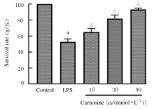
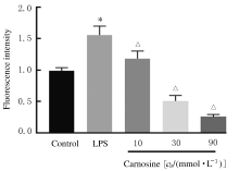
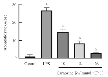
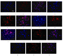
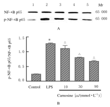
  • Received:2020-07-11 Online:2021-03-28 Published:2021-03-25
  • Contact: Jing YANG E-mail:yangjing@jzmu.edu.cn

摘要: 目的

探讨肌肽对脂多糖(LPS)诱导的星形胶质细胞(AS)炎症因子释放的影响,初步阐明其作用机制。

方法

体外培养新生大鼠大脑皮质AS并纯化,随机分为对照组、LPS组和LPS+肌肽组(10、30和90 mmol·L-1肌肽)。对照组只加入完全培养基,LPS组培养基中加入终浓度为1 mg·L-1 LPS,体外刺激细胞 24 h构建炎症损伤模型,LPS+肌肽组培养基中预先分别加入10、30和90 mmol·L-1肌肽,培养1 h后再加入终浓度为1 mg·L-1 的LPS。MTT法检测各组AS存活率,ELISA法检测各组AS培养液中白细胞介素1β(IL-1β)和肿瘤坏死因子α(TNF-α)水平,DCFH-DA探针法检测各组AS中活性氧(ROS)水平,Hoechst 33258荧光染色法检测各组AS凋亡率,免疫荧光染色法测定各组AS中磷酸化核转录因子κB p65(p-NF-κB p65)蛋白表达情况和p-NF-κB p65阳性细胞数,采用Western blotting 法检测各组AS中p-NF-κB p65蛋白表达水平。

结果

与对照组比较,LPS组AS存活率明显降低(P<0.05),AS培养液中IL-1β和TNF-α水平升高(P<0.05),AS中ROS水平、AS凋亡率和p-NF-κB p65阳性细胞数均明显升高(P<0.05),AS中p-NF-κB p65蛋白表达水平升高(P<0.05)。与LPS组比较,LPS+肌肽组AS存活率明显升高(P<0.05),AS培养液中IL-1β和TNF-α水平降低(P<0.05),AS中ROS水平、AS凋亡率和p-NF-κB p65阳性细胞数均明显降低(P<0.05),AS中p-NF-κB p65蛋白表达水平降低(P<0.05)。

结论

肌肽可抑制LPS引起的AS中炎症因子IL-1β和TNF-α释放,其机制可能与肌肽抑制LPS诱导AS中ROS生成、进而抑制NF-κB p65激活有关。

关键词: 肌肽, 脂多糖, 星形胶质细胞, 炎症因子, 核转录因子κB p65

Abstract: Objective

To investigate the effect of carnosine on the inflammatory cytokine release in the astrocytes (AS) induced by lipopolysaccharide (LPS), and to clarify its mechanism.

Methods

The neonatal rat cerebral cortex AS were cultured and purified in vitro. The AS were randomly divided into control group, LPS group and LPS+carnosine groups (10,30,and 90 mmol·L-1 carnosine ).The cells in control group were only cultured with complete medium; the cells in LPS group were stimulated in vitro with 1 mg·L-1 LPS for 24 h to construct the inflammatory injury model; the cells in LPS+carnosine groups were cultured with 10,30, and 90 mmol·L-1carnosine, respectively; in complete medium for 1 h, then treated with LPS with a final concentration of 1 mg·L-1 for 24 h.The survival rates of AS in various groups were determined by MTT assay. The levels of interleukin-1β(IL-1β) and tumor necrosis factor-α(TNF-α) in the AS culture medium were detected by ELISA. DCFH-DA fluorescence probe was used to detect the reactive oxygen species(ROS) levels in AS in various groups; Hoechst 33258 fluorescence staining was used to detect the apoptotic rates of AS in various groups. The expressions of phosphorylated nuclear transcription factor-κB p65(p-NF-κB p65) in the AS and the number of p-NF-κB p65 positive cells in various groups were determined by immunofluorescence staining. The expression levels of p-NF-κB p65 protein in the AS in various groups were detected by Western blotting method.

Results

Compared with control group, the survival rate of AS in LPS group was significantly decreased(P<0.05), the levels of IL-1β and TNF-α in AS culture medium were increased(P<0.05), the ROS level in AS was significantly increased(P<0.05),the apoptotic rate and the number of p-NF-κB p65 positive AS were increased significantly(P<0.05),and the expression level of p-NF-κB p65 protein in the AS was increased(P<0.05).Compared with LPS group, the survival rates of AS in LPS+carnosine groups were significantly increased(P<0.05), the levels of IL-1β and TNF-α in AS culture medium were decreased(P<0.05), the ROS levels in the AS were decreased(P<0.05), the apoptotic rates and the number of p-NF-κB p65 positive AS were decreased significantly(P<0.05),and the expression levels of p-NF-κB p65 protein in the AS were decreased(P<0.05).

Conclusion

Carnosine can inhibit the release of the inflammatory factors IL-1β and TNF-α in the AS induced by LPS, and its mechanism may be related to inhibiting the ROS production in AS induced by LPS and then inhibiting the activation of NF-κB p65.

Key words: carnosine, lipopolysaccharide, astrocyte, inflammatory factor, nuclear transcription factor κB p65

中图分类号: 

  • Q78