吉林大学学报(医学版) ›› 2021, Vol. 47 ›› Issue (4): 826-833.doi: 10.13481/j.1671-587X.20210402

• 基础研究 • 上一篇    下一篇

解毒通络方通过TGF-β1/Smad和ROCK通路对大鼠心肌纤维化的改善作用

张丹1,2,惠菊1,郭家娟1()   

  1. 1.长春中医药大学附属医院心病中心,吉林 长春 130021
    2.吉林省长春市中心医院心内一科,吉林 长春 130051
  • 收稿日期:2020-12-21 出版日期:2021-07-28 发布日期:2021-07-22
  • 通讯作者: 郭家娟 E-mail:gjj-2005@163.com
  • 作者简介:张 丹(1976-),女,吉林省德惠市人,副主任医师,医学硕士,主要从事心血管疾病诊治、心肌纤维化发生机制和治疗方面研究。
  • 基金资助:
    国家自然科学基金青年科学基金项目(81503544);吉林省科技厅自然科学基金项目(20180101108JC)

Improvement effect of Jiedu Tongluo Decoction on myocardial fibrosis in rats through TGF-β1/Smad2/3 and ROCK pathways

Dan ZHANG1,2,Ju HUI1,Jiajuan GUO1()   

  1. 1.Department of Cardiovascular Disease,School of Chinese Medicine,Changchun University of Chinese Traditional Medicine,Changchun 130021,China
    2.Department of Cardiology,Changchun Central Hospital,Jilin Changchun 130051,China
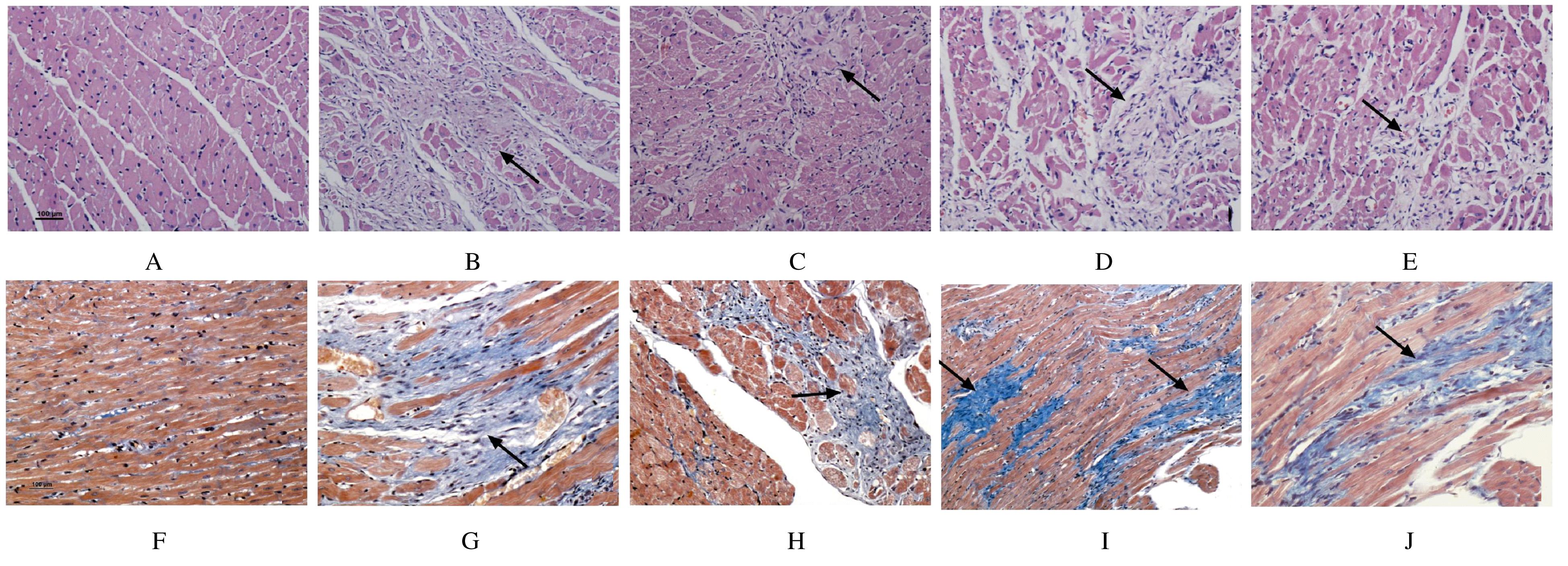
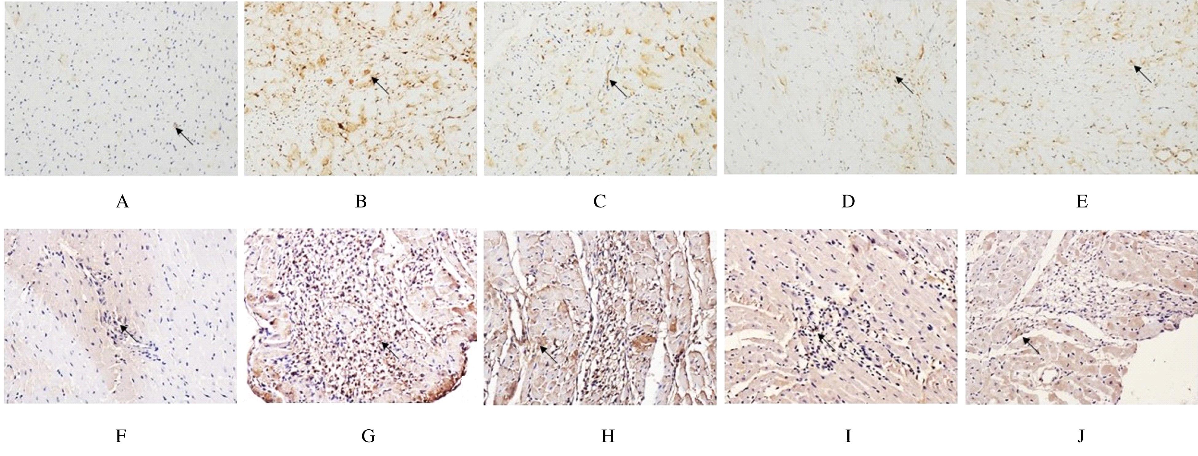
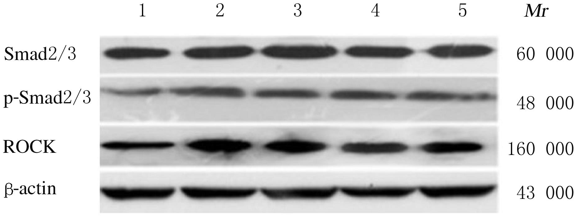
  • Received:2020-12-21 Online:2021-07-28 Published:2021-07-22
  • Contact: Jiajuan GUO E-mail:gjj-2005@163.com

摘要: 目的

探讨解毒通络方在大鼠心肌纤维化进程中的作用,并阐明其作用机制。

方法

30只SPF级Wistar大鼠随机分为正常对照组、模型组、卡托普利组、低剂量解毒通络方组和高剂量解毒通络方组(n=6),以5 mg·kg-1盐酸异丙肾上腺素皮下注射法复制大鼠心肌纤维化模型,卡托普利组、低剂量解毒通络方组和高剂量解毒通络方组大鼠分别用0.005 g·kg -1·d -1卡托普利和1、5 g·kg-1·d -1解毒通络方灌胃28 d,对照组和模型组大鼠灌胃给予等体积蒸馏水,苏木素-伊红(HE)和Masson三色染色观察大鼠心肌组织病理形态表现并计算胶原纤维面积百分率,碱水解法检测大鼠心肌组织中羟脯氨酸水平,酶联免疫吸附测定法(ELISA)检测大鼠心肌组织中血管紧张素Ⅱ(AngⅡ)水平,免疫组织化学法检测转化生长因子β1(TGF-β1)和LIM激酶(LIMK)蛋白表达水平,实时荧光定量PCR(RT-qPCR)法检测大鼠心肌组织中Smad2、Smad3和Rho相关蛋白激酶(ROCK) mRNA表达水平,Western blotting法检测大鼠心肌组织中磷酸化Smad2/3(p-Smad2/3)和ROCK蛋白表达水平。

结果

注射异丙肾上腺素28 d后,大鼠心肌组织呈典型纤维化改变,与正常对照组比较,模型组大鼠心肌组织中胶原纤维面积百分率及羟脯氨酸和Ang Ⅱ水平明显升高(P<0.05),TGF-β1和LIMK蛋白表达水平明显升高(P<0.05),Smad2、Smad3和ROCK mRNA表达水平明显升高(P<0.05),p-Smad2/3和ROCK 蛋白表达水平明显升高(P<0.05);与模型组比较,卡托普利组、低剂量解毒通络方组和高剂量解毒通络方组大鼠心肌组织中胶原纤维面积百分率及羟脯氨酸和Ang Ⅱ水平明显降低(P<0.05),TGF-β1和LIMK蛋白表达水平明显降低(P<0.05),Smad2、Smad3和ROCK mRNA表达水平明显降低(P<0.05),p-Smad2/3和ROCK蛋白表达水平明显降低(P<0.05)。与卡托普利组比较,不同剂量解毒通络方组大鼠上述指标差异均无统计学意义(P>0.05)。

结论

解毒通络方可通过抑制TGF-β1/Smad和ROCK通路延缓大鼠心肌纤维化进程。

关键词: 解毒通络, 心肌纤维化, 转化生长因子β1, Smad2, Smad3, Rho相关蛋白激酶

Abstract: Objective

To explore the effect of Jiedu Tongluo Decoction on the myocardial fibrosis in the rats, and to clarify its mechanism.

Methods

A Total of 30 SPF Wistar rats were randomly divided into normal control group, model group, captopril group,low and high doses of Jiedu Tongluo Decoction groups (n=6). The myocardial fibrosis model was established by subcutaneous injection of isoproterenol hydrochloride (5 mg·kg-1). The rats in captopril group and low and high doses of Jiedu Tongluo Decoction groups were treated with 0.005 g·kg-1·d-1 captopril and 1, 5 g·kg-1·d-1 Jiedu Tongluo Recipe for 28 d,and the rats in control group and model group were given the equal volume of distilled water by gavage. Hematoxylin-eosin (HE) staining and Masson tri-chrome staining were applied to observe the pathomorphology of myocardium tissue of the rats in various groups,and the percentages of collagen fiber areas were calculated.The hydroxyproline levels in myocardium tissue of the rats were determined by alkaline hydrolysis method, and the angiotensin Ⅱ(Ang Ⅱ) levels in myocardium tissue of the rats were detected by enzyme-linked immunosorbent assay (ELISA);the expression levels of transforming growth factor-β1 (TGF-β1) and the LIM kinase (LIMK)proteins were detected by immunohistochemistry; the expression levels of Smad2, Smad3, and Rho-associated protein kinase (ROCK) mRNA in myocardium tissue of the rats were detected by Real-time fluorescence quantitative PCR (RT-qPCR),and the expression levels of Smad2/3, phosphorylated Smad2/3 (p-Smad2/3) and ROCK proteins in myocardium tissue of the rats were analyzed by Western blotting method.

Results

The myocardium tissue showed the typical fibrosis changes 28 d after isoproterenol injection; compared with normal control group, the percentage of collagen fiber area and the levels of hydroxyproline and Ang Ⅱ in myocardium tissue of the rats in model group were significantly increased (P<0.05),the expression levels of TGF-β1,and LIMK were increased(P<0.05), the expression levels of Smad2, Smad3, and ROCK mRNA were increased(P<0.05), and the expression levels of p-Smad2/3 and ROCK proteins were significantly increased (P<0.05). Compared with model control group, the percentages of collagen fiber areas and the levels of hydroxyproline and Ang Ⅱ in myocardium tissue of the rats in captopril group and low and high doses of Jiedu Tongluo Decoction groups were significantly decreased (P<0.05),and the expression levels of TGF-β1 and LIMK proteins were decreased(P<0.05),the expression levels of Smad2, Smad3, and ROCK mRNA were decreased(P<0.05),and the expression levels of p-Smad2/3 and RCOK proteins of the rats were significantly decreased(P<0.05).There were no significant differences in the above indexes of the rats between different doses of Jiedu Tongluo Decoction groups and captopril group(P>0.05).

Conclusion

Jiedu Tongluo Decoction can inhibit the process of myocardial fibrosis by inhibiting the TGF-β1/Smad2/3 and ROCK pathway.

Key words: Jiedu Tongluo, myocardial fibrosis, transforning growth factor-β1, Smad2/3 pathway, Rho-associated protein kinase

中图分类号: 

  • R285.5