吉林大学学报(医学版) ›› 2021, Vol. 47 ›› Issue (6): 1422-1428.doi: 10.13481/j.1671-587X.20210611

• 基础研究 • 上一篇    下一篇

载奥沙利铂细胞膜囊泡纳米药物的制备及其对小鼠结肠癌细胞的杀伤作用

黄莉莉,刘宇轩,方楷漪,穆业腾,胡楠楠,郭冲,杨馥旭,关新刚()   

  1. 北华大学医学技术学院医药生物工程重点实验室,吉林 吉林 132013
  • 收稿日期:2021-04-16 出版日期:2021-11-28 发布日期:2021-12-14
  • 通讯作者: 关新刚 E-mail:guanxg@ciac.ac.cn
  • 作者简介:黄莉莉(1996-),女,浙江省杭州市人,在读医学硕士,主要从事肿瘤免疫治疗机制方面的研究。
  • 基金资助:
    吉林省科技厅科技发展计划项目(20180101213JC);吉林省教育厅科学技术研究项目(JJKH20200033KJ);吉林省卫健委卫生与健康技术创新项目(2020J023)

Preparation of oxaliplatin-loaded cell membrane nanodrugs and its killing effect on colon cancer cells of mice

Lili HUANG,Yuxuan LIU,Kaiyi FANG,Yeteng MU,Nannan HU,Chong GUO,Fuxu YANF,Xingang GUAN()   

  1. Key Laboratory of Pharmaceutical Biotechnology,School of Medical Technology,Beihua University,Jilin 132013,China
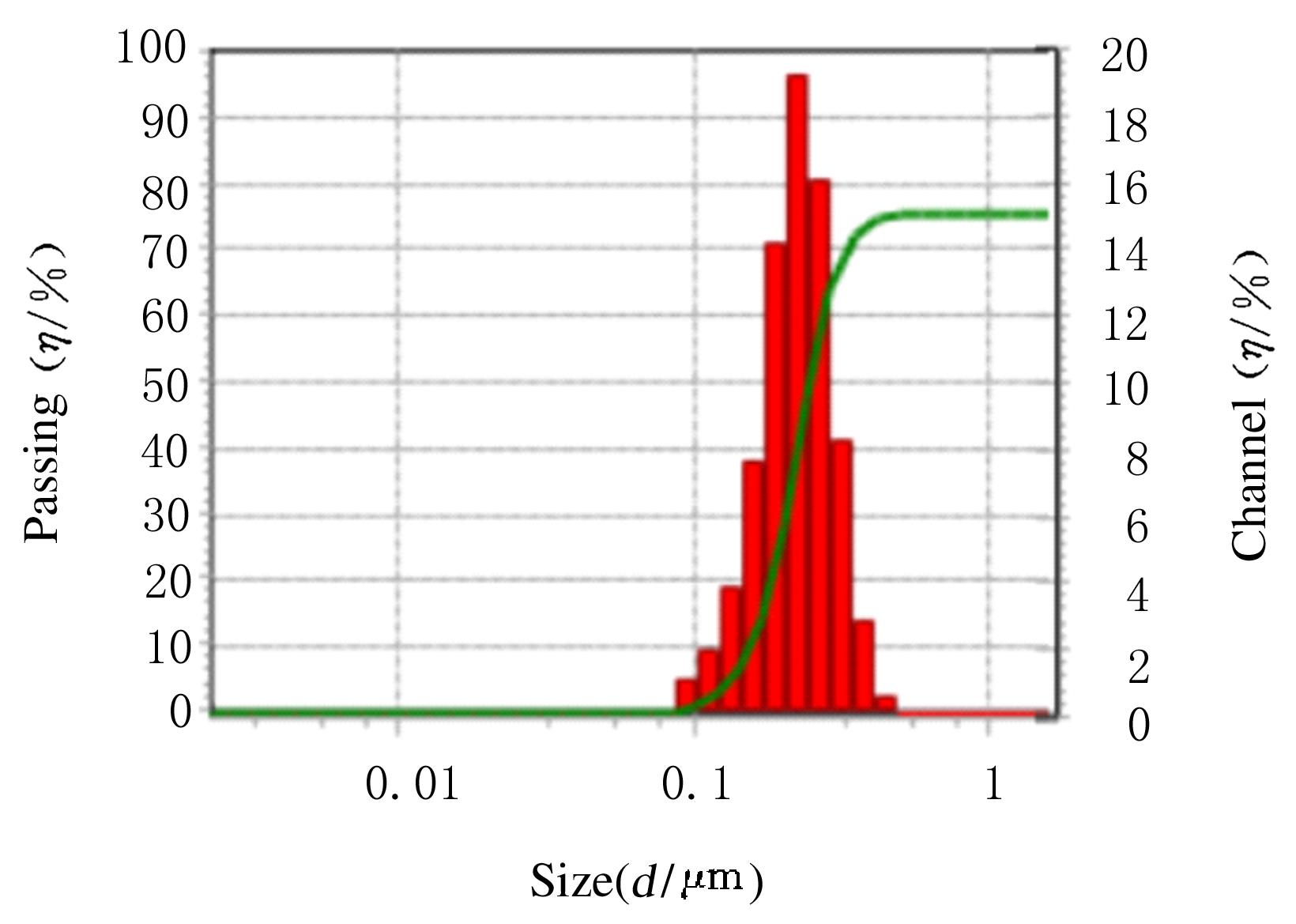
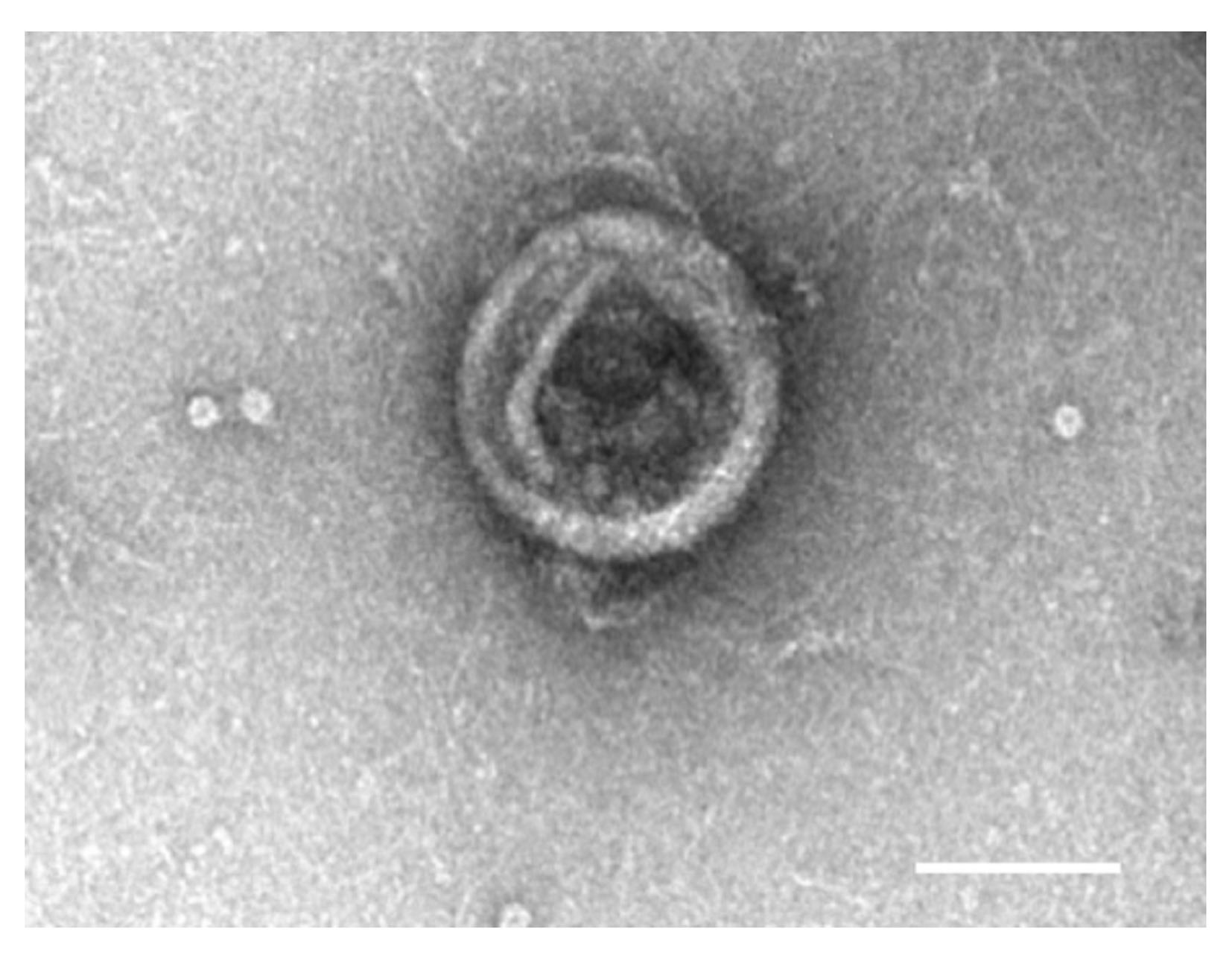
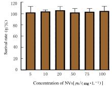
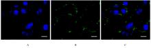
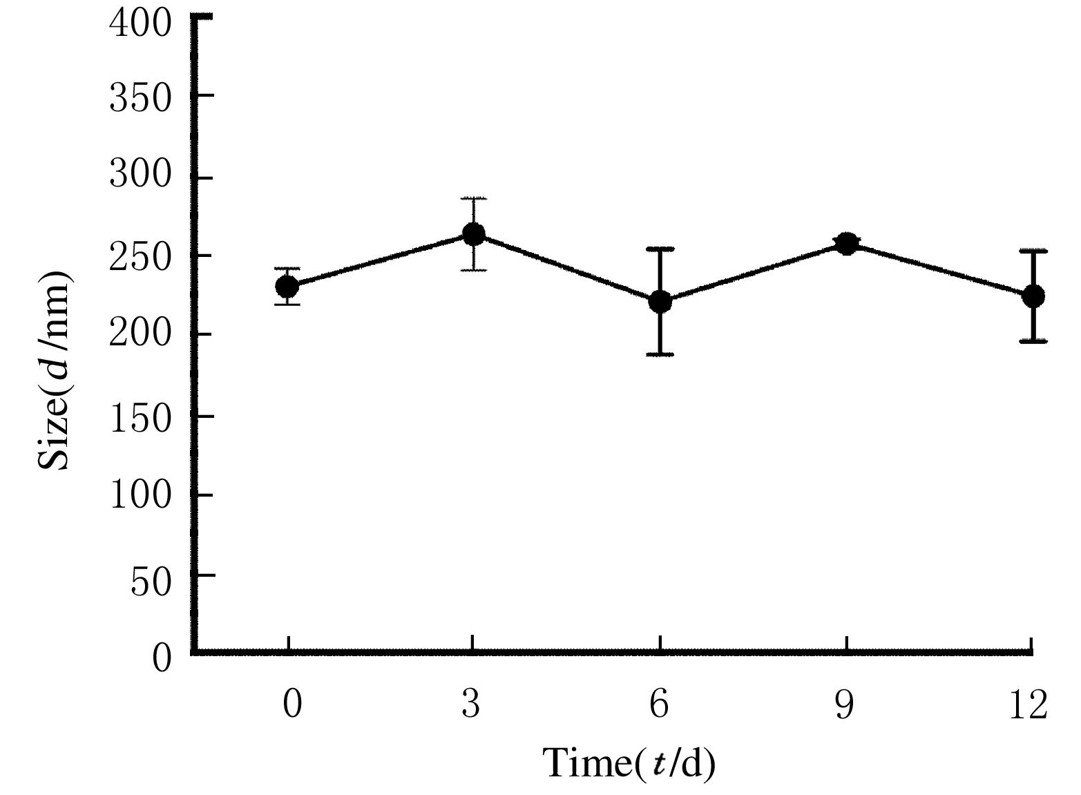
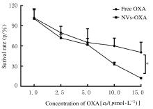
  • Received:2021-04-16 Online:2021-11-28 Published:2021-12-14
  • Contact: Xingang GUAN E-mail:guanxg@ciac.ac.cn

摘要: 目的

制备内腔担载奥沙利铂(OXA)的细胞膜纳米囊泡(NVs),得到纳米药物NVs@OXA,探讨NVs@OXA对小鼠结肠癌细胞的内吞和杀伤效应。

方法

利用超速离心法分离HEK-293T细胞的细胞膜,通过脂质体挤出仪制备NVs,动态光散射检测NVs粒径,透射电子显微镜下观察NVs的超微结构。检测含有不同浓度(5、10、20、50 、75和 100 mg·L-1)NVs作用后小鼠树突状DC2.4细胞的存活率。激光共聚焦荧光显微镜成像分析囊泡的细胞内吞情况。采用电击法或孵育法将OXA装载于囊泡内腔,获得纳米药物 NVs@OXA。检测纳米药物NVs@OXA的装载效率和粒径变化。以游离OXA(游离OXA组)作为对照,MTT法检测含有不同浓度(1.0、2.5、5.0、10.0和15.0 μmol·L-1)NVs@OXA(NVs@OXA组)结肠癌CT26细胞的存活率,流式细胞术检测各组小鼠结肠癌CT26细胞凋亡率。

结果

采用细胞膜制备平均粒径为222.2 nm的NVs。细胞相容性检测,所有 NVs 处理的小鼠树突状DC2.4细胞存活率均>100%;电击法制备的OXA装载效率高于孵育法;纳米药物制备12 d内OXA粒径尺寸未见明显变化;激光共聚焦荧光显微镜下,NVs@OXA能成功进入结肠癌CT26细胞内;MTT法检测,在OXA 浓度为10 和15 μmol·L-1时, NVs@OXA组结肠癌细胞的存活率低于游离OXA组(P<0.05)。流式细胞术检测,NVs@OXA组结肠癌CT26细胞凋亡率高于游离NVs组(P<0.05)。

结论

利用NVs成功制备了内腔载有OXA的纳米药物NVs@OXA,电击法较孵育法制备的OXA装载效率更高,纳米药物可以被CT26结肠癌细胞高效内吞;纳米药物具有较游离OXA更强的肿瘤杀伤作用。

关键词: 细胞膜, 纳米囊泡, 奥沙利铂, 细胞毒效应, 内吞

Abstract: Objective

To prepare the intracellular membrane vesicles (NVs) loaded with oxaliplatin (OXA) and obtain the nano drugs NVs@OXA, and to discuss the endocytosis and killing effects of NVs@OXA on colon cancer cells of the mice.

Methods

The cell membrane of HEK-293T cells was separated by ultracentrifugation,and the NVs were prepared by liposome extrusion instrument; the particle size of NVs was detected by dynamic light scattering, and the ultrastructure of NVs was observed under transmission electron microscope. The surival rates of dendritic DC2.4 cells of the mice after treated with different concentrations (5, 10, 20, 50, 75 and 100 mg·L-1)of NVs were detected. The endocytosis of the vesicles was analyzed under laser confocal fluorescence microscope. The OXA was loaded into the lumen of the vesicles by electrioporation or incubation to obtain the nano drugs NVs@OXA.The loading efficiency and changes of particle sizes of the nano drugs NVs@OXA were detected.The free OXA was selected as control(free OXA group),the survival rates of colon cancer CT26 cells treated with different concentrations (1.0, 2.5, 5.0, 10.0 and 15.0 μmol·L-1) of NVs@OXA (NVs@OXA groups)were detected by MTT method, and the apoptotic rates of the colon cancer CT26 cells in various groups was detected by flow cytometry.

Results

The NVs with an average particle size of 222.2 nm were prepared by cell membrane.The cytocompatibility test results showed that the survival rates of all the dendritic DC2.4 cells of the mice treated with NVs were >100%; the loading efficiency of OXA prepared by electrioporation method was higher than that prepared by incubation method. There was no significant change in the particle size of OXA during 12 d after the preparation of nano drugs. Under laser confocal fluorescence microscope, the NVs@OXA could successfully enter the colon cancer CT26 cells. The MTT detection results showed that the survival rates of the colon cancer cells in NVs@OXA groups were lower than that in free OXA group when the concentrations of OXA were 10 and 15 μ mol·L-1P<0.05).The flow cytometry results showed that the apoptotic rates of the colon cancer cells in NVs@OXA group was higher than that in free NVs group (P<0.05).

Conclusion

The OXA-loaded nanomedicine NVs@OXA is successfully prepared by NVs.The electroporation method has a higher OXA-loading efficiency than the incubation method. NVs@OXA can be internalized into the CT26 cells. NVs@OXA shows a stronger tumor killing effect than the free OXA.

Key words: cell membrane, nanovesicles, oxaliplatin, cytotoxicity, endocytosis

中图分类号: 

  • R735.35