吉林大学学报(医学版) ›› 2022, Vol. 48 ›› Issue (3): 684-691.doi: 10.13481/j.1671-587X.20220317

• 基础研究 • 上一篇    

施万细胞样细胞对大鼠背根神经节细胞突起生长和神经生长因子表达的影响及其机制

杜元良1,任旺2,刘琳2,胡朔丹2,刘茗宇2,杜鹏飞2,付秀美2,3()   

  1. 1.承德医学院附属医院骨外科,河北 承德 067000
    2.承德医学基础医学院人体解剖学教研室,河北 承德 067000
    3.河北省神经损伤与修复重点实验室,河北 承德 067000
  • 收稿日期:2021-10-24 出版日期:2022-05-28 发布日期:2022-06-21
  • 通讯作者: 付秀美 E-mail:fuxiumei2012@163.com
  • 作者简介:杜元良(1981-),男,河北省廊坊市人,副主任医师,医学硕士,主要从事神经损伤和修复方面的研究。
  • 基金资助:
    河北省科技厅自然科学基金项目(H20214066056);河北省科技厅“技术创新引导专项-科技工作会商”资助项目(2020年);河北省高等学校科学技术研究项目(ZD2020178);承德医学院国家级大学生创新创业训练计划项目(2018001)

Effect of Schwann cell-like cells on growth of neurites and expression of nerve growth factor in dorsal root ganglion cells of rats and their mechanisms

Yuanliang DU1,Wang REN2,Lin LIU2,Shuodan HU2,Mingyu LIU2,Pengfei DU2,Xiumei FU2,3()   

  1. 1.Department of Bone Surgery,Affiliated Hospital,Chengde Medical University,Chengde 067000,China
    2.Department of Human Anatomy,School of Basic Medical Sciences,Chengde Medical University,Chengde 067000,China
    3.Key Laboratory of Nerve Injury and Repair,Hebei Province,Chengde 067000,China
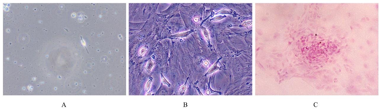
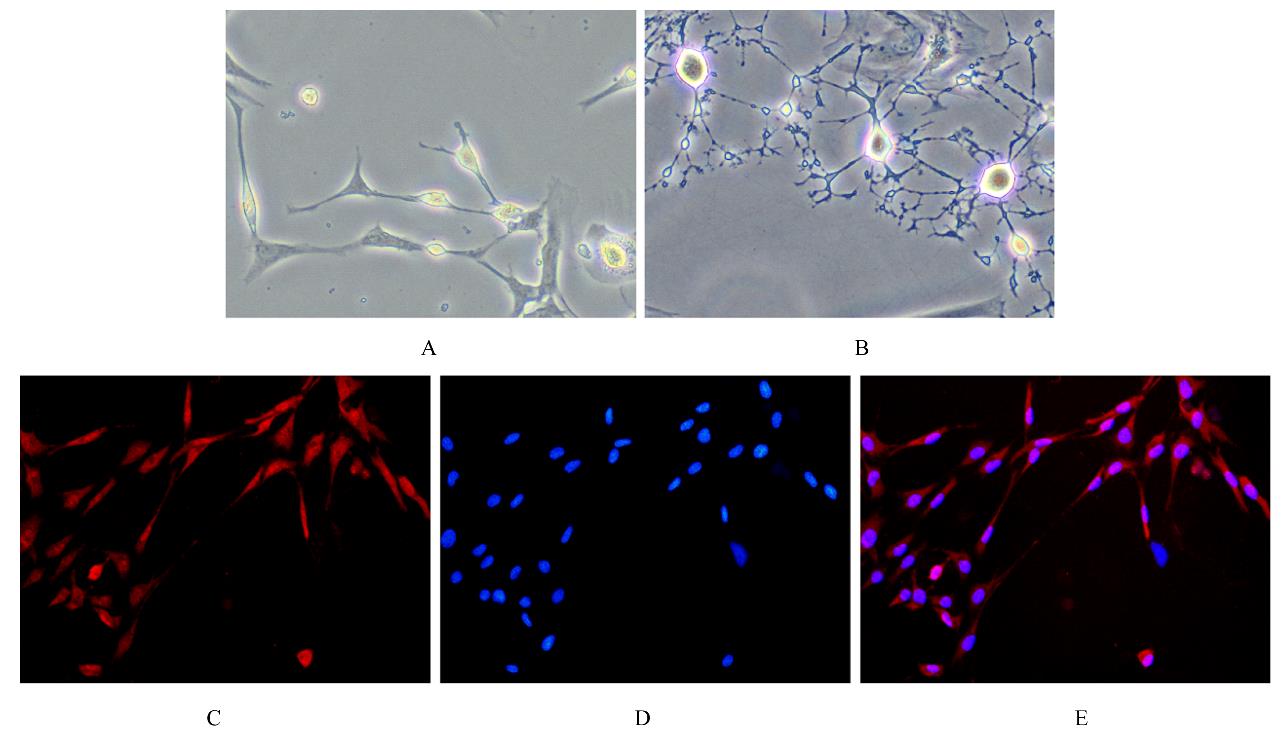
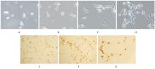
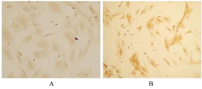
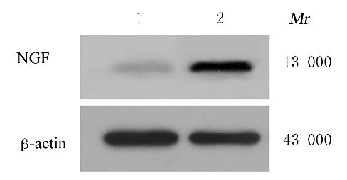
  • Received:2021-10-24 Online:2022-05-28 Published:2022-06-21
  • Contact: Xiumei FU E-mail:fuxiumei2012@163.com

摘要: 目的

探讨施万细胞样细胞(SCLCs)对大鼠背根神经节(DRG)细胞突起生长和神经生长因子(NGF)表达的影响,阐明SCLCs促进DRG细胞生长的作用及其机制。

方法

分离培养脂肪源性干细胞(ADSCs),茜素红染色检测细胞中钙化结节从而观察其分化能力,将其诱导分化为SCLCs,免疫荧光法检测SCLCs 中S100蛋白的表达。分离培养DRG细胞,免疫组织化学染色检测DRG细胞中神经元核抗原(NeuN)蛋白的表达。建立SCLCs与DRG细胞共培养体系,将细胞分为单培养组和共培养组。HE染色观察2组DRG细胞的形态结构,计数2组DRG细胞数和DRG细胞突起数,免疫组织化学染色检测2组DRG细胞中NGF蛋白的表达,Western blotting法检测2组DRG细胞中NGF蛋白表达水平。

结果

原代ADSCs培养7 d,倒置显微镜下可见数量较多、体积较大、呈长梭形的细胞类似旋涡状或铺路石样排列。茜素红染色,镜下可见钙化结节,即所培养细胞具有分化能力。免疫荧光染色,大多数细胞均呈S100染色阳性,即ADSCs可成功诱导分化为SCLCs。培养72 h后,DRG细胞突起细而长、呈花环样排列,7~8 d后,细胞数量进一步增加,胞体较大,呈圆形,突起细长,相互连接似栅栏样。免疫组织化学染色,几乎所有细胞核均被标记为棕褐色,呈NeuN染色阳性。与单培养组比较,共培养组DRG细胞数及细胞突起数均明显增加(P<0.05),共培养组DRG细胞中NGF蛋白表达水平明显升高(P<0.05)。

结论

共培养条件下SCLCs可增加DRG细胞数和细胞突起数,提高DRG细胞中NGF蛋白的表达,发挥促进DRG细胞生长的作用。

关键词: 施万细胞样细胞, 背根神经节细胞, 共培养, 神经生长因子, 神经元核抗原

Abstract: Objective

To investigate the effect of Schwann cell-like cells (SCLCs) on the growth of neurites and the expression of nerve growth factor (NGF) of dorsal root ganglion (DRG) cells in the rats, and to clarify the role of SCLCs on promoting the growth in the DRG cells and its mechanism.

Methods

The adipose-derived stem cells (ADSCs) were isolated and cultured,and the calcified nodules in the cells were detected by Alizarin red staining to observe their differentiation ability, and the ADSCs were induced to differentiate into the SCLCs.The DRG cells were isolated and cultured, and the expression of neuronal nuclear antigen (NeuN) protein in the DRG cells was detected by immunohistochemical staining. The co-culture system of SCLCs and DRG cells was established. The cells were divided into single culture group and co-culture group. HE staining was used to observe the morphology of the DRG cells in two groups, the number of DRG cells and the number of DRG cell processes in two groups were counted, immunohistochemical staining was used to detect the expression of NGF protein in the DRG cells in two groups, and Western blotting method was used to detect the expression level of NGF protein in the DRG cells in two groups.

Results

The primary ADSCs were cultured for 7 d, under the inverted microscope, a large number of cells with large volume and long spindle shape could be seen, which were similar to the arrangement of vortex or paving stone.The Alizarin red staining results showed that the calcified nodules can be seen under the microscope, and the cultured cells had the ability of differentiation. The immunofluorescence staining results showed that most cells were S100 staining positive, and the ADSCs could be successfully induced into the SCLCs. After cultured for 72 h, the processes of DRG cells were thin and long, arranged in a wreath-like arrangement. After 7-8 d, the number of cells was increased, the body of cells was large and round, and the processes were slender and connected like a fence. The immunohistochemical staining results showed that almost all the nuclei were marked as brown and NeuN staining positive. Compared with single culture group, the number of DRG cells and the number of DRG cell processes in co-culture group were increased significantly (P<0.05), and the expression level of NGF protein in the DRG cells in co-culture group was increased significantly (P<0.05).

Conclusion

SCLCs could promote the growth of DRG cells through increasing the number of DRG cells and neurites and upregulating the expression of NGF protein.

Key words: Schwann cell-like cells, Dorsal root ganglion cells, Co-cultivation, Nerve growth factor, Neuronal nuclear antigen

中图分类号: 

  • R322.8