吉林大学学报(医学版) ›› 2022, Vol. 48 ›› Issue (3): 796-800.doi: 10.13481/j.1671-587X.20220330

• 临床医学 • 上一篇    

以肺部影像学改变为首发表现的血管免疫母细胞性T细胞淋巴瘤1例报告及文献复习

李倩,苑静怡,周佳奇,赵敏,王珂()   

  1. 吉林大学第二医院呼吸与危重症医学科,吉林 长春 130041
  • 收稿日期:2021-08-27 出版日期:2022-05-28 发布日期:2022-06-21
  • 通讯作者: 王珂 E-mail:kewangm1@hotmail.com
  • 作者简介:李 倩(1994-),女,安徽省亳州市人,在读硕士研究生,主要从事肺部感染性疾病和肺癌诊治方面的研究。
  • 基金资助:
    吉林省科技厅科技发展计划项目(20200708083YY)

Pulmonary imaging changes as first manifestation of angioimmunoblastic T-cell lymphoma: A case report and literature review

Qian LI,Jingyi YUAN,Jiaqi ZHOU,Min ZHAO,Ke WANG()   

  1. Department of Respiratory and Critical Care Medicine,Second Hospital,Jilin University,Changchun 130041,China
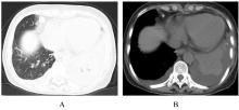
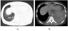
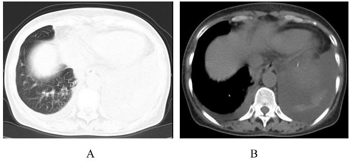
  • Received:2021-08-27 Online:2022-05-28 Published:2022-06-21
  • Contact: Ke WANG E-mail:kewangm1@hotmail.com

摘要: 目的

探讨血管免疫母细胞性T细胞淋巴瘤(AITL)的临床特点、诊断过程和治疗方法,提高临床医生对该病的认识。

方法

收集1例AITL患者的临床资料、影像学表现、支气管镜和病理检查结果,分析上述资料,并进行相关文献复习。

结果

患者,女性,66岁,因“发热伴咳嗽和咳痰15 d”入院。患者四肢散在红色皮疹,胸廓无畸形,气管居中,叩诊左下肺浊音,左下肺呼吸音减弱,无其他明显阳性体征。胸部CT检查结果显示双肺下叶见多发条片状高密度影,以左下肺为著,纵隔和双侧腋窝淋巴结肿大。初步考虑双肺炎症可能性大,待除外占位性病变。经支气管镜肺活检术(TBLB)、纵隔淋巴结支气管穿刺针吸活检术(TBNA)和胸水脱落细胞检测,结果均排除肺恶性病变。先后行抗感染治疗和实验性抗结核治疗,患者仍反复发热,多次复查胸部CT结果显示左下肺高密度影范围增大和实变。全面的实验室检查和影像学检查均未见异常,患者具体发热原因不明确。患者查体见躯干和四肢多发淡红色皮疹,双侧锁骨上淋巴结肿大,经右侧锁骨上淋巴结穿刺活组织检查,最终病理回报AITL。

结论

有肺部影像学改变的不明原因发热,且伴有皮疹及淋巴结肿大的患者,如系统抗感染治疗效果不好,应考虑AITL。

关键词: 外周血T细胞淋巴瘤, 血管免疫母细胞性T细胞淋巴瘤, 影像学表现, 病例报告

Abstract: Objective

To explore the clinical characteristics, diagnostic process and treatment of angioimmunoblastic T-cell lymphoma (AITL), and to improve clinicians’ understandings of the disease.

Methods

The clinical data, imaging findings, bronchoscope and pathological findings of one patient with AITL were collected, the above data were analyzed, and the relevant literatures were reviewed.

Results

A 66-year-old female patient was admitted with fever accompanying with cough and expectoration for 15 d. The physical examination results showed that there was a red rash scattered on all four limbs, no abnormality of the thorax, and the trachea was in the center. Dullness of the left lower lung was found by percussion, and the breath sounds of the left lower lung were weakened, without other obvious positive signs.The chest CT results showed the multiple patchy high-density shadows in the lower lobe of both lungs, especially in the left lower lung, and the mediastinum and bilateral axillary lymph nodes were enlarged. The possibility of double pneumonia was considered initially, and the space-occupying lesions were excluded. The results of bronchoscopic lung biopsy (TBLB),mediastinal lymph node transbronchial needle aspiration biopsy (TBNA), and exfoliated pleural cells all excluded pulmonary malignancy. After anti-infection treatment and experimental anti-tuberculosis treatment, the patient still had repeated fever. The chest CT results showed that the high-density shadow range of the left lower lung was increased and consolidated. The comprehensive laboratory examination and imaging examination results showed no abnormalities, and the specific cause of the patient’s fever was unclear. The physical examination of the patient showed the multiple light red rashes on the trunk and limbs and the bilateral supraclavicular lymph node enlargement. Biopsy of right supraclavicular lymph node was performed, and AITL was finally reported pathologically.

Conclusion

AITL should be considered in the patients with unexplained fever with pulmonary imaging changes, rash and lymph node enlargement, if systemic anti-infective therapy does not respond well.

Key words: Peripheral T-cell lymphoma, Angioimmunoblastic T-cell lymphoma, Imaging findings, Case report

中图分类号: 

  • R733.4