吉林大学学报(医学版) ›› 2023, Vol. 49 ›› Issue (6): 1561-1568.doi: 10.13481/j.1671-587X.20230620

• 临床研究 • 上一篇    下一篇

CBCT测量不同骨面型患者下颌骨结构特征和磨牙后间隙的影响因素

孙芸芸1,姚玉光2,张晗1,黎涵懿1,朱宪春1()   

  1. 1.吉林大学口腔医院正畸科,吉林 长春 130021
    2.河北省唐山市曹妃甸区医院口腔科,河北 唐山 063299
  • 收稿日期:2022-12-08 出版日期:2023-11-28 发布日期:2023-12-22
  • 通讯作者: 朱宪春 E-mail:zhuxc@jlu.edu.cn
  • 作者简介:孙芸芸(1991-),女,辽宁省朝阳市人,在读硕士研究生,主要从事口腔正畸学方面的研究。
  • 基金资助:
    吉林省科技厅科技发展计划项目(20210203064SF)

Structural characteristics of mandible in patients with different osteofacial types measured by CBCT and influencing factors of mandibular retromolar space

Yunyun SUN1,Yuguang YAO2,Han ZHANG1,Hanyi LI1,Xianchun ZHU1()   

  1. 1.Department of Orthodontics,Stomatology Hospital,Jilin University,Changchun 130021,China
    2.Department of Stomatology,Hospital of Caofeidian District,Tangshan City,Hebei Province,Tangshan 063299,China
  • Received:2022-12-08 Online:2023-11-28 Published:2023-12-22
  • Contact: Xianchun ZHU E-mail:zhuxc@jlu.edu.cn

摘要:

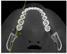
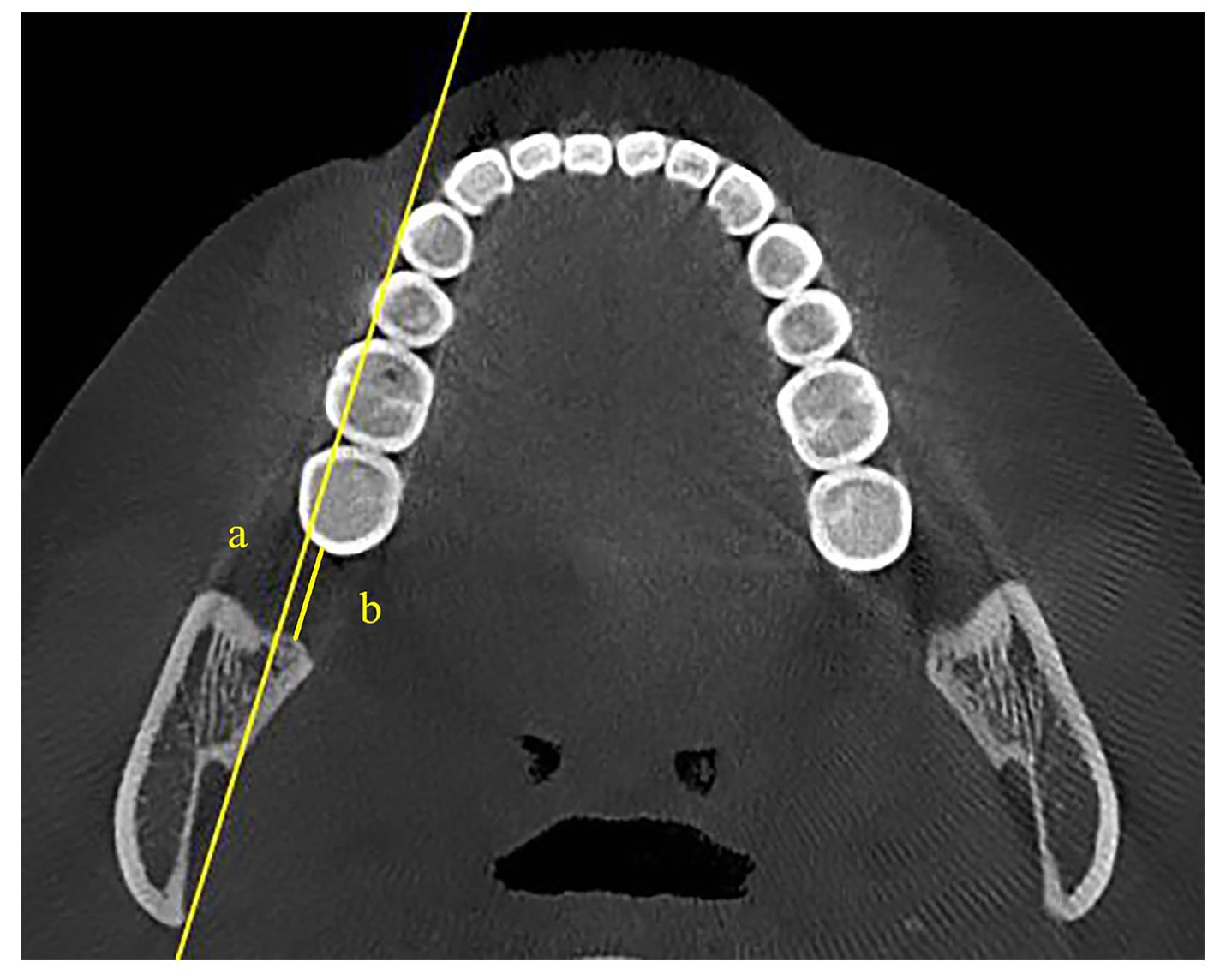
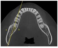
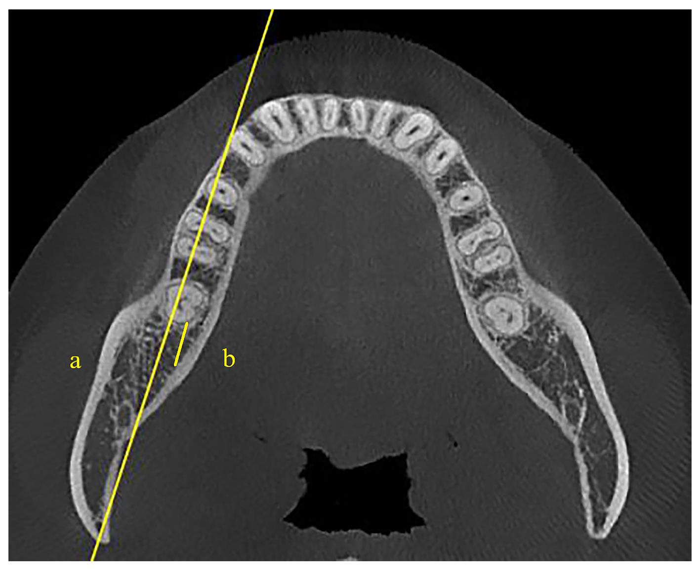
目的 探讨不同骨面型患者磨牙后间隙的影响因素,分析下颌骨结构特征和磨牙后间隙与颅面部结构的相关性。 方法 选取除第三磨牙外下颌牙列完整且拥挤量<5 mm的成年正畸患者200例,分为骨性Ⅰ类低角组、骨性Ⅰ类均角组、骨性Ⅰ类高角组、骨性Ⅱ类均角组和骨性Ⅲ类均角组。测量各组患者蝶窦A点角(SNA)、蝶窦B点角(SNB)、上下齿槽座角(ANB)、下颌平面角(SN-MP)、面高指数(FHI)、下颌升支长度(Go-Co)和下颌体长度(Go-Gn)。采用锥形束计算机断层扫描技术(CBCT)测量在水平面上与牙尖线平行的下颌平面及其根方2 mm平面下颌第二磨牙至下颌升支前缘的最短距离C0和C2,釉牙骨质界根方2、4、6、8和10 mm平面的牙根至下颌骨舌侧内层骨皮质的最短距离R2、R4、R6、R8和R10,并采用Spearman相关分析法分析其相关性。 结果 与骨性Ⅰ类低角组比较,骨性Ⅰ类均角组和骨性Ⅰ类高角组患者SN-MP均明显增大(P<0.01),FHI均明显减小(P<0.01)。与骨性Ⅰ类均角组比较,骨性Ⅰ类高角组患者SN-MP明显增大(P<0.01),FHI明显减小(P<0.01)。与骨性Ⅰ类均角组比较,骨性Ⅱ类均角组患者ANB明显增大(P<0.01),骨性Ⅲ类均角组患者ANB明显减小(P<0.01)。与骨性Ⅱ类均角组比较,骨性Ⅲ类均角组患者ANB明显减小(P<0.01)。在C2、R2、R4、R6、R8、R10平面下和Go-Co及Go-Gn中,与骨性Ⅰ类低角组比较,骨性Ⅰ类均角组和骨性Ⅰ类高角组患者下颌磨牙后间隙明显减小(P<0.01);与骨性Ⅰ类均角组比较,骨性Ⅰ类高角组患者下颌磨牙后间隙明显减小(P<0.01)。在C0平面下,与骨性Ⅰ类低角组和骨性Ⅰ类均角组比较,骨性Ⅰ类高角组患者下颌磨牙后间隙明显减小(P<0.01)。在C0、R2、R4、R6、R8和R10平面下,与骨性Ⅰ类均角组比较,骨性Ⅱ类均角组患者下颌磨牙后间隙明显减小(P<0.01),骨性Ⅲ类均角组患者下颌磨牙后间隙明显增大(P<0.01);与骨性Ⅱ类均角组比较,骨性Ⅲ类均角组患者下颌磨牙后间隙明显增大(P<0.01)。在C2平面下和Go-Co及Go-Gn中,与骨性Ⅰ类均角组和骨性Ⅱ类均角组比较,骨性Ⅲ类均角组患者下颌磨牙后间隙明显增大(P<0.01)。骨性Ⅰ类低角组、骨性Ⅰ类均角组和骨性Ⅰ类高角组患者在各平面下颌磨牙后间隙及Go-Co和Go-Gn与SNB和FHI均呈正相关关系(P<0.01),与SN-MP呈负相关关系(P<0.01);各平面下颌磨牙后间隙与Go-Co呈正相关关系(P<0.01);除C2平面,其余各平面下颌磨牙后间隙与Go-Gn呈正相关关系(P<0.01);Go-Co与Go-Gn呈正相关关系(P<0.01);Go-Gn与ANB呈负相关关系(P<0.01)。骨性Ⅰ类均角组、骨性Ⅱ类均角组和骨性Ⅲ类均角组患者在各平面下颌磨牙后间隙及Go-Co和Go-Gn与SNB均呈正相关关系(P<0.01);各平面下颌磨牙后间隙与Go-Co和Go-Gn均呈正相关关系(P<0.01);Go-Co与Go-Gn呈正相关关系(P<0.01);Go-Co与FHI呈正相关关系(P<0.01)。 结论 骨面型是影响下颌磨牙后间隙的重要因素,在正畸制订磨牙远移方案时应参考患者的骨面型,采用CBCT以评估下颌磨牙远移的可用空间。

关键词: 磨牙后间隙, 磨牙远移, 垂直骨面型, 矢状骨面型, 锥形束计算机断层扫描技术

Abstract:

Objective To discuss the influence factors of retromolar space in the patients with different osteofacial types, and to analyze the correlation between the mandibular bone structure and retromolar space and craniofacial structure. Methods A total of 200 adult orthodontic patients with fully erupted dentition except the third molars and crowding less than 5 mm were chosen and divided into skeletal type Ⅰ low angle group, skeletal type Ⅰ average angle group, skeletal type Ⅰ high angle group, skeletal type Ⅱ average angle group, and skeletal type Ⅲ average angle group. The sella nasion A point angle(SNA), sella nasion B point angle(SNB), AB plane angle(ANB), skull base anterior-mandibular plane angle(SN-MP),facial height index(FHI),mandibular ramus length(Go-Co),and mandibular body length(Go-Gn) of all the patients were detected. Cone-beam computed tomography (CBCT) technology was used to detect the shortest distances C0 and C2 from the mandibular second molar to the anterior border of the ramus at the planes parallel to the cuspid line with a depth of 0 and 2 mm form the occlusal plane, and the shortest distances R2, R4, R6, R8, and R10 from the tooth root to the lingual cortical bone of the mandible at the enamel-bone junction in the root direction 2, 4, 6, 8, and 10 mm planes.The correlation was analyzed by using Spearman correlation analysis. Results Compared with skeletal type Ⅰ low angle group, the SN-MP of the patients in skeletal type Ⅰ average angle group and skeletal type Ⅰ high angle group was increased significantly (P<0.01), and the FHI was decreased significantly (P<0.01). Compared with skeletal type Ⅰ average angle group, the SN-MP of the patients in skeletal type I high angle group was increased significantly (P<0.01),FHI was decreased significantly (P<0.01). Compared with skeletal type Ⅰ average angle group, the ANB of patients in skeletal type Ⅱaverage angle group was significantly increased (P<0.01), while the ANB of the patients in skeletal type Ⅲ average angle group were significantly decreased (P<0.01). Compared with skeletal type Ⅱ average angle group, the ANB of the patients in skeletal type Ⅲ average angle group was significantly decreased (P<0.01). Under the C2, R2, R4, R6, R8, R10 planes and in the Go-Co and Go-Gn, compared with skeletal type Ⅰ low angle group, the retromolar spaces of the patients in skeletal type Ⅰ average angle group and the skeletal type Ⅰ high angle group were significantly decreased (P<0.01); compared with skeletal type Ⅰ average angle group, the retromolar space of the patients in skeletal type Ⅰ high angle group was significantly decreased (P<0.01). Under the C0 plane, compared with skeletal type Ⅰ low angle group and the skeletal type Ⅰ average angle group, the retromolar space of the patients in skeletal type Ⅰ high angle group was decreased (P<0.01). Under the C0, R2, R4, R6, R8, and R10 planes, compared with skeletal type Ⅰ average angle group, the retromolar space of the patients in skeletal type Ⅱ average angle group was significantly decreased (P<0.01), the retromolar space of the patients in skeletal type Ⅲ average angle group was increased (P<0.01); compared with skeletal type Ⅱ average angle group, the retromolar space of the patients in skeletal type Ⅲ average angle group was increased (P<0.01).Under the C2 plane and in the Go-Co and Go-Gn, compared with skeletal type Ⅰ average angle group and skeletal type Ⅱ average angle group, the retromolar space of the patients in skeletal type Ⅲ average angle group was increased (P<0.01). The retromolar space,Go-Co and Go-Gn of the patients in skeletal type Ⅰ low angle group, skeletal type Ⅰ average angle group, and skeletal type Ⅰ high angle group under all planes had positive correlations with the SNB and FHI (P<0.01), and a negative correlation with SN-MP (P<0.01);the retromolar space at all levels had a positive correlation with Go-Co (P<0.01); except for the C2 plane, the remaining retromolar space under all planes had positive correlations with Go-Gn (P<0.01);there was a positive correlation between Go-Co and Go-Gn (P<0.01);there was a negative correlation between Go-Gn and ANB((P<0.01).The retromolar space and Go-Co and Go-Gn of the patients in skeletal type Ⅰ average angle group, skeletal type Ⅱ average angle group, and skeletal type Ⅲ average angle group under all planes had positive correlations with SNB (P<0.01),and the retromolar space had positive correlations with Go-Co and Go-Gn (P<0.01); there was a positive correlation between Go-Co and Go-Gn (P<0.01);there was a positive correlation between Go-Co and FHI(P<0.01). Conclusion The skeletal type is an important influence factor of the retromolar space of mandible, when the molar distalization plan is formulated, the skeletal type of the patient should be considered; CBCT should be used to evaluate the available space for mandibular molar distalization.

Key words: Retromolar space, Molar distalization, Vertical osteofacial type, Sagittal osteofacial type, Cone-beam computed tomography

中图分类号: 

  • R783.5