吉林大学学报(医学版) ›› 2024, Vol. 50 ›› Issue (4): 939-946.doi: 10.13481/j.1671-587X.20240407

• 基础研究 • 上一篇    下一篇

PTMC/PVP温控收缩性纳米纤维膜与小鼠成纤维细胞的生物相容性及其对大鼠皮肤全层缺损的修复作用

刘丽萍1,李驰宇2,杨韬1,王少如1,刘昀1,刘国民2,程志强3,罗云纲4(),刘志辉1()   

  1. 1.吉林大学口腔医院修复科,吉林 长春 130021
    2.吉林大学第二医院骨科,吉林 长春 130041
    3.吉林农业大学资源与环境学院,吉林 长春 130118
    4.吉林大学第一医院 净月分院口腔医学中心,吉林 长春 130118
  • 收稿日期:2023-07-22 出版日期:2024-07-28 发布日期:2024-08-01
  • 通讯作者: 罗云纲,刘志辉 E-mail:luoygjlu@sina.com;liu_zh@jlu.edu.cn
  • 作者简介:刘丽萍(1997-),女,山东省潍坊市人,在读硕士研究生,主要从事纳米纤维临床应用方面的研究。
  • 基金资助:
    吉林省科技厅科技发展计划项目(20220204124YY)

Biocompatibility of PTMC/PVP temperature-controlled shrinkage nanofiber membrane with mouse fibroblasts and its repairment effect on full-thickness skin defects in rats

Liping LIU1,Chiyu LI2,Tao YANG1,Shaoru WANG1,Yun LIU1,Guomin LIU2,Zhiqiang CHENG3,Yungang LUO4(),Zhihui LIU1()   

  1. 1.Department of Prosthodontics, Stomatology Hospital, Jilin University, Changchun 130021, China
    2.Department of Orthopedics, Second Hospital, Jilin University, Changchun 130041, China
    3.School of Resources and Environment, Jilin Agricultural University, Changchun 130118, China
    4.Stomatology Center, Jingyue Campus, First Hospital, Jilin University, Changchun 130118, China
  • Received:2023-07-22 Online:2024-07-28 Published:2024-08-01
  • Contact: Yungang LUO,Zhihui LIU E-mail:luoygjlu@sina.com;liu_zh@jlu.edu.cn

摘要:

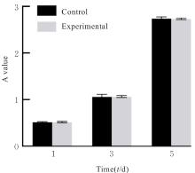
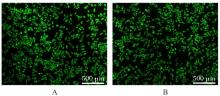
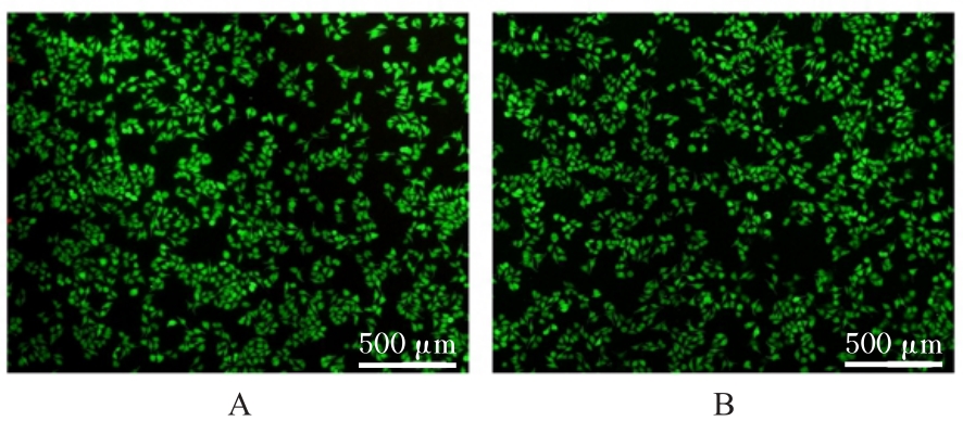
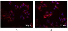
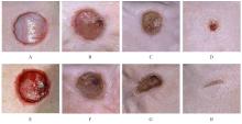
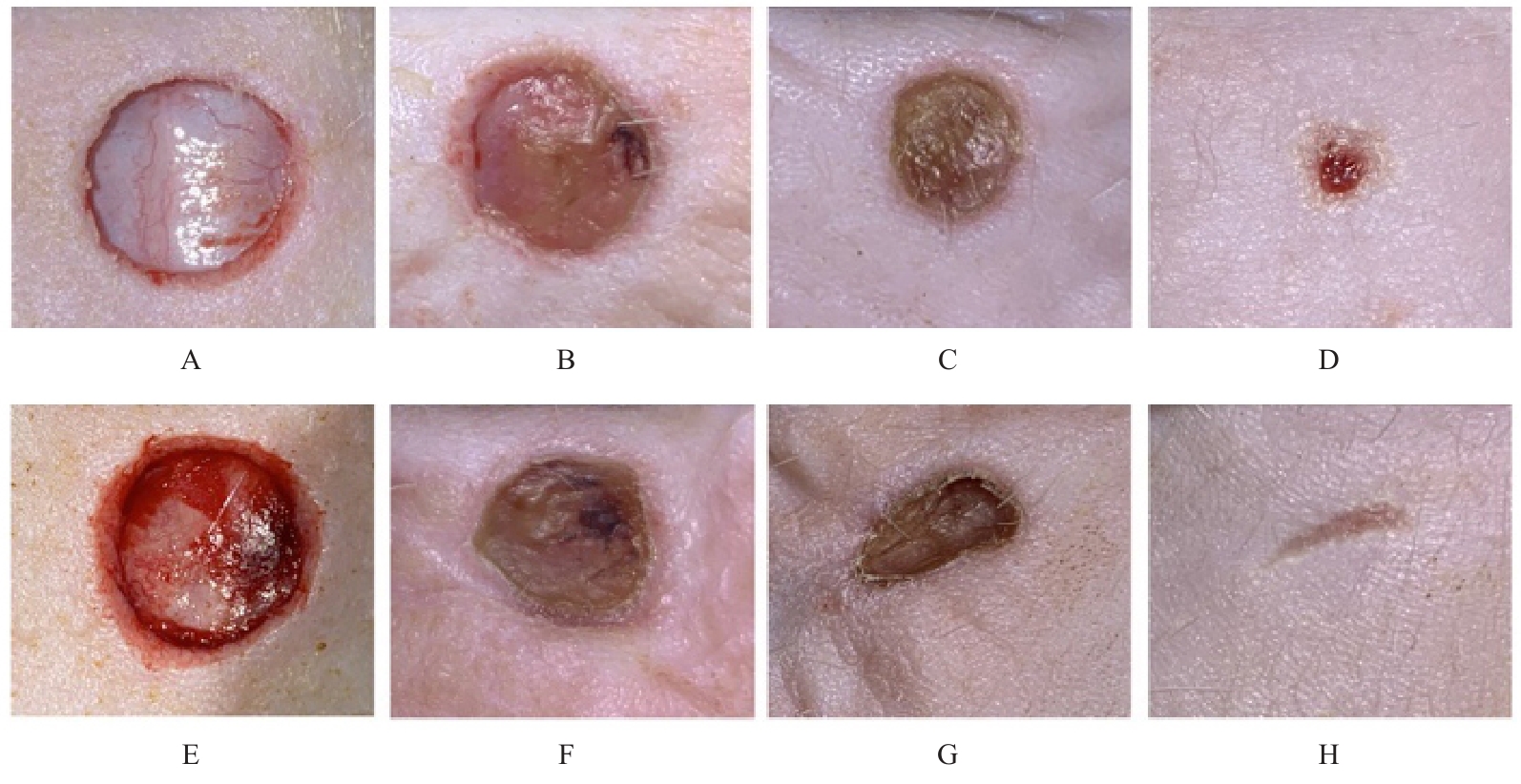
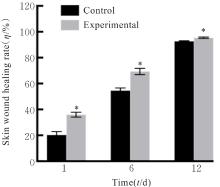
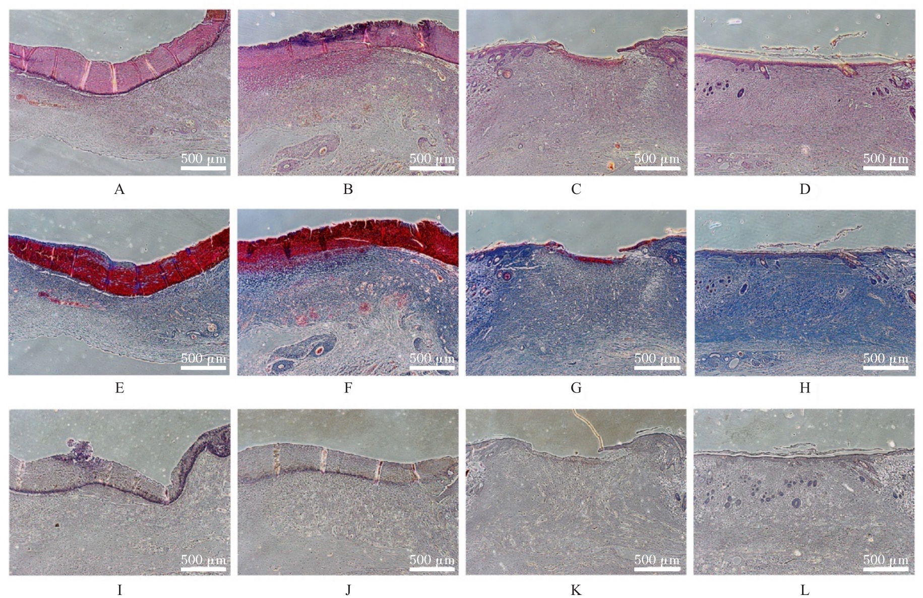
目的 探讨温控收缩性聚三亚甲基碳酸酯(PTMC)/聚乙烯吡咯烷酮(PVP)纳米纤维膜对小鼠成纤维细胞生物学行为的影响及对大鼠全层皮肤缺损的修复作用,并阐明其可能的作用机制。 方法 体外实验选用小鼠成纤维L929细胞,分为对照组和实验组(采用PTMC/PVP纳米纤维膜处理),CCK-8法检测2组细胞增殖活性,活/死细胞染色实验观察2组细胞中活/死细胞数量,细胞骨架染色实验观察2组细胞形态表现。体内实验选用12只6周龄雄性SD大鼠,随机分为对照组和实验组,每组6只,建立全层皮肤缺损模型,实验组大鼠采用PTMC/PVP纳米纤维膜治疗,术后拍照观察,第0、3、6和12天时计算2组大鼠创面愈合率,术后第6和12天切取2组大鼠皮肤创面和周围组织,采用HE染色观察皮肤创面和周围组织病理形态表现,Masson三色染色观察2组大鼠皮肤创面组织中胶原蛋白沉积情况,CD31免疫组织化学染色检测2组大鼠皮肤创面组织中血管形成数量。 结果 CCK-8实验,实验组细胞增殖活性在第1、3和5天均呈升高趋势,与对照组比较差异无统计学意义(P>0.05)。活/死细胞染色实验,与对照组比较,实验组细胞密度和数量无明显变化且以活细胞为主。细胞骨架染色实验,实验组和对照组细胞均呈梭形且细胞伸展。体内实验,第3、6和12 天时,与对照组比较,实验组大鼠创面愈合率升高(P<0.01),12 d时实验组大鼠创面愈合率为95.45%,创面基本愈合。HE染色,与对照组比较,第12天实验组大鼠创面皮肤结构更接近正常皮肤,有丰富的肉芽组织、规则的表皮结构和新血管生成。Masson三色染色,与对照组比较,实验组大鼠创面组织中胶原蛋白沉积量更多。免疫组织化学染色,与对照组比较,实验组大鼠创面组织中CD31表达增多,表明血管形成数量增加。 结论 PTMC/PVP温控收缩性纳米纤维膜具有良好的生物相容性,且能促进大鼠皮肤全层缺损的修复,其作用机制可能与增强基底细胞增殖活性有关。

关键词: 伤口敷料, 机械力学, 温控收缩性, 聚三亚甲基碳酸酯/聚乙烯吡咯烷酮, 纳米纤维, 静电纺丝

Abstract:

Objective To discuss the effect of temperature-controlled shrinkage polytrimethylene carbonate (PTMC)/polyvinylpyrrolidone (PVP) nanofiber membrane on the biological behavior of mouse fibroblasts and the repairment effect on full-thickness skin defects in the rats, and to clarify the potential mechanism. Methods The murine L929 fibroblast cells were used in the in vitro experiments and were divided into control group and experimental group (treated with PTMC/PVP nanofiber membranes). The proliferation activities of the cells in two groups were detected by CCK-8 assay; the numbers of live/dead cells in two groups were observed by live/dead cell staining; the morphology of the cells was observed by cytoskeletal staining. A total of 12 six-week-old male SD rats were selected in the in vivo experiment, and were randomly divided into control group and experimental group, and there were six rats in each group. The full-thickness skin defect model was established, and the rats in experimental group were treated with PTMC/PVP nanofiber membranes. The photographs were taken after operation, and the wound healing rates of the cells in two groups were calculated on the 0, 3rd, 6th, and 12th days. On the 6th and 12th days after operation, the skin samples around the wound of the rats in two groups were taken, and the histopathology of the would skin and adjacent tissue was detected by HE staining; the collagen deposition in wound skin tissue of the rats in two groups was observed by Masson trichrome staining; the numbers of angiogenesis in wound skin tissue of the rats were detected by CD31 immunohistochemical staining. Results The CCK-8 assay results showed that the proliferation activity of the cells in experimental group showed an increasing trend on the 1st, 3st, and 5st days, and there was no significant difference in the proliferation activities of the cells bewteen experimental group and control group (P>0.05). The live/dead cell staining experiment results showed that compared with control group, the cell density and number of the cells in experimental group had no significant changes, and were predominantly live cells. The cytoskeletal staining results showed that the cells in experimental and control groups appeared spindle-shaped and well-spread. In the in vivo experiments, on the 3rd, 6th, and 12th days, compared with control group, the wound healing rates of the cells in experimental group were increased (P<0.01), and the wound healing rate of the cells was 95.45% on the 12th day, indicating nearly complete healing of the wound. The HE staining showed that on the 12th day, the wound skin structure of the cells in experimental group was more similar to the normal skin, and there was abundant granulation tissue, regular epidermal structure, and new blood vessel formation. The Masson trichrome staining results showed that compared with control group, the collagen deposition in wound tissue of the rats in experimental group was increased. The immunohistochemical staining results showed that the expression of CD31 in wound tissue of the rats in experimental group was increased, indicating the increasing of the number of angiogenesis. Conclusion The PTMC/PVP thermoresponsive nanofiber membranes exhibit good biocompatibility and can promote the repairment of full-thickness skin defects in the rats; its mechanism may be related to the enhancement of proliferation activity of the basal cells.

Key words: Wound dressing, Mechanical mechanics, Temperature-controlled shrinkage, Polytrimethylene carbonate /polyvinylpyrrolidone, Nanofiber, Electrospinning

中图分类号: 

  • R318.08