吉林大学学报(医学版) ›› 2024, Vol. 50 ›› Issue (4): 1026-1034.doi: 10.13481/j.1671-587X.20240417

• 基础研究 • 上一篇    下一篇

SENP-1/HIF-1α通路对慢性间歇性低氧诱导大鼠血管内皮损伤的影响

贾远航,江义霞,何振华,陈林,周芳()   

  1. 南华大学衡阳医学院附属第二医院呼吸与危重症医学科,湖南 衡阳 421001
  • 收稿日期:2023-07-19 出版日期:2024-07-28 发布日期:2024-08-01
  • 通讯作者: 周芳 E-mail:932659965@qq.com
  • 作者简介:贾远航(1983-),男,湖南省邵阳市人,主治医师,医学硕士,主要从事阻塞性睡眠呼吸暂停低通气综合征发病机制方面的研究。
  • 基金资助:
    湖南省卫健委科研基金项目(202203024555)

Effect of SENP-1/HIF-1α pathway on vascular endothelial injury in rats with chronic intermittent hypoxia

Yuanhang JIA,Yixia JIANG,Zhenhua HE,Lin CHEN,Fang ZHOU()   

  1. Department of Respiratory and Critical Care Medicine,Second Affiliated Hospital,Hengyang Medical School,University of South China,Hengyang 421001,China
  • Received:2023-07-19 Online:2024-07-28 Published:2024-08-01
  • Contact: Fang ZHOU E-mail:932659965@qq.com

摘要:

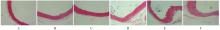
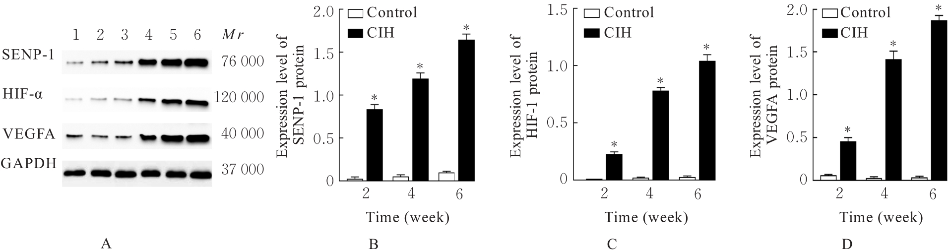
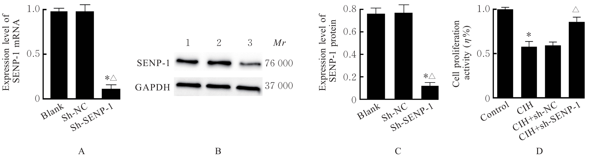
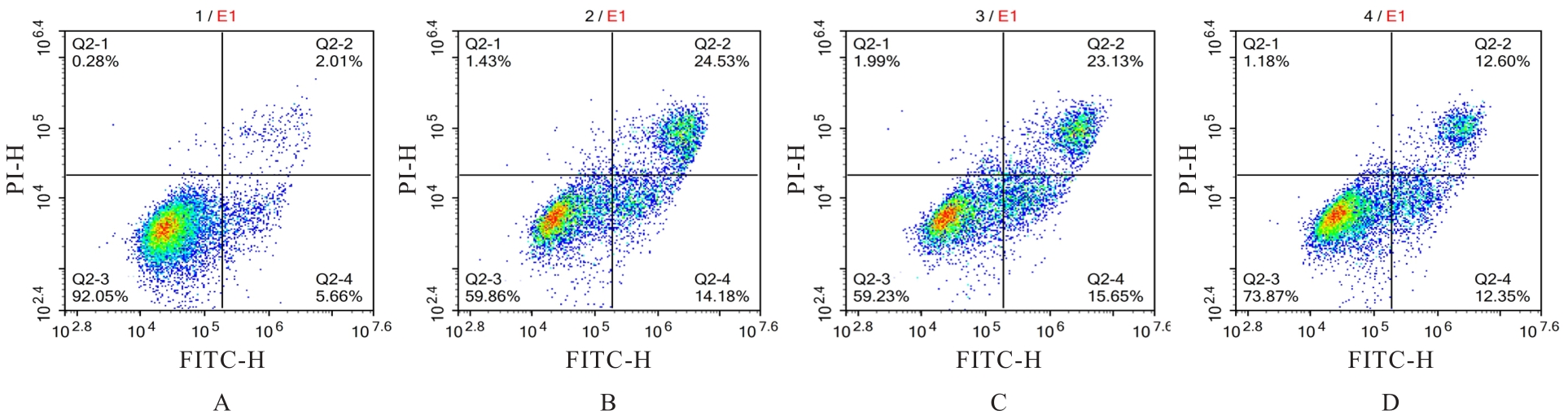
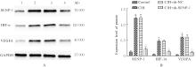
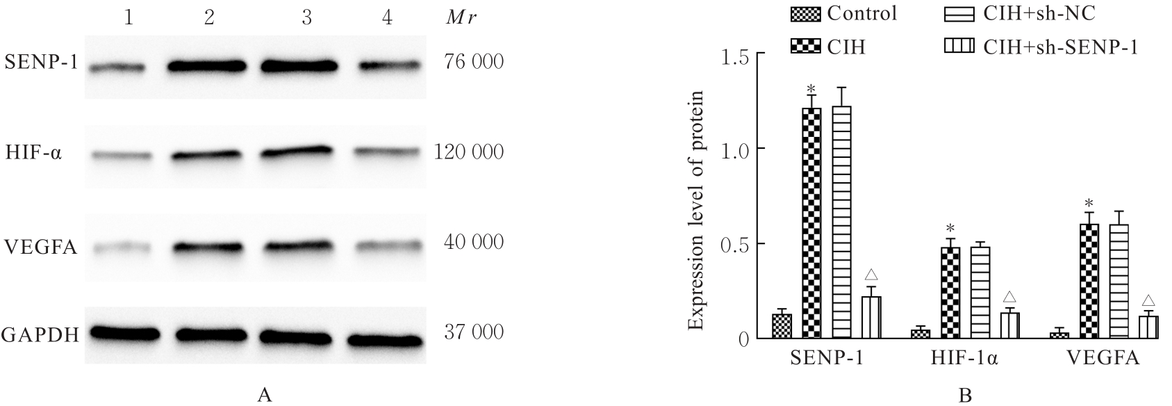
目的 探讨小泛素样修饰特异性蛋白酶1(SENP-1)/低氧诱导因子1α(HIF-1α)通路对慢性间歇性低氧(CIH)诱导大鼠血管内皮损伤的影响,阐明其相关作用机制。 方法 SD大鼠随机分为对照组和CIH组,再将每组分为2、4和6周3个时间点亚组,每亚组8只。CIH组大鼠暴露于CIH舱中进行CIH诱导,制备阻塞性睡眠呼吸暂停低通气综合征(OSAHS)模型,对照组大鼠暴露于常氧环境中。于各时间点收集各组大鼠血清和胸主动脉组织。HE染色观察各组大鼠胸主动脉血管损伤情况,采用酶联免疫吸附试验(ELISA)法检测各组大鼠血清中一氧化氮(NO)、内皮素1(ET-1)、血管性血友病因子(vWF)和血栓调节蛋白(TM)水平,Western blotting法检测各组大鼠胸主动脉组织中SENP-1、HIF-1α和血管内皮生长因子A(VEGFA)蛋白表达水平。体外培养大鼠主动脉内皮细胞(rAECs),经SENP-1 shRNA腺病毒(sh-SENP-1)感染构建SENP-1基因低表达的rAECs 细胞株,采用 CIH 诱导建立血管内皮细胞损伤模型,分为 CIH 组、CIH+sh-NC 组和 CIH+sh-SENP-1组,另设对照组。CCK-8检测各组细胞增殖活性,ELISA法检测各组细胞培养上清中乳酸脱氢酶(LDH)活性及细胞中NO、ET-1、丙二醛(MDA)水平和超氧化物歧化酶(SOD)活性,流式细胞术检测各组细胞凋亡率,Western blotting法检测各组细胞中SENP-1、HIF-1α和VEGFA蛋白表达水平。 结果 随着CIH诱导时间的延长,与对照组比较,CIH组大鼠胸主动脉内膜逐渐粗糙并明显增厚,血清中NO水平逐渐减低(P<0.05),血清中ET-1、vWF和TM水平及胸主动脉组织中SENP-1、HIF-1α和VEGFA蛋白表达水平逐渐升高(P<0.05)。与对照组比较,CIH组细胞增殖活性降低(P<0.05),细胞培养上清中LDH活性及细胞中ET-1、MDA水平和细胞凋亡率升高(P<0.05),细胞中NO水平和SOD活性降低(P<0.05),SENP-1、HIF-1α和VEGFA蛋白表达水平升高(P<0.05);与CIH组比较,CIH+sh-SENP-1组细胞增殖活性升高(P<0.05),细胞培养上清中LDH活性及细胞中ET-1、MDA水平和细胞凋亡率降低(P<0.05),细胞中NO水平和SOD活性升高(P<0.05),SENP-1、HIF-1α和VEGFA蛋白表达水平降低(P<0.05)。 结论 SENP-1/HIF-α通路在CIH诱导的大鼠胸主动脉损伤组织中高度活化,沉默SENP-1表达可减轻CIH诱导的血管内皮细胞损伤,其作用机制可能与下调SENP-1/HIF-α通路活化水平有关。

关键词: 慢性间歇性低氧, 胸主动脉, 小泛素样修饰特异性蛋白酶1, 低氧诱导因子1α, 血管内皮损伤

Abstract:

Objective To discuss the effect of the small ubiquitin-like modifier-specific protease 1 (SENP-1)/hypoxia-inducible factor 1α (HIF-1α) pathway on chronic intermittent hypoxia (CIH)-induced vascular endothelial injury in the rats, and to clarify the related mechanism. Methods The SD rats were randomly divided into control group and CIH group, and then the rats in each group were further divided into 2, 4, and 6-week subgroups, and there were 8 rats in each subgroup. The rats in CIH group were exposed to CIH in a CIH chamber to induce CIH and create the obstructive sleep apnea hypopnea syndrome (OSAHS) models, while the rats in control group were exposed to normoxic conditions.The serum and thoracic aorta tissue of the rats in various groups were collected at each time point. HE staining was used to observe the thoracic aorta vascular injury of the rats in various groups; ELISA method was used to detect the levels of nitric oxide (NO), endothelin-1 (ET-1), von Willebrand factor (vWF), and thrombomodulin (TM) in serum of the rats in various groups; Western blotting method was used to detect the expression levels of SENP-1, HIF-1α, and vascular endothelial growth factor A (VEGFA) proteins in thoracic aorta tissue of the rats in various groups.In vitro, the aortic endothelial cells (rAECs) of the rats were cultured and infected with SENP-1 shRNA adenovirus (sh-SENP-1) to construct the cell line with low expression of SENP-1. The CIH was used to induce the vascular endothelial cell injury, and the cells were divided into CIH group, CIH+sh-NC group, and CIH+sh-SENP-1 group; control group was set up separately. CCK-8 method was used to detect the proliferation activities of the cells in various groups; ELISA method was used to detect the activities of lactate dehydrogenase (LDH) in the supernatant and the levels of NO, ET-1, malondialdehyde (MDA), and activities of superoxide dismutase (SOD) in the cells in various groups; flow cytometry was used to detect the apoptotic rates of the cells in various groups; Western blotting method was used to detect the expression levels of SENP-1, HIF-1α, and VEGFA proteins in the cells in various groups. Results With the extension of CIH induction time, compared with control group, the thoracic aorta endothelium in CIH group gradually became rough and significantly thickened, the level of serum NO of the rats in CIH group was decreased (P<0.05), and the levels of serum ET-1, vWF, and TM, and the expression levels of SENP-1, HIF-1α, and VEGFA proteins in thoracic aorta tissue were increased (P<0.05). Compared with control group, the proliferation activity of the cells in CIH group was decreased (P<0.05), the LDH activity in the supernatant, the levels of ET-1, MDA, and the apoptotic rate in the cells were increased (P<0.05), while the levels of NO and activity of SOD in the cells were decreased (P<0.05), and the expression levels of SENP-1, HIF-1α, and VEGFA proteins in the cells were increased (P<0.05). Compared with CIH group, the proliferation activity of cells in CIH+sh-SENP-1 group was increased (P<0.05), the activity of LDH in the supernatant, the levels of ET-1, MDA, and the apoptotic rate of the cells were decreased (P<0.05), while the level of NO and activity of SOD in the cells were increased (P<0.05), and the expression levels of SENP-1, HIF-1α, and VEGFA proteins were decreased (P<0.05). Conclusion The SENP-1/HIF-1α pathway is highly activated in the thoracic aorta injury tissue of the rats induced by CIH. Silencing SENP-1 expression can reduce CIH-induced vascular endothelial cell injury, and its mechanism may be related to downregulating the activation level of SENP-1/HIF-1α pathway.

Key words: Chronic intermittent hypoxia, Thoracic aorta, Small ubiquitin-like modified specific protease 1, Hypoxic inducible factor 1α, Vascular endothelial injury

中图分类号: 

  • R364.4