吉林大学学报(医学版) ›› 2025, Vol. 51 ›› Issue (1): 133-142.doi: 10.13481/j.1671-587X.20250116

• 临床研究 • 上一篇    

漆酚底涂剂对脱矿牙本质的再矿化及粘接性能的影响

柏婷婷,魏飞,孙广迪,陈雪,朱松()   

  1. 吉林大学口腔医院修复科,吉林 长春 130021
  • 收稿日期:2024-02-26 接受日期:2024-05-14 出版日期:2025-01-28 发布日期:2025-03-06
  • 通讯作者: 朱松 E-mail:zhusong1965@163.com
  • 作者简介:柏婷婷(1996-),女,辽宁省本溪市人,在读硕士研究生,主要从事口腔修复学相关方面的研究。
  • 基金资助:
    国家自然科学基金项目(82071163)

Effect of urushiol primer on remineralization and adhesion properties of demineralized dentin

Tingting BAI,Fei WEI,Guangdi SUN,Xue CHEN,Song ZHU()   

  1. Department of Prosthodontics,Stomatology Hospital,Jilin University,Changchun 130021,China
  • Received:2024-02-26 Accepted:2024-05-14 Online:2025-01-28 Published:2025-03-06
  • Contact: Song ZHU E-mail:zhusong1965@163.com

摘要:

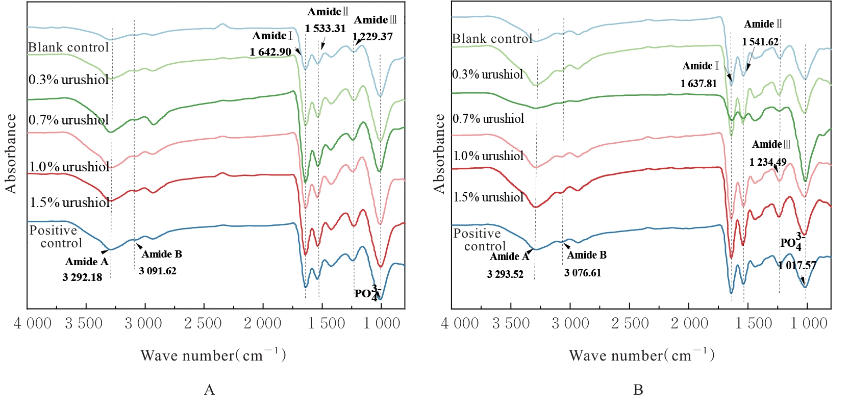
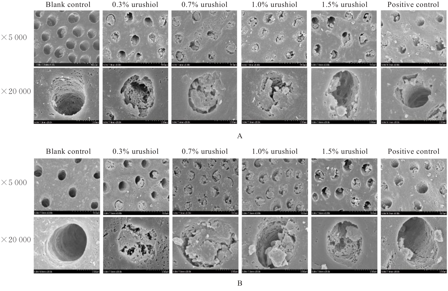
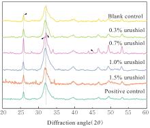
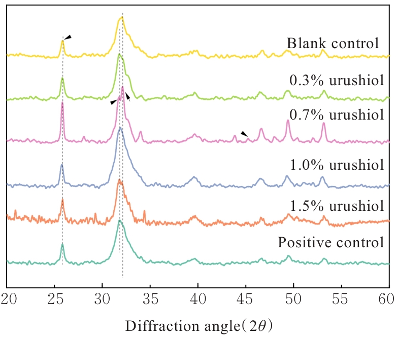
目的 探讨漆酚底涂剂应用于酸蚀-冲洗类粘接剂对脱矿牙本质再矿化作用的促进作用,阐明漆酚底涂剂对牙本质粘接耐久性的影响。 方法 选用96颗新鲜无龋第三磨牙制备成牙本质样本,采用37%磷酸凝胶酸蚀后进行预处理,随机分为空白对照组,0.3%、0.7%、1.0%和1.5%漆酚组及阳性对照(丙酮溶液)组,处理后的样本置于改良模拟体液中再矿化14和28 d。采用衰减全反射傅里叶变换红外光谱(ATR-FTIR)检测各组牙本质小管内矿物的相对质量,X射线衍射仪(XRD)和X射线能谱仪(EDS)分析各组牙本质表面物质成分,扫描电子显微镜(SEM)观察各组样本表面形态,维氏硬度仪测量各组样本表面的显微硬度,微拉伸强度(μTBS)测试检测各组样本粘接强度。 结果 粘接剂双键转化率(DC),与空白对照组比较,阳性对照组底涂剂对粘接剂DC降低,但差异无统计学意义(P>0.05),0.3%、0.7%、1.0%和1.5%漆酚组底涂剂对粘接剂DC均明显升高(P<0.05)。XRD和EDS检测,0.3%、0.7%、1.0%和1.5%漆酚组牙本质小管内新形成的矿物质为羟基磷灰石(HA)晶体。SEM检测,再矿化14和28 d后,与空白对照组比较,阳性对照组牙本质小管内见少量膜状物;0.3%漆酚组牙本质小管矿化物较少;0.7%漆酚组牙本质小管内可见大量松散矿物颗粒沉积堵塞管口;1.0%漆酚组牙本质小管内可见小块矿物沉淀;1.5%漆酚组中牙本质小管较空。显微硬度测试,再矿化14 d后,与空白对照组比较,阳性对照组牙本质显微硬度未见明显提升(P>0.05),而与0.3%、0.7%、1.0%和1.5% 漆酚组比较差异有统计学意义(P<0.05);再矿化14 d后,与阳性对照组比较,0.7%和1.0%漆酚组牙本质显微硬度提升明显(P<0.05);再矿化28 d后,与空白对照组和阳性对照组比较,各漆酚组牙本质显微硬度明显升高(P<0.05),其中0.7%和1.5%漆酚组牙本质显微硬度明显增强(P<0.05)。μTBS测试,再矿化14 d后,与阳性对照组比较,0.3%、0.7%、1.0%和1.5%漆酚组的μTBS明显增强(P<0.05);再矿化28 d后,空白对照组μTBS最低,且与空白对照组比较,阳性对照组μTBS差异无统计学意义(P>0.05);与阳性对照组比较,0.3%、0.7%、1.0%和1.5%漆酚组μTBS升高(P<0.05),0.7%、1.0%和1.5%漆酚组μTBS升高更为明显(P<0.05)。 结论 天然来源的漆酚作为一种新型底涂剂对脱矿的牙本质基质进行预处理,可以促进牙本质胶原的交联,裸露的胶原可通过漆酚吸引钙离子和磷离子,包裹牙本质胶原纤维促进再矿化,因而提高树脂-牙本质粘接界面的强度。

关键词: 漆酚, 牙本质, 粘接, 再矿化, 羟基磷灰石

Abstract:

Objective To explore the promotion effect on remineralization of demineralized dentin of urushiol primer applicated in acid-etch-rinse adhesives, and to clarify its impact on the longevity of dentin adhesion. Methods Ninety-six freshly extracted,caries-free third molars were selected to prepare the dentin specimens. Following acid etching with 37% phosphoric acid gel, the specimens were randomly divided into blank control group, 0.3%, 0.7%, 1.0%, and 1.5% urushiol groups, and positive control group(acetone solvent). The treated samples were placed in modified simulated body fluids for remineralization for 14 and 28 d. Attenuated total reflection Fourier transform infrared spectroscopy(ATR-FTIR) was used to detect the relative mineralization mass of minerals in the dentinal tubules in various groups, and X-ray Diffractometery and Energy Dispersive spectrometer were used to analyze the dentin surface material compositions in various groups. Scanning electron microscope(SEM) was used to observe the surface morphology of the specimens in various groups, Vickers hardness tester was used to measure the microhardness of the dentin surface in various groups, and microtensile strength (μTBS) was used to examine the effect of the bond strengthes in various groups. Results Compared with blank control group, the conversion rate of adhesive double bonds by primer in positive control group was decreased, but the difference was not significant (P>0.05); but the conversion rates of adhesive double bonds by primer in 0.3%, 0.7%, 1.0%, and 1.5% urushiol groups were increased (P<0.05). The SEM results revealed that at 14 and 28 d, compared with bland control group, a minimal membranous deposit in dentinal tubules was seen in positive control group, minimal mineralization displayed in 0.3 % urushiol group, significant deposition of loose mineral particles with blocking the tubule orifices was found in 0.7% urushiol group, noticeable mineral precipitates exhibited in 1.0% urushiol group, and relatively empty dentinal tubules were seen in 1.5% urushiol group. The microhardness results showed that at 14 d after remineralization, compared with blank control group, the microhardness in positive control group showed no significant improvement(P>0.05), while the differences in 0.3%, 0.7%, 1.0% and 1.5% urushiol groups were statistically significant (P<0.05); at 14 d after remineralization compared with positive control group, the microhardness of dertin in 0.7 % and 1.0% urushiol groups were increased(P<0.05); at 28 d after remineralization, compared with blank control and postive control groups, the microhardness in urushiol groups were significantly increased (P<0.05), espectially in 0.7% to 1.5% urushiol groups (P<0.05). In the μTBS test, at 14 d after remineralization, compared with postive control group, the μTBS in 0.3%, 0.7%, 1.0% and 1.5% urushiol groups were increased(P<0.05); at 28 d after remineralization, the μTBS in blank control group was the lowest; compared with blank control group, there was no significant difference in the μTBS in postive control group(P>0.05); compared with positive control group, the μTBS in 0.3%, 0.7%, 1.0%, and 1.5% urushiol groups were increased (P<0.05), espectially in 0.7%,1.0, and 1.5% urushiol groups (P<0.05). Conclusion Natural-derived urushiol, as a novel primer, can pretreat the demineralized dentin substrates, and facilitate collagen cross-linking within the dentin matrix; moreover, it leverages the phenolic hydroxyl groups within its structure to attact calcium and phosphate ions, envelope dentin collagen fibers to promote remineralization, in order to enhance the strength of the resin-dentin bonding interface.

Key words: Urushiol, Dentin, Adhesion, Remineralization, Hydroxyapatite

中图分类号: 

  • R783.1