吉林大学学报(医学版) ›› 2024, Vol. 50 ›› Issue (3): 697-707.doi: 10.13481/j.1671-587X.20240314

• 基础研究 • 上一篇    

血必净对抗NMDA受体脑炎模型小鼠脑组织损伤及脑脊液中Th17/Treg免疫失衡的改善作用

陈琳,闫丽敏,邢槐杰,陈敏,黎晓艳,曾超胜()   

  1. 海南医学院第二附属医院神经内科,海南 海口 570311
  • 收稿日期:2023-05-25 出版日期:2024-05-28 发布日期:2024-07-01
  • 通讯作者: 曾超胜 E-mail:zengchaosheng176@sina.com
  • 作者简介:陈 琳(1985-),女,江苏省南京市人,医学硕士,副主任医师,主要从事神经免疫性疾病方面的研究。
  • 基金资助:
    海南省科技厅社会发展类重点研发项目(ZDYF2022SHFZ291)

Improvement effect of Xuebijing on brain tissue injury and Th17/Treg immune imbalance in cerebrospinal fluid in NMDA receptor encephalitis model mice

Lin CHEN,Limin YAN,Huaijie XING,Min CHEN,Xiaoyan LI,Chaosheng ZENG()   

  1. Department of Neurology,Second Affiliated Hospital,Hainan Medical College,Haikou 570311,China
  • Received:2023-05-25 Online:2024-05-28 Published:2024-07-01
  • Contact: Chaosheng ZENG E-mail:zengchaosheng176@sina.com

摘要:

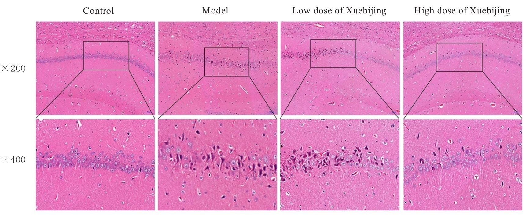
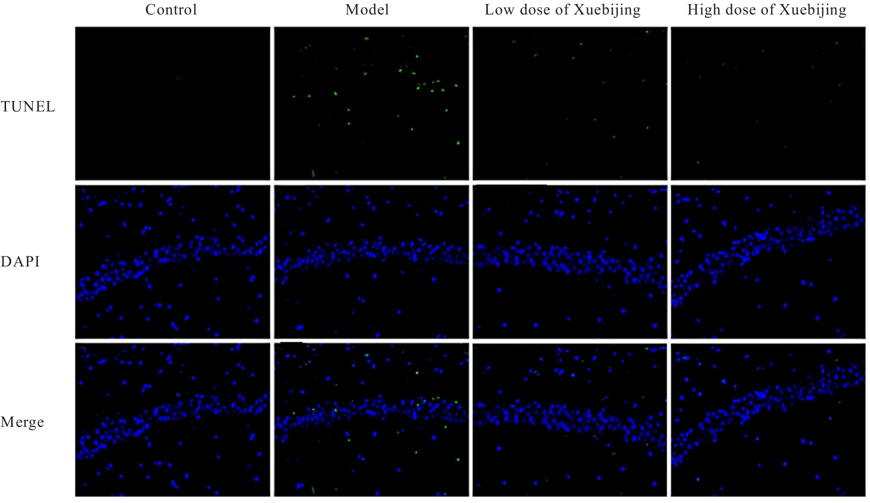
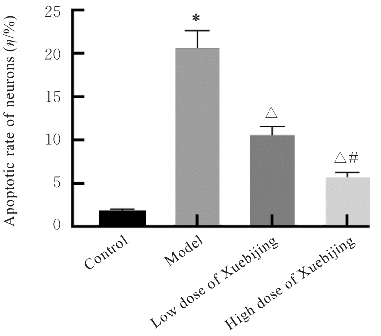
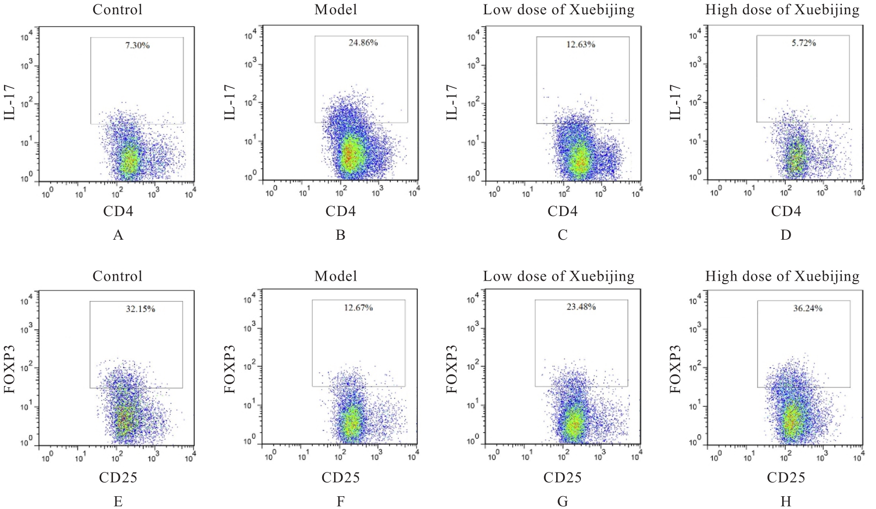
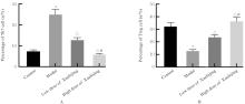
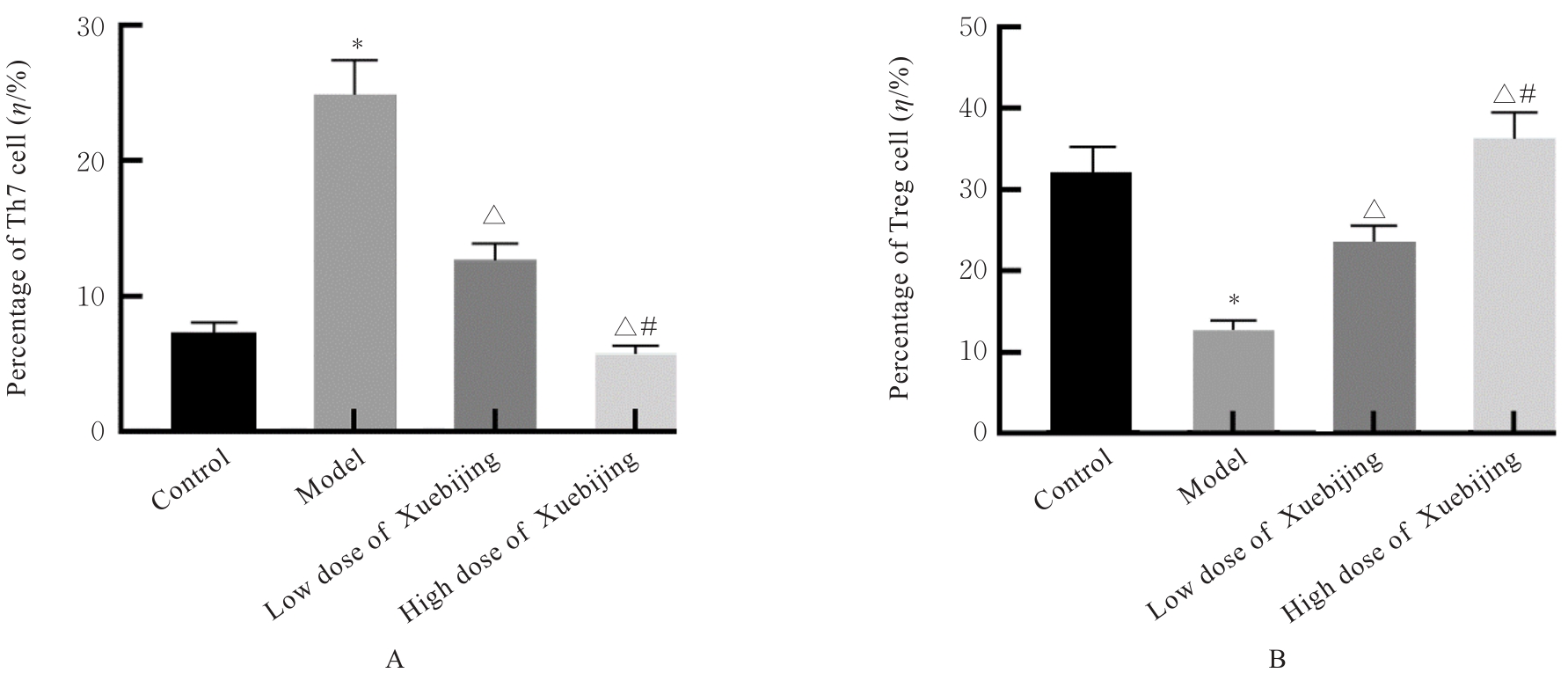
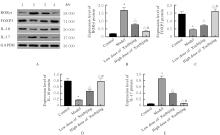
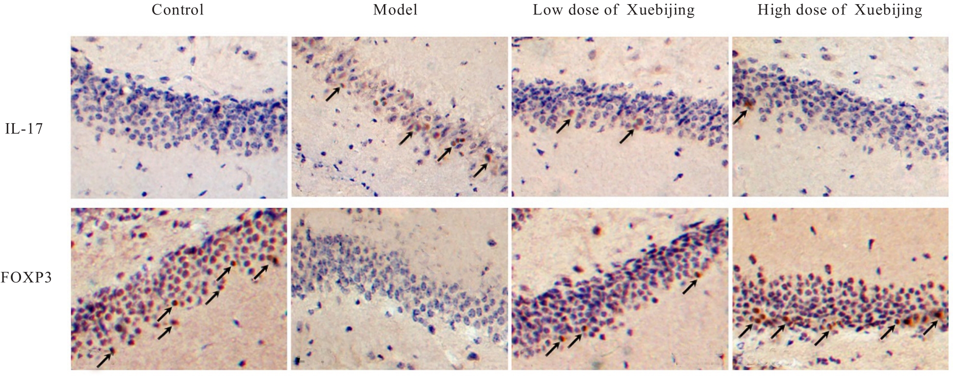
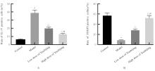
目的 探讨血必净对抗N-甲基-D-天门冬氨酸(NMDA)受体脑炎模型小鼠脑组织损伤及脑脊液(CSF)中辅助性T淋巴细胞17(Th17)/调节性T淋巴细胞(Treg)免疫失衡的影响,阐明其治疗作用。 方法 60只健康雄性C57BL/6J小鼠随机分为对照组、模型组、低剂量血必净组和高剂量血必净组,每组15只。除对照组外,其余3组小鼠均给予抗原注射与免疫刺激结合法建立抗NMDA受体脑炎模型,低和高剂量血必净组小鼠分别给予腹腔注射5和10 mL·kg-1血必净注射液。采用HE染色观察各组小鼠脑组织病理形态表现,TUNEL法检测各组小鼠脑组织海马CA1区神经元凋亡率,酶联免疫吸附试验(ELISA)法检测各组小鼠血清中白细胞介素(IL)-6、IL-10、IL-17和转化生长因子β(TGF-β)水平,流式细胞术检测各组小鼠CSF中Th17和Treg细胞百分率,Western blotting法检测各组小鼠脑组织中维甲酸相关核孤儿受体(RORγt)、叉头状转录因子3(Foxp3)、IL-10和 IL-17 蛋白表达水平, 免疫组织化学染色法检测各组小鼠脑组织中 IL-17 和 Foxp3 阳性细胞率。 结果 HE染色,对照组小鼠脑组织海马 CA1 区结构清晰,未见明显病变;与对照组比较,模型组小鼠脑组织海马CA1区部分锥体细胞呈三角形固缩浓染,顶树突拉长,少数神经细胞脱失,组织稀疏;与模型组比较,低和高剂量血必净组小鼠脑组织海马CA1区细胞损伤减小,形态恢复正常,排列较为整齐,且高剂量血必净组小鼠脑组织海马CA1区损伤的改善情况更明显。TUNEL法,与对照组比较,模型组小鼠脑组织海马CA1区神经元凋亡率明显升高(P<0.05);与模型组比较,低和高剂量血必净组小鼠脑组织海马CA1区神经元凋亡率明显降低(P<0.05);与低剂量血必净组比较,高剂量血必净组小鼠脑组织海马CA1区神经元凋亡率明显降低(P<0.05)。ELISA法,与对照组比较,模型组小鼠血清中IL-6和IL-17水平明显升高(P<0.05),IL-10和TGF-β水平明显降低(P<0.05);与模型组比较,低和高剂量血必净组小鼠血清中IL-6和IL-17水平明显降低(P<0.05),IL-10和TGF-β水平明显升高(P<0.05);与低剂量血必净组比较,高剂量血必净组小鼠血清中IL-6和IL-17水平明显降低(P<0.05),IL-10和TGF-β水平明显升高(P<0.05)。流式细胞术,与对照组比较,模型组小鼠CSF中CD4+IL-17A+Th17细胞百分率明显升高(P<0.05),CD25+Foxp3+Treg细胞百分率明显降低(P<0.05);与模型组比较,低和高剂量血必净组小鼠CSF中CD4+IL-17A+Th17细胞百分率明显降低(P<0.05),CD25+Foxp3+Treg细胞百分率明显升高(P<0.05);与低剂量血必净组比较,高剂量血必净组小鼠CSF中CD4+IL-17A+Th17细胞百分率明显降低(P<0.05),CD25+Foxp3+Treg细胞百分率明显升高(P<0.05)。Western blotting法,与对照组比较,模型组小鼠脑组织中RORγt和IL-17蛋白表达水平明显升高(P<0.05),Foxp3和IL-10蛋白表达水平明显降低(P<0.05);与模型组比较,低和高剂量血必净组小鼠脑组织中RORγt及IL-17蛋白表达水平明显降低(P<0.05),Foxp3和IL-10蛋白表达水平明显升高(P<0.05);与低剂量血必净组比较,高剂量血必净组小鼠脑组织中RORγt和IL-17蛋白表达水平明显降低(P<0.05),Foxp3和IL-10蛋白表达水平明显升高(P<0.05)。免疫组织化学染色法,与对照组比较,模型组小鼠脑组织中IL-17阳性细胞率明显升高(P<0.05),Foxp3阳性细胞率明显降低(P<0.05);与模型组比较,低和高剂量血必净组小鼠脑组织中IL-17阳性细胞率明显降低(P<0.05),Foxp3阳性细胞率明显升高(P<0.05);与低剂量血必净组比较,高剂量血必净组小鼠脑组织中IL-17阳性细胞率明显降低(P<0.05),Foxp3阳性细胞率明显升高(P<0.05)。 结论 血必净能够有效改善抗NMDA受体脑炎小鼠脑组织损伤,调控细胞因子水平,干预抗NMDA受体脑炎小鼠Th17/Treg免疫失衡现象。

关键词: 抗N-甲基-D-天门冬氨酸受体脑炎, 血必净, 神经元凋亡, 辅助性T淋巴细胞17, 调节性T淋巴细胞, 免疫失衡

Abstract:

Objective To discuss the effect of Xuebijing on brain tissue damage and immune imbalance of helper T lymphocyte 17 (Th17)/regulatory T lymphocyte (Treg) in cerebrospinal fluid (CSF) of the N-methyl-D-aspartate (NMDA) receptor encephalitis model mice, and to clarify its therapeutic effect. Methods Sixty healthy male C57BL/6J mice were randomly divided into control group, model group, low dose of Xuebijing group, and high dose of Xuebijing group, and there were 15 mice in each group. Except for control group, the mice in the other three groups were injected with the antigen combined with immunostimulation to establish the NMDA receptor encephalitis models. The mice in low and high doses of Xuebijing groups were injected intraperitoneally with 5 and 10 mL·kg-1 of Xuebijing injection, respectively. HE staining was used to observe the pathomorphology of brain tissue of the mice in various groups; TUNEL assay was used to detect the apoptotic rates of the neurons in hippocampus CA1 region of brain tissue of the mice in various groups;enzyme-linked immunosorbent assay (ELISA) method was used to detect the levels of interleukin (IL)-6, IL-10, IL-17, and transforming growth factor β (TGF-β) in serum of the mice in various groups; flow cytometry was used to detect the percentages of Th17 and Treg cells in CSF of the mice in various groups; Western blotting method was used to detect the expression levels of retinoic acid-related orphan receptor γt (RORγt), forkhead box protein 3 (Foxp3), IL-10, and IL-17 proteins in brain tissue of the mice in various groups;immunohistochemistry method was used to detect the rates of IL-17 and Foxp3 positive cells in brain tissue of the mice in various groups. Results The HE staining results showed that the hippocampus CA1 region of brain tissue of the mice in control group had a clear structure without obvious lesions; compared with control group, the mice in model group showed partial pyramidal cell shrinkage, elongation of apical dendrites, loss of a few neurons, and sparse tissue in the hippocampus CA1 region of brain tissue; compared with model group, the mice in low and high doses of Xuebijing groups showed that the damage of the cells in the hippocampus CA1 region of brain tissue was decreased, and the morphological recovery, more orderly arrangement, and more significant improvement could be seen in hippocampus CA1 region of the mice in high dose of Xuebijing group. The TUNEL assay results showed that compared with control group, the apoptotic rate of the neurons in hippocampus CA1 region of brain tissue of the mice in model group was significantly increased (P<0.05); compared with model group, the apoptotic rate of the neurons in hippocampus CA1 region of brain tissue of the mice in low and high doses of Xuebijing groups were significantly decreased (P<0.05); compared with low dose of Xuebijing group, the apoptotic rate of the neurons in hippocampus CA1 region of brain tissue of the mice in high dose of Xuebijing group was significantly decreased (P<0.05). The ELISA results showed that compared with control group, the levels of IL-6 and IL-17 in serum of the mice in model group were significantly increased (P<0.05), while the levels of IL-10 and TGF-β were significantly decreased (P<0.05); compared with model group, the levels of IL-6 and IL-17 in serum of the mice in low and high doses of Xuebijing groups were significantly decreased (P<0.05), while the levels of IL-10 and TGF-β were significantly increased (P<0.05); compared with low dose of Xuebijing group, the levels of IL-6 and IL-17 in serum of the mice in high dose of Xuebijing group were significantly decreased (P<0.05), while the levels of IL-10 and TGF-β were significantly increased (P<0.05). The flow cytometry results showed that compared with control group, the percentage of CD4+IL-17A+ Th17 cells in CSF of the mice in model group was significantly increased (P<0.05), while the percentage of CD25+Foxp3+ Treg cells was significantly decreased (P<0.05); compared with model group, the percentages of CD4+IL-17A+ Th17 cells in CSF of the mice in low and high doses of Xuebijing groups were significantly decreased (P<0.05), while the percentage of CD25+Foxp3+ Treg cells was significantly increased (P<0.05); compared with low dose of Xuebijing group, the percentage of CD4+IL-17A+ Th17 cells in CSF of the mice in high dose of Xuebijing group was significantly decreased (P<0.05), while the percentage of CD25+Foxp3+ Treg cells was significantly increased (P<0.05). The Western blotting results showed that compared with control group, the expression levels of RORγt and IL-17 proteins in brain tissue of the mice in model group were significantly increased (P<0.05), while the expression levels of Foxp3 and IL-10 proteins were significantly decreased (P<0.05); compared with model group, the expression levels of RORγt and IL-17 proteins in brain tissue of the mice in low and high doses of Xuebijing groups were significantly decreased (P<0.05), while the expression levels of Foxp3 and IL-10 proteins were significantly increased (P<0.05); compared with low dose of Xuebijing group, the expression levels of RORγt and IL-17 proteins in brain tissue of the mice in high dose of Xuebijing group were significantly decreased (P<0.05), while the expression levels of Foxp3 and IL-10 proteins were significantly increased (P<0.05). The immunohistochemistry results showed that compared with control group, the rate of IL-17 positive cells in brain tissue of the mice in model group was significantly increased (P<0.05), while the rate of Foxp3 positive cells was significantly decreased (P<0.05); compared with model group, the rates of IL-17 positive cells in brain tissue of the mice in low and high doses of Xuebijing groups were significantly decreased (P<0.05), while the rates of Foxp3 positive cells were significantly increased (P<0.05); compared with low dose of Xuebijing group, the rate of IL-17 positive cells in brain tissue of the mice in high dose of Xuebijing group was significantly decreased (P<0.05), while the rate of Foxp3 positive cells was significantly increased (P<0.05). Conclusion Xuebijing can effectively ameliorate the brain tissue injury, regulate the cytokine levels, and intervene in immune imbalance of Th17/Treg in the mice with anti-NMDA receptor encephalitis.

Key words: Anti-N-methyl-D-aspartate receptor encephalitis, Xuebijing, Neuronal apoptosis, Helper T lymphocyte 17, Regulatory T lymphocyte, Immune imbalance

中图分类号: 

  • R512.3