吉林大学学报(医学版) ›› 2024, Vol. 50 ›› Issue (6): 1535-1546.doi: 10.13481/j.1671-587X.20240607

• 基础研究 • 上一篇    

浓缩生长因子联合骨髓间充质干细胞膜片的生物学性能及其在骨缺损修复中的作用

石剑虹1,2,田原野1,陈楷1,孙高1,吴国民1()   

  1. 1.吉林大学口腔医院口腔颌面外一科与口腔整形美容外科,吉林 长春 130021
    2.吉林大学口腔医院 吉林省牙发育及颌骨重塑与再生重点实验室,吉林 长春 130021
  • 收稿日期:2023-12-28 出版日期:2024-11-28 发布日期:2024-12-10
  • 通讯作者: 吴国民 E-mail:guominwu2006@sina.com
  • 作者简介:石剑虹(1997-),女,山西省朔州市人,在读硕士研究生,主要从事口腔正颌和口腔颌面外科方面的研究。
  • 基金资助:
    吉林省财政厅医疗卫生人才建设项目(JCSZ2023481-30)

Biological properties of concentrated growth factor combined with bone marrow mesenchymal stem cell sheet and its effect on bone defect repairment

Jianhong SHI1,2,Yuanye TIAN1,Kai CHEN1,Gao SUN1,Guomin WU1()   

  1. 1.Department of Oral and Maxillofacial Surgery and oral Plastic Surgery,Stomatology Hospital,Jilin University,Changchun 130021,China
    2.Jilin Provincal Key Laboratory of Tooth Development and Bone Remodeling and Regeneration,Stomatology Hospital,Jilin University,Changchun 130021,China
  • Received:2023-12-28 Online:2024-11-28 Published:2024-12-10
  • Contact: Guomin WU E-mail:guominwu2006@sina.com

摘要:

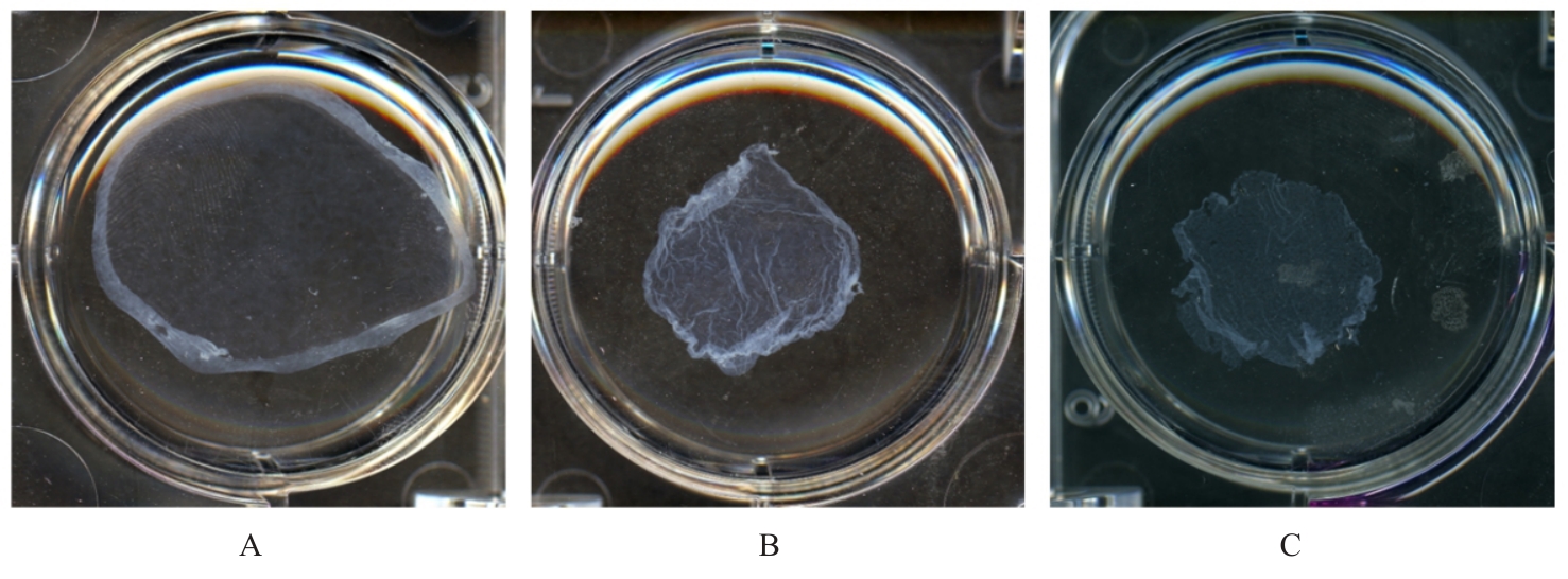
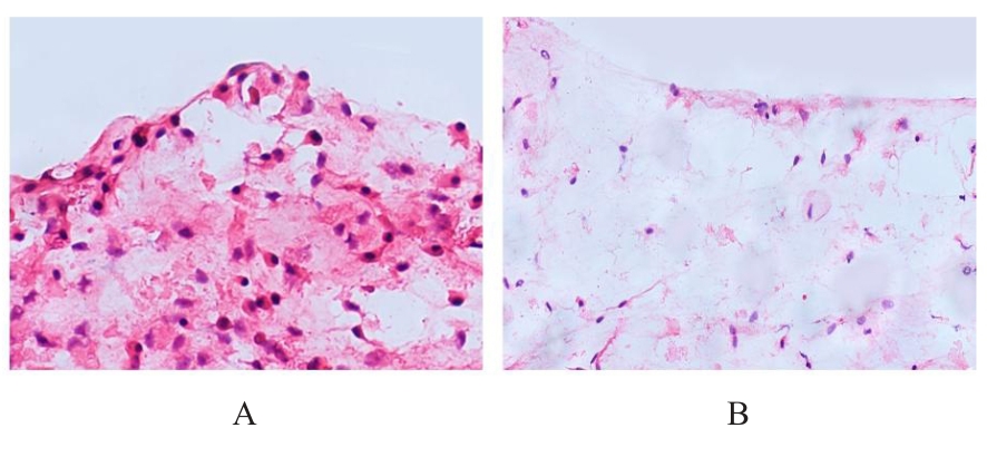
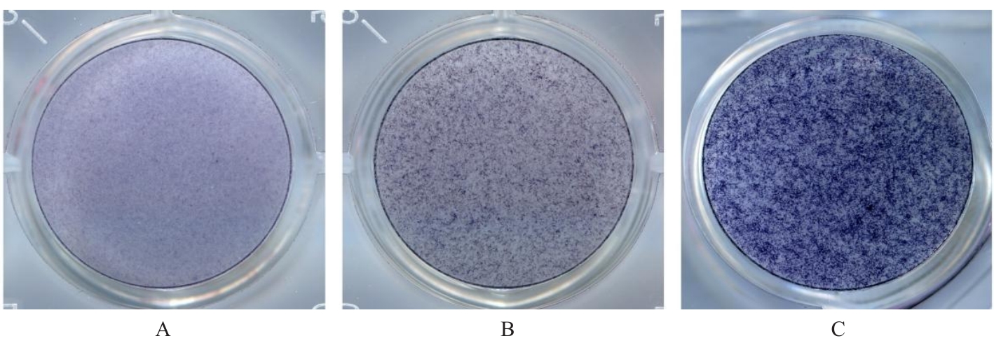
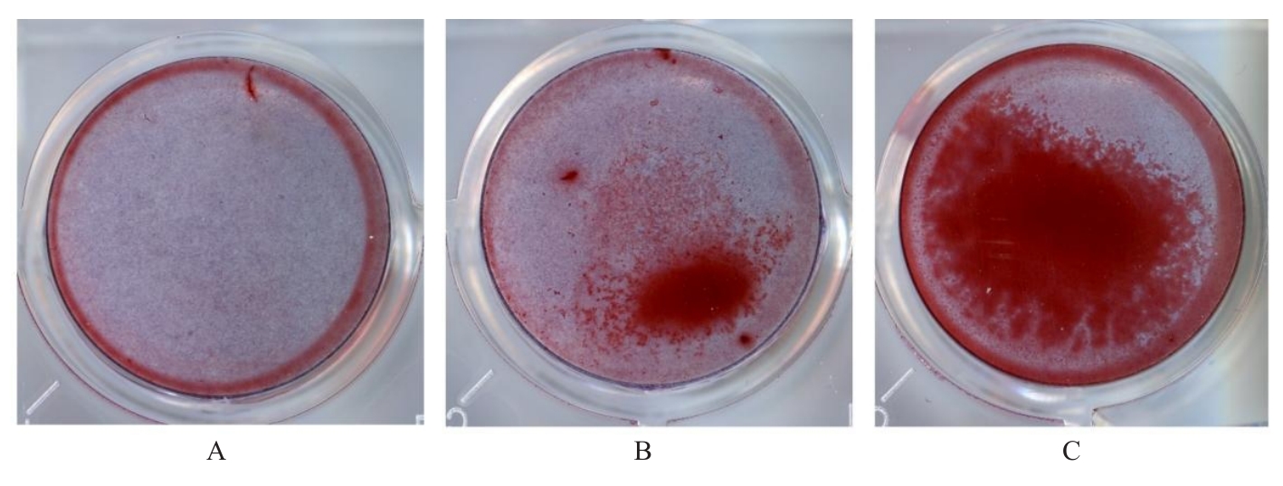
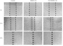
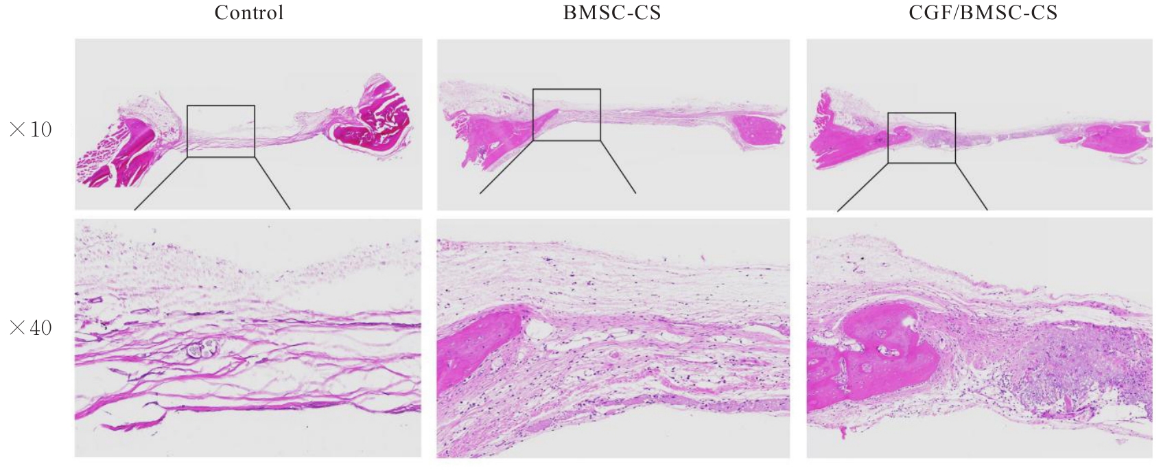
目的 探讨浓缩生长因子(CGF)对骨髓间充质干细胞(BMSCs)膜片性能的影响,并阐明含CGF复合细胞膜片(CS)在骨缺损修复中的作用。 方法 体外实验,选取2只3周龄SD大鼠,分离培养获得BMSCs,茜素红和油红O染色鉴定BMSCs成骨和成脂能力。选取3只3周龄SD大鼠,制备CGF液态提取物(CGFe),将细胞分为对照组、传统CS(BMSC-CS)组和含CGF复合CS(CGF/BMSC-CS)组。HE染色观察2组CS形态表现,茜素红和碱性磷酸酶(ALP)染色检测各组CS体外成骨情况,细胞划痕实验检测各组细胞迁移能力,实时荧光定量PCR(RT-qPCR)法检测各组细胞中ALP、胶原酶Ⅰ型(COL-1)、Runt相关转录因子2(RUNX2)和骨钙蛋白(OCN)mRNA表达水平。体内实验,选取15只SD大鼠随机分为对照组、BMSC-CS组和CGF/BMSC-CS组,显微计算机断层扫描(Micro-CT)检测各组大鼠颅骨缺损处骨形成参数,HE染色和Masson染色观察各组大鼠颅骨缺损组织形态表现。 结果 第3代BMSCs为梭形,排列紧密,呈漩涡团簇状生长;茜素红染色有明显的钙结节生成,油红O染色有红色脂滴形成,证实细胞具有良好的成骨和成脂分化的能力。CS为白色半透明状,边缘轻微卷曲,剥离后的CS卷曲皱缩为不规则状。与BMSC-CS组比较,CGF/BMSC-CS组CS白色更深,透明程度较低,在厚度和延展性方面明显增加,不易破损,有一定黏性和可塑性。HE染色观察,与BMSC-CS组比较,CGF/BMSC-CS组CS细胞数增加,排列密集,细胞外基质(ECM)更丰富,包裹连接细胞形成一个整体的片状结构。茜素红和ALP染色检测,与对照组比较,BMSC-CS组CS的ALP活性和矿化提升值均明显升高(P<0.05);与对照组和BMSC-CS组比较,CGF/BMSC-CS组CS成骨细胞及红色矿化结节数明显增多,染色明显加深,阳性面积增大,ALP活性和矿化提升值均明显升高(P<0.05)。细胞划痕实验检测,培养24 h,与对照组比较,BMSC-CS组和CGF/BMSC-CS组细胞迁移率均明显升高(P<0.05);与BMSC-CS组比较,CGF/BMSC-CS组细胞迁移率明显升高(P<0.01);培养48 h,与对照组比较,CGF/BMSC-CS组细胞迁移率明显升高(P<0.05)。RT-qPCR法检测,与对照组比较,BMSC-CS组细胞中COL-1和OCN mRNA表达水平均明显升高(P<0.01),CGF/BMSC-CS组细胞中ALP、COL-1、OCN和RUNX2 mRNA表达水平均明显升高(P<0.01);与BMSC-CS组比较,CGF/BMSC-CS组细胞中ALP、COL-1和OCN mRNA表达水平均明显升高(P<0.01)。Micro-CT检测,对照组大鼠颅骨缺损区域边界清晰,几乎无新骨生成;BMSC-CS组大鼠颅骨仅在骨缺损边缘有少量新骨形成,缺损中心区域有明显空缺;CGF/BMSC-CS组大鼠颅骨新骨沿骨缺损边缘向中心区域生成,修复大部分骨缺损;与对照组比较,BMSC-CS组大鼠颅骨骨体积分数[骨体积(BV)/组织体积(TV)]和骨小梁间距(Tb.N)均明显升高(P<0.05),CGF/BMSC-CS组大鼠颅骨BV、BV/TV、骨小梁数目(Tb.Th)和Tb.N均明显升高(P<0.05);与BMSC-CS组比较,CGF/BMSC-CS组大鼠颅骨BV、BV/TV、Tb.Th和Tb.N均明显升高(P<0.01)。HE和Masson染色观察,对照组大鼠颅骨缺损组织几乎无新骨生成,仅见大量胶原纤维连接两侧骨断端;BMSC-CS组大鼠颅骨缺损组织仅在骨缺损边缘有少量新骨形成,中央为致密的胶原纤维与缺损边缘的新生骨连接;CGF/BMSC-CS组大鼠颅骨缺损组织除在骨缺损边缘可以看到新生骨组织外,缺损中央亦有骨岛形成,骨岛周围可见骨细胞及大量的胶原纤维。Masson染色观察,细胞质和类骨质呈红色,胶原呈蓝色;CGF/BMSC-CS组大鼠颅骨缺损组织中可见明显的新形成的类骨质,新骨形成量最高。 结论 CGF可以促进BMSCs膜片的成骨分化和ECM的丰富度,含CGF复合CS可以高效修复大鼠颅骨缺损,是一种理想和安全的促骨再生材料。

关键词: 浓缩生长因子, 细胞膜片, 骨髓间充质干细胞, 骨缺损再生修复

Abstract:

Objective To discuss the effect of concentrated growth factor (CGF) on the performance of bone marrow mesenchymal stem cells (BMSCs) sheets, and to clarify the role of CGF-containing composite cell sheets(CS) in the bone defect repairment. Methods In in vitro experiments, the BMSCs were isolated and cultured from two 3-week-old SD rats; Alizarin Red S and Oil Red O staining were used to identify the osteogenic and adipogenic capabilities of BMSCs; CGF liquid extracts (CGFe) was prepared from three 3-week-old SD rats. The cells were divided into control group, traditional CS (BMSC-CS) group, and CGF-containing composite CS (CGF/BMSC-CS) group. The morphology of the CS in two groups was observed by HE staining. Alizarin Red and alkaline phosphatase (ALP) staining were used to detect the osteogenic differentiation of the CS in various groups; cell scratch assay was used to detect the migration abilities of the cells in various groups; real-time fluorescence quantitative PCR(RT-qPCR) method was used to detect the mRNA expression levels of ALP, collagen are type 1 (COL-1), Runt-related transcription factor 2 (RUNX2), and osteocalcin (OCN) in the cells in various groups. In in vivo experiments, 15 SD rats were randomly divided into control group, BMSC-CS group, and CGF/BMSC-CS group; micro computed tomography (Micro-CT) was used to detect the bone formation parameters in skull defects of the rats in various groups; HE staining and Masson staining were used to observe the morphology of skull defect tissue of the rats in various groups. Results The third-generation BMSCs were spindle-shaped, closely arranged, and grew in a vortex cluster. The Alizarin red staining results showed obvious calcium nodules, and the Oil red O staining showed red lipid droplets, confirming the cells’ ability to undergo osteogenic and adipogenic differentiation. The CS were white and semi-transparent, with slightly curled edges. The peeled CS were irregularly curled and wrinkled. Compared with BMSC-CS group, the CS in CGF/BMSC-CS group were whiter, less transparent, significantly increased in thickness and extensibility, less prone to breakage, and had a certain degree of stickiness and plasticity. The HE staining results showed that compared with BMSC-CS group, the number of the cells of CS in CGF/BMSC-CS group was increased, with denser arrangement and more abundant extracellular matrix (ECM), which wrapped and connected the cells to form an integral sheet-like structure. The Alizarin red and ALP staining results showed that compared with control group, the ALP activity and mineralization uplift value of CS in BMSC-CS group were significantly increased (P<0.05); compared with control group and BMSC-CS group, the number of osteoblasts and red mineralized nodules in the CS in CGF/BMSC-CS group was significantly increased, with obvious deepening of the staining, increased positive area, and the ALP activity and mineralization uplift value were significantly increased (P<0.05). Compared with BMSC-CS group, the ALP activity and mineralization uplif value of the CS in CGF/DMSC-CS group were increased (P<0.05). The cell scratch assay results showed that after 24 of culture, compared with control group, the migration rates of the cells in BMSC-CS group and CGF/BMSC-CS group were significantly increased (P<0.05). Compared with BMSC-CS group, the migration rate of the cells in CGF/BMSC-CS group was significantly increased (P<0.01). After 48 h of culture, compared with control group, the migration rate of the cells in CGF/BMSC-CS group was significantly increased (P<0.05). The RT-qPCR results showed that compared with control group, the expression levels of COL-1 and OCN mRNA in the cells in BMSC-CS group were significantly increased (P<0.01), and the expression levels of ALP, COL-1, OCN, and RUNX2 mRNA in the cells in CGF/BMSC-CS group were significantly increased (P<0.01). Compared with BMSC-CS group, the expression levels of ALP, COL-1, and OCN mRNA in the cells in CGF/BMSC-CS group were significantly increased(P<0.01). The Micro-CT detection results showed that in control group, the boundary of the rat skull defect area was clear, with almost no new bone formation. In BMSC-CS group, a small amount of new bone formed only at the edge of the bone defect in skull of the rats, with a significant gap in the central area of the defect. In CGF/BMSC-CS group, new bone formed along the edge of the bone defect towards the central area in skull of the rats, repairing most of the bone defect. Compared with control group, the bone volume (BV) and trabecular number (Tb.N) of the rats in BMSC-CS group were significantly increased (P<0.05); the bone volume (BV), bone volume fraction [BV/tissue volume (TV)], trabecular thickness (Tb.Th), and trabecular number (Tb.N) in skull of the rats in CGF/BMSC-CS group,were significantly increased (P<0.05). Compared with BMSC-CS group, the BV, BV/TV, Tb.Th, and Tb.N in skull of the rats in CGF/BMSC-CS group were significantly increased (P<0.01). The HE and Masson staining observation showed that in control group, almost no new bone formed in the skull defect tissue of the rats, with only a large amount of collagen fibers connecting the two sides of the bone ends. In BMSC-CS group, a small amount of new bone formed only at the edge of the bone defect in skull tissue of the rats, with the central area of the defect containing dense collagen fibers connected to the newly formed bone at the defect edge. In CGF/BMSC-CS group, new bone tissue could be seen at the edge of the bone defect, and bone islands formed in the central area of the defect, surrounded by osteocytes and a large amount of collagen fibers. The Masson staining observation results showed that the cytoplasm and osteoid were red, and the collagen was blue. In CGF/BMSC-CS group, newly formed osteoid was observed in skull defect tissue of the rats, with the highest amount of new bone formation. Conclusion CGF can promote the osteogenic differentiation and increase the richness of ECM in BMSCs sheets. CGF-containing composite CS can efficiently repair skull defects of the rats and serve as an ideal and safe material for promoting the bone regeneration.

Key words: Concentrated growth factor, Cell sheet, Bone marrow mesenchymal stem cell, Bone defect regeneration and repairment

中图分类号: 

  • R78