吉林大学学报(医学版) ›› 2025, Vol. 51 ›› Issue (4): 958-967.doi: 10.13481/j.1671-587X.20250412

• 基础研究 • 上一篇    下一篇

肺癌A549细胞源性外泌体中lncRNA DUXAP8对肺癌细胞生长和肿瘤免疫逃逸的作用及其机制

何小双1,徐丽娜1,崔梅2,赵宇3,王蓓1,黄征1,王玉超1,辛雯艳1,邬超1()   

  1. 1.石河子大学第一附属医院呼吸与危重症医学科,新疆 石河子 832099
    2.新疆维吾尔自治区人民 医院病理科,新疆 石河子 830001
    3.石河子大学第一附属医院胃肠外科,新疆 石河子 832099
  • 收稿日期:2024-12-24 接受日期:2025-02-17 出版日期:2025-07-28 发布日期:2025-08-25
  • 通讯作者: 邬超 E-mail:mail@healthy.vip
  • 作者简介:何小双(1988-),女,四川省南充市人,医学硕士,主治医师,主要从事呼吸系统疾病诊治方面的研究。
  • 基金资助:
    新疆维吾尔自治区科技厅“天山英才”培养计划项目(2022TSYCCX0036)

Effects of lncRNA DUXAP8 in lung cancer A549 cells-derived exosomes on lung cancer cell growth and its mechnism

Xiaoshuang HE1,Lina XU1,Mei CUI2,Yu ZHAO3,Bei WANG1,Zheng HUANG1,Yuchao WANG1,Wenyan XIN1,Chao WU1()   

  1. 1.Department of Respiratory and Critical Care Medicine,First Affiliated Hospital,Shihezi University,Shihezi 832099,China
    2.Department of Pathology,People’s Hospital,Xinjiang Uygur Autonomous Region,Shihezi 830001,China
    3.Department of Gastroenterology,First Affiliated Hospital,Shihezi University,Shihezi 832099,China
  • Received:2024-12-24 Accepted:2025-02-17 Online:2025-07-28 Published:2025-08-25
  • Contact: Chao WU E-mail:mail@healthy.vip

摘要:

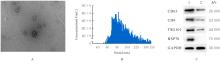
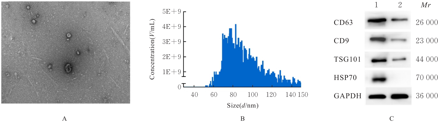
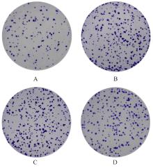
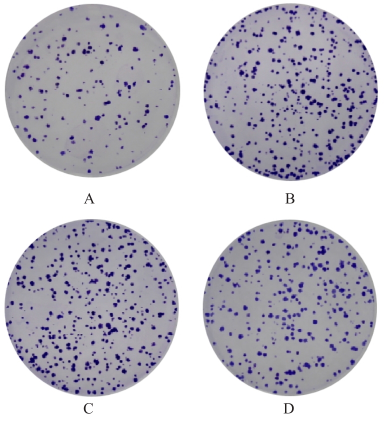
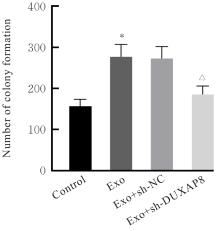
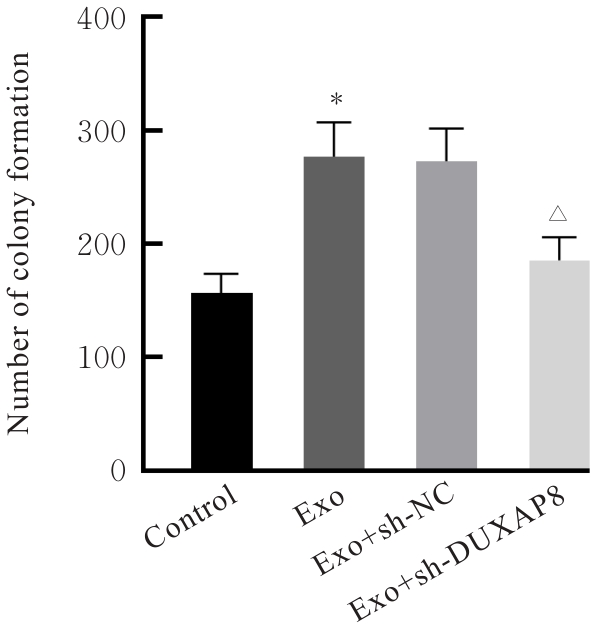
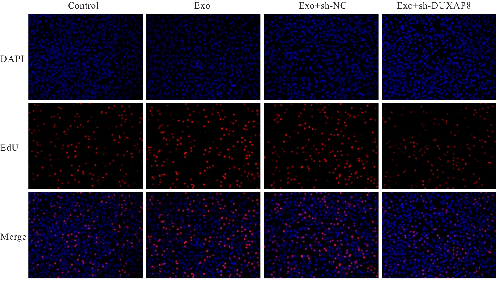
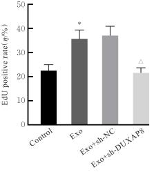
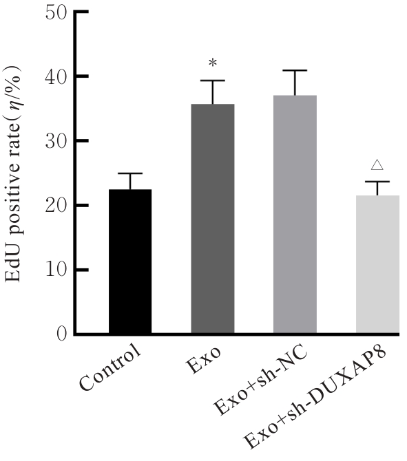
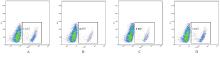
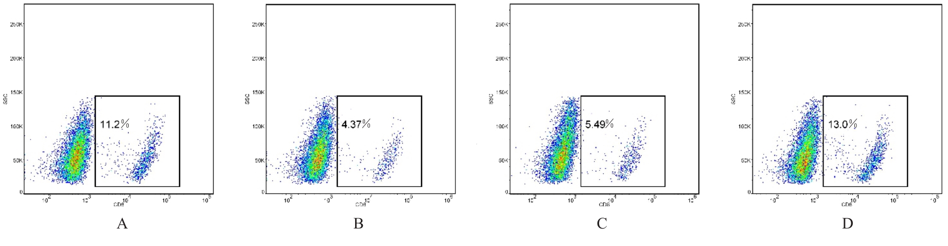
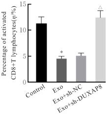
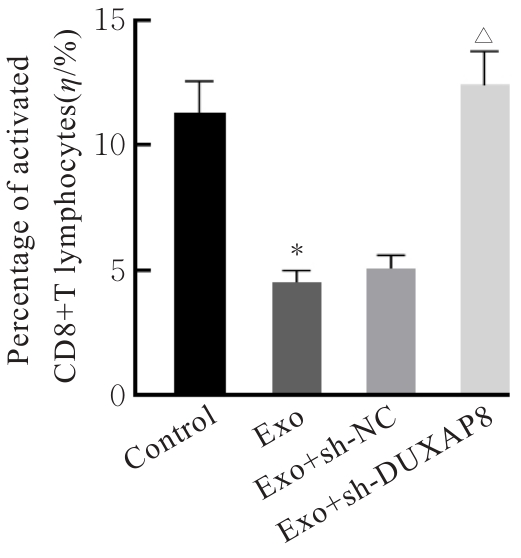
目的 探讨肺癌A549细胞源性外泌体(Exo)中长链非编码 RNA(lncRNA)DUXAP8对肺癌细胞生长和免疫逃逸的影响,并阐明其作用机制。 方法 培养人肺癌细胞系A549细胞,提取其Exo并鉴定。采用PKH67标记的Exo处理A549细胞,观察A549细胞摄取Exo情况。实时荧光定量PCR(RT-qPCR)法检测Exo处理前后A549细胞中lncRNA DUXAP8表达水平。将A549细胞分为对照组(不处理)、Exo组(Exo处理A549细胞)、Exo+sh-NC组(Exo处理A549细胞后,转染sh-NC至A549细胞)和Exo+sh-DUXAP8组(Exo处理A549细胞后,转染sh-DUXAP8至A549细胞)。RT-qPCR法检测各组A549细胞中lncRNA DUXAP8表达水平,平板集落形成实验检测各组A549细胞克隆形成能力,5-乙炔基-2'-脱氧尿苷(EdU)法检测各组A549细胞增殖能力。各组A549细胞与人外周血淋巴细胞共培养后,流式细胞术检测各组人外周血淋巴细胞中活化的CD8+T淋巴细胞百分率,3-(4,5-二甲基噻唑-2)-2,5-二苯基四氮唑溴盐(MTT)法检测各组人外周血淋巴细胞对A549细胞的杀伤率。 结果 Exo囊泡直径为50~150 nm,且分化簇63(CD63)、分化簇9(CD9)、肿瘤易感基因101(TSG101)和热休克蛋白70(HSP70)外泌体特异性标志物蛋白表达阳性,说明Exo提取成功。A549细胞能够很好地摄取PKH67标记的Exo。RT-qPCR法,与单独培养的A549细胞比较,Exo处理后,A549细胞中lncRNA DUXAP8表达水平升高(P<0.05)。与对照组比较,Exo组A549细胞中lncRNA DUXAP8表达水平升高(P<0.05);与Exo组比较,Exo+sh-DUXAP8组A549细胞中lncRNA DUXAP8表达水平降低(P<0.05),Exo+sh-NC组lncRNA DUXAP8表达水平差异无统计学意义(P>0.05)。平板集落形成实验,与对照组比较,Exo组A549细胞中集落形成数增加(P<0.05);与Exo组比较,Exo+sh-DUXAP8组A549细胞中集落形成数减少(P<0.05),Exo+sh-NC组A549细胞中集落形成数差异无统计学意义(P>0.05)。EdU染色,与对照组比较,Exo组A549细胞中EdU阳性细胞率升高(P<0.05);与Exo组比较,Exo+sh-DUXAP8组A549细胞中EdU阳性细胞率降低(P<0.05),Exo+sh-NC组A549细胞中EdU阳性细胞率差异无统计学意义(P>0.05)。流式细胞术,与对照组比较,Exo组人外周血淋巴细胞中活化CD8+T淋巴细胞百分率降低(P<0.05);与Exo组比较,Exo+sh-DUXAP8组人外周血淋巴细胞中活化CD8+T淋巴细胞百分率升高(P<0.05),Exo+sh-NC组人外周血淋巴细胞中活化的CD8+T淋巴细胞百分率差异无统计学意义(P>0.05)。MTT法,与对照组比较,Exo组人外周血淋巴细胞对 A549细胞的杀伤率降低(P<0.05);与Exo组比较,Exo+sh-DUXAP8组人外周血淋巴细胞对 A549细胞的杀伤率升高(P<0.05),Exo+sh-NC组人外周血淋巴细胞对 A549细胞的杀伤率差异无统计学意义(P>0.05)。 结论 肺癌A549细胞源性Exo lncRNA DUXAP8促进肺癌细胞增殖和肿瘤免疫逃逸。

关键词: 肺肿瘤, 外泌体, 长链非编码 RNA, DUXAP8, 细胞增殖, 免疫逃逸

Abstract:

Objective To discuss the effect of long non-coding RNA (lncRNA) DUXAP8 in exosomes (Exo) derived from the lung cancer A549 cells on the growth and immune escape of the lung cancer cells, and to clarify the mechanism. Methods The human lung cancer cell line A549 was cultured, and its exosomes were extracted and identified. The A549 cells were treated with PKH67-labeled Exo to observe the uptake of Exo by A549 cells. Real-time fluorescence quantitative PCR (RT-qPCR) method was used to detect the expression level of lncRNA DUXAP8 in A549 cells before and after Exo treatment. The A549 cells were divided into control group (no treatment), Exo group (A549 cells treated with Exo), Exo+sh-NC group (A549 cells treated with Exo and then transfected with sh-NC), and Exo+ sh-DUXAP8 group (A549 cells treated with Exo and then transfected with sh-DUXAP8). RT-qPCR method was used to detect the expression level of lncRNA DUXAP8 in A549 cells in various groups; colony formation assay was used to detect the colony formation abilities of the A549 cells in various groups; 5-ethynyl-2'-deoxyuridine (EdU) staining method was used to detect the proliferation abilities of the A549 cells in various groups. After co-culturing A549 cells in various groups with human peripheral blood lymphocytes, flow cytometry was used to detect the percentages of activated CD8+ T lymphocytes in the human peripheral blood lymphocytes in various groups; 3-(4,5-dimethylthiazol-2-yl)-2, 5-diphenyltetrazolium bromide (MTT) method was used to detect the killing rates of human peripheral blood lymphocytes on the A549 cells in various groups. Results The diameter of Exo vesicles was 50-150 nm, and the exosome-specific marker proteins cluster of differentiation 63(CD63), cluster of differentiation 9(CD9), tumor susceptibility gene 101(TSG101), and heat shock protein 70(HSP70) were positively expressed, indicating successful exosome extraction. A549 cells efficiently took up PKH67-labeled Exo. The RT-PCR results showed that compared with A549 cells cultured alone, the expression level of lncRNA DUXAP8 in the A549 cells was increased after treatment with Exo derived from A549 cells (P<0.05). compared with control group, the expression level of lncRNA DUXAP8 in the A549 cells in Exo group was increased (P<0.05); compared with Exo group, the expression level of lncRNA DUXAP8 in the A549 cells in Exo+sh-DUXAP8 group was decreased (P<0.05), while there were no significant difference in the expression level of IncRNA DUXAP8 in the cells in Exo+sh-NC group (P>0.05). The colony formation assay results showed that compared with control group, the number of colony formation of the A549 cells in Exo group was increased (P<0.05); compared with Exo group, the number of colony formation of the A549 cells in Exo+sh-DUXAP8 group was decreased (P<0.05), while there was no significant difference in the number of colony formation of the A549 cells in Exo+sh-NC group (P>0.05). The EdU staining results showed that compared with control group, the EdU-positive rate of the A549 cells in Exo group was increased (P<0.05); compared with Exo group, the EdU-positive rate in A549 cells in Exo+sh-DUXAP8 group was decreased (P<0.05), while there was no significant difference in the EDU-positive rate in the cells in Exo+sh-NC group (P>0.05). The flow cytometry results showed that compared with control group, the percentage of activated CD8+T lymphocytes in the human peripheral blood lymphocytes in Exo group was decreased (P<0.05); compared with Exo group, the percentage of activated CD8+T lymphocytes in the human peripheral blood lymphocytes in Exo+ sh-DUXAP8 group was increased (P<0.05), while there was no significant difference in the percentage of activated CD8+T lymphaytes in Exo+sh-NC group (P>0.05). The MTT assay results showed that compared with control group, the killing rate of human peripheral blood lymphocytes on the A549 cells in Exo group was decreased (P<0.05); compared with Exo group, the killing rate of human peripheral blood lymphocytes on A549 cells in Exo+sh-DUXAP8 group was increased (P<0.05), while no significant difference was observed in Exo+sh-NC group (P>0.05). Conclusion The lncRNA DUXAP8 in exosomes derived from the lung cancer A549 cells promotes the proliferation of lung cancer cells and tumor immune escape.

Key words: Lung neoplasms, Exosomes, Long non-coding RNA, DUXAP8, Cell proliferation, Immune escape

中图分类号: 

  • R734.2