吉林大学学报(医学版) ›› 2025, Vol. 51 ›› Issue (2): 284-295.doi: 10.13481/j.1671-587X.20250202

• 基础研究 • 上一篇    

糖脂转运蛋白对胰腺癌PANC-1细胞增殖、迁移和侵袭的影响及其机制

卢梦云1,2,韩语诚1,2,胡溢洪1,2,何旻蕙1,2,张艳群3,邹先琼1,2()   

  1. 1.桂林医学院科学实验中心,广西 桂林 541199
    2.桂林医学院基础医学院免疫学教研室,广西 桂林 541199
    3.中南大学湘雅医院肿瘤科,湖南 长沙 410008
  • 收稿日期:2024-04-09 接受日期:2024-06-03 出版日期:2025-03-28 发布日期:2025-04-22
  • 通讯作者: 邹先琼 E-mail:zouxq019@glmc.edu.cn
  • 作者简介:卢梦云(1995-),女,河南省商丘市人,医学硕士,主要从事黏膜免疫方面的研究。
  • 基金资助:
    国家自然科学基金项目(82160185);广西壮族自治区科技厅自然科学基金项目(2020GXNSFDA238026)

Effects of glycolipid transfer protein on proliferation, migration,and invasion of pancreatic cancer PANC-1 cells and their mechanisms

Mengyun LU1,2,Yucheng HAN1,2,Yihong HU1,2,Minhui HE1,2,Yanqun ZHANG3,Xianqiong ZOU1,2()   

  1. 1.Scientific Research Center,Guilin Medical University,Guilin 541199,China
    2.Department of Immunology,College of Basic Medical Science,Guilin Medical University,Guilin 541199,China
    3.Department of Oncology,Xiangya Hospital,Central South University,Changsha 410008,China
  • Received:2024-04-09 Accepted:2024-06-03 Online:2025-03-28 Published:2025-04-22
  • Contact: Xianqiong ZOU E-mail:zouxq019@glmc.edu.cn

摘要:

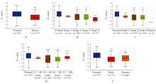
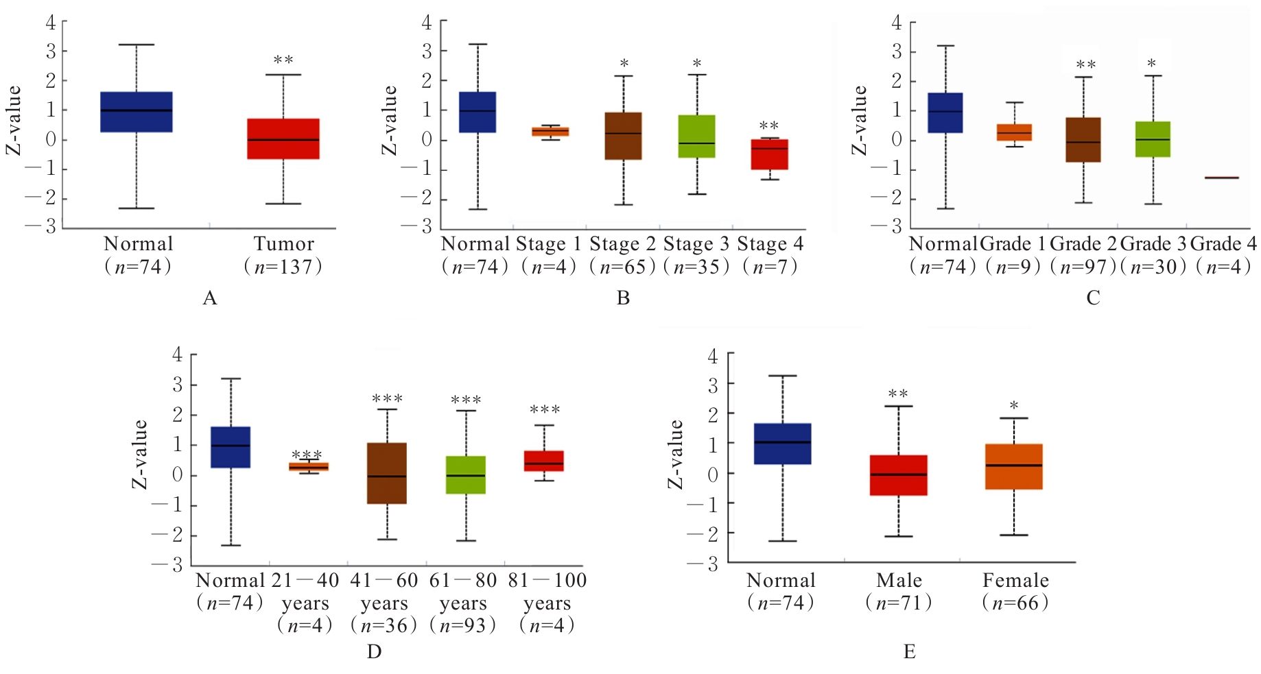
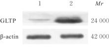
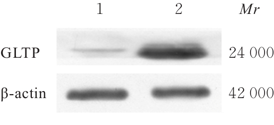
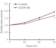
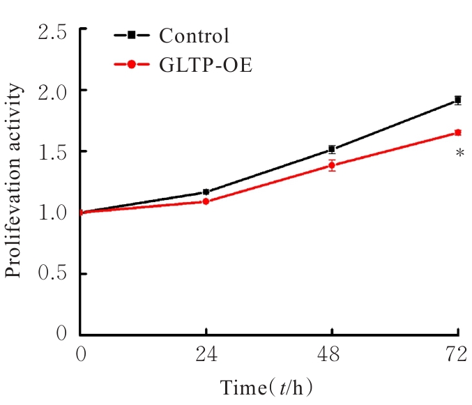
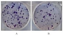
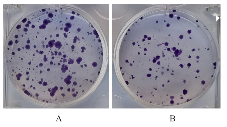
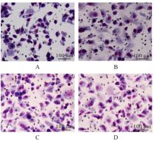
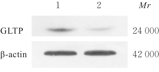
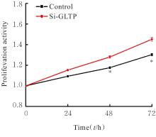
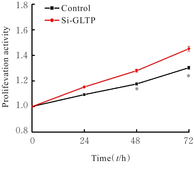
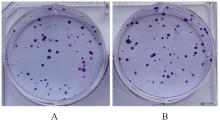
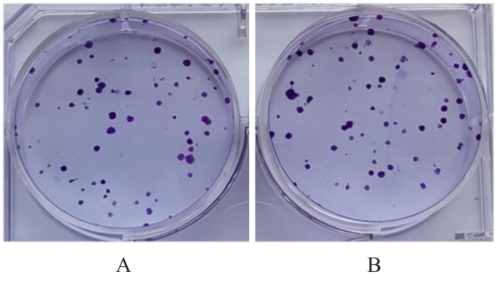
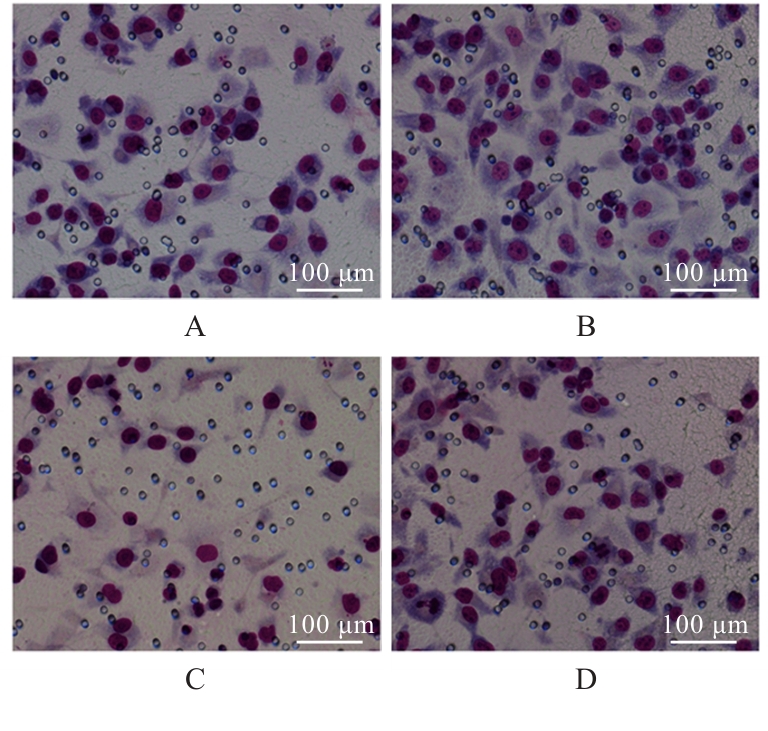
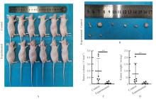
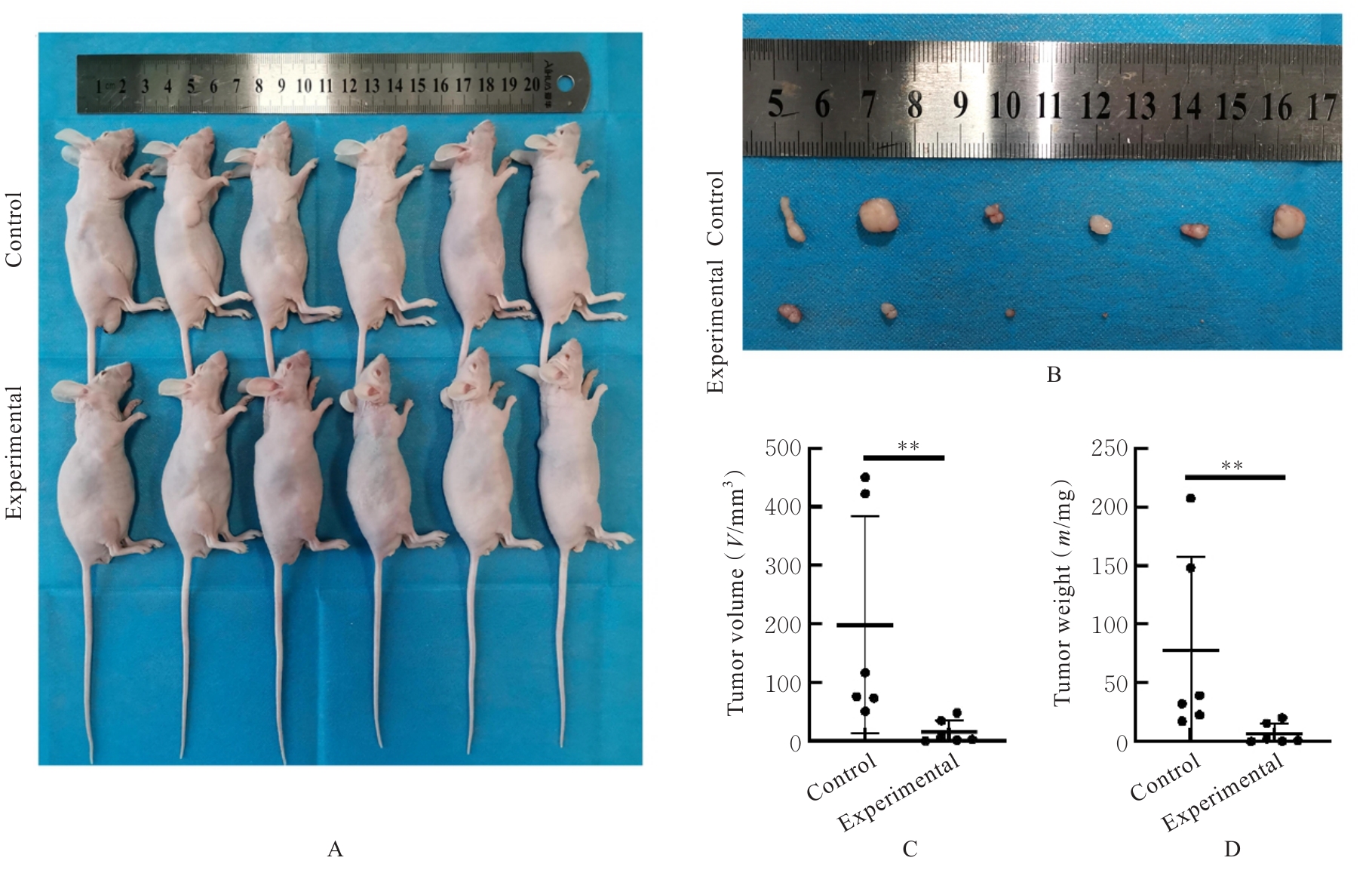
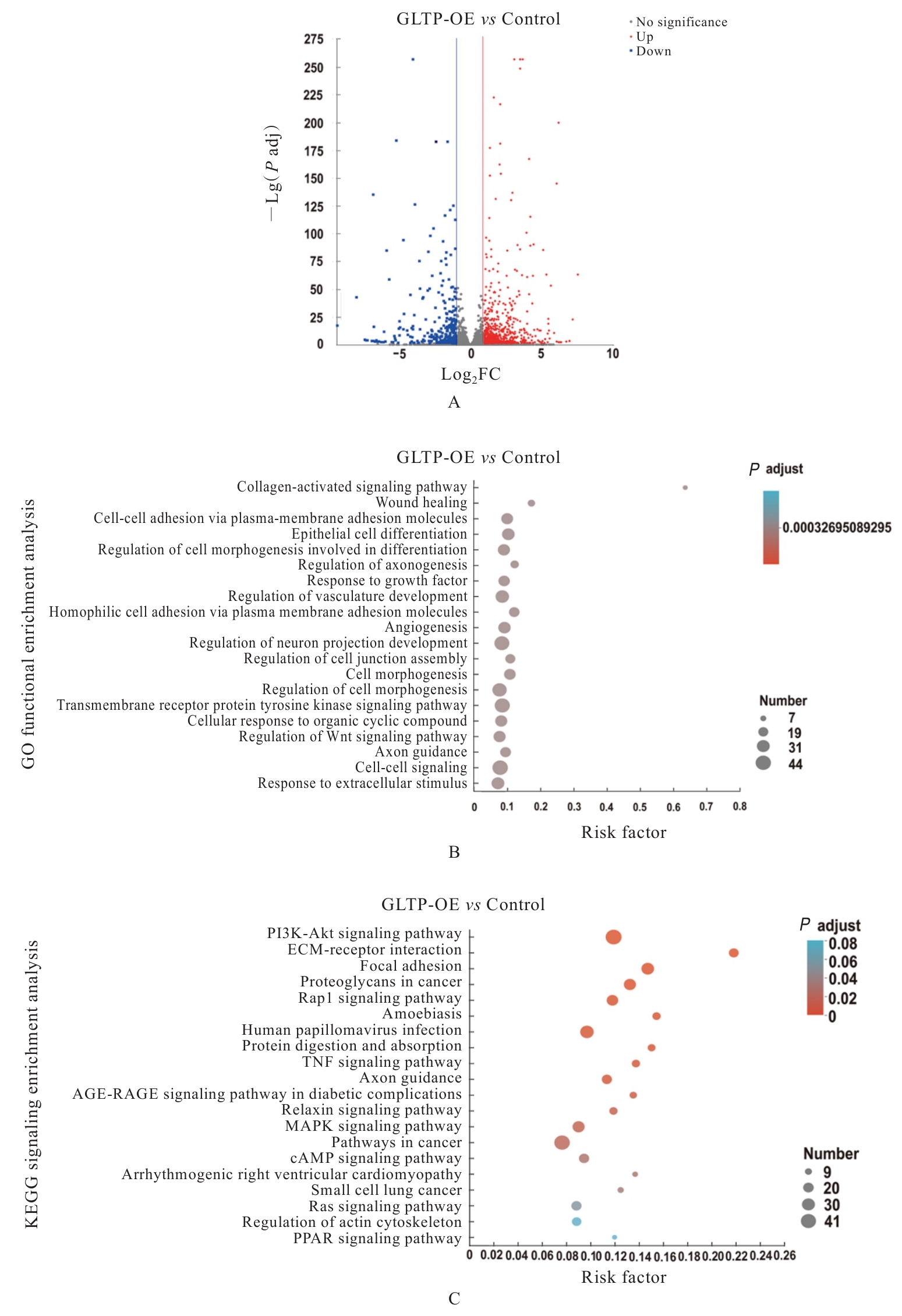
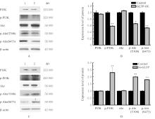
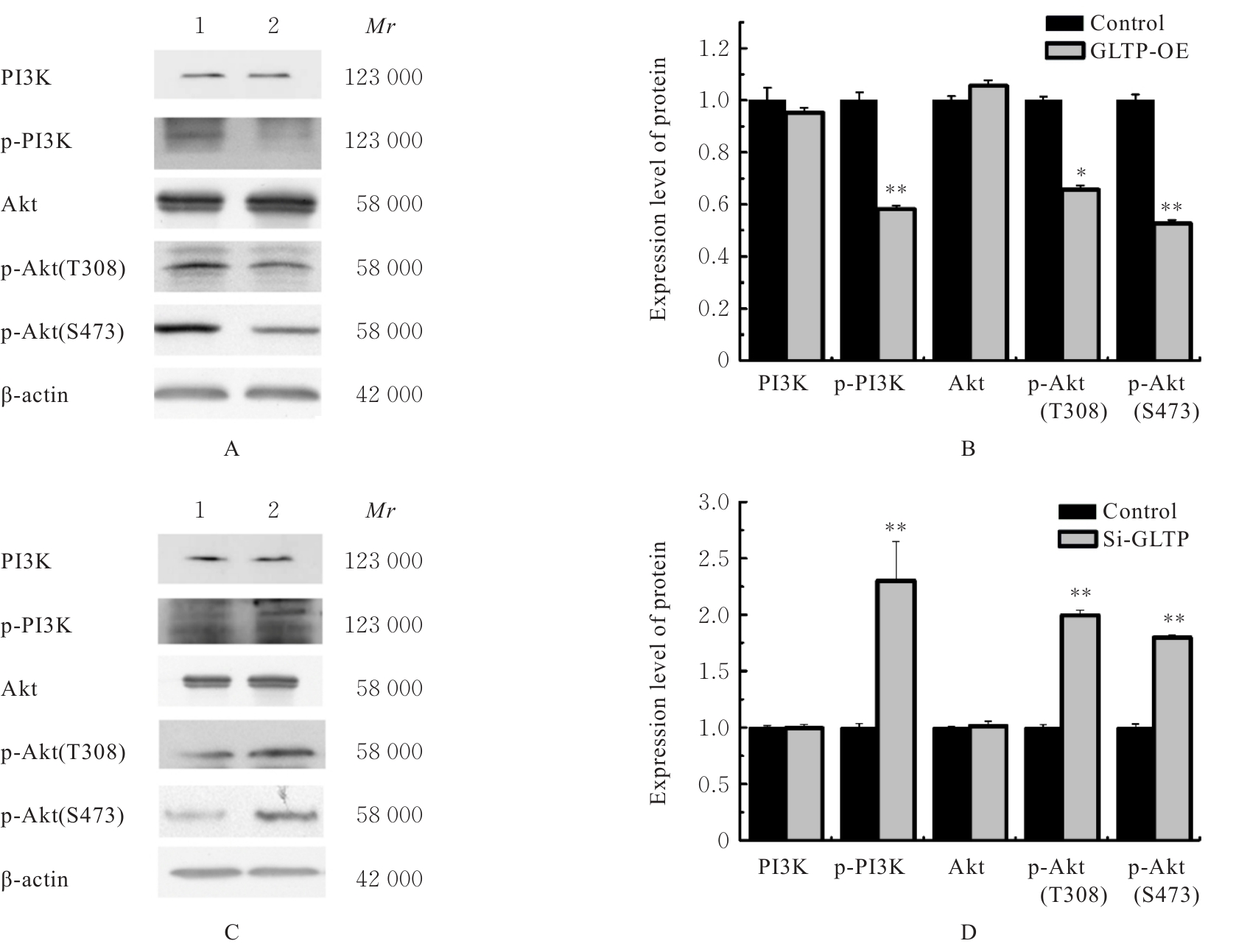
目的 探讨人类糖脂转运蛋白(GLTP)对胰腺癌(PC)细胞增殖、迁移和侵袭的影响,并阐明其作用机制。 方法 采用阿拉巴马大学伯明翰分校癌症数据分析平台(UALCAN)数据库分析PC组织和正常胰腺组织中GLTP蛋白表达差异,以及不同临床病理特征PC患者PC组织和正常胰腺组织中GLTP蛋白表达的差异。体外培养人PC细胞PANC-1细胞,分为对照组(转染pFLAG-CMV4质粒)和GLTP过表达(GLTP-OE)组(转染pFLAG-GLTP质粒),采用抗生素筛选法建立稳定转染GLTP的细胞;敲低实验采用非特异siRNA转染PANC-1细胞作为对照组,si-GLTP转染PANC-1细胞作为si-GLTP组。采用Western blotting法检测细胞中GLTP蛋白表达情况,细胞计数试剂盒8(CCK-8)法检测PANC-1细胞的增殖活性,克隆形成实验检测PANC-1细胞克隆形成数,Transwell小室实验检测PANC-1细胞的迁移及侵袭细胞数;转录组测序分析GLTP影响PANC-1细胞的潜在作用机制;Western blotting法检测各组PANC-1细胞中磷脂酰肌醇3-激酶(PI3K)、磷酸化PI3K(p-PI3K)、蛋白激酶B(Akt)和磷酸化Akt(p-Akt)蛋白表达水平,实时荧光定量PCR(RT-qPCR)法检测各组细胞中双调蛋白(AREG)和激酶插入域受体(KDR)mRNA表达水平。小鼠随机分为对照组(注射pFLAG-CMV4转染的PANC-1细胞)和实验组(注射pFLAG-GLTP稳定转染的PANC-1细胞),制备小鼠皮下移植瘤模型,检测2组小鼠移植瘤体积和质量。 结果 UALCAN数据库分析,PC组织中GLTP蛋白表达水平低于正常胰腺组织(P<0.01),且不同癌症分期(P<0.05)、肿瘤分级(P<0.05)、年龄(P<0.001)和性别(P<0.05)PC患者PC组织与正常胰腺组织中GLTP蛋白表达水平比较差异有统计学意义。与对照组比较,GLTP-OE组细胞增殖活性(P<0.01)和克隆形成数(P<0.001)明显降低,迁移细胞数(P<0.001)和侵袭细胞数(P<0.01)明显降低。敲低实验,与对照组比较,si-GLTP组细胞增殖活性(P<0.01)和克隆形成数(P<0.05)明显升高,迁移细胞数(P<0.001)和侵袭细胞数(P<0.001)明显升高。与对照组比较,实验组小鼠注入肿瘤细胞4周后,肿瘤质量和体积降低(P<0.01)。过表达实验,与对照组比较,GLTP-OE组细胞中p-PI3K(P<0.01)、p-Akt-S473(P<0.01)和p-Akt-T308(P<0.05)蛋白表达水平降低,AREG(P<0.01)和KDR(P<0.01)mRNA表达水平降低。敲低实验,与对照组比较,si-GLTP组细胞中p-PI3K(P<0.01)、p-Akt-S473(P<0.01)和p-Akt-T308(P<0.01)蛋白表达水平升高,AREG(P<0.01)和KDR(P<0.05)mRNA表达水平升高。 结论 GLTP在PC组织中呈低表达。GLTP过表达能抑制PANC-1细胞增殖、迁移和侵袭能力,并抑制裸鼠皮下移植瘤生长,其可能的作用机制与降低PI3K/Akt信号通路活性有关。

关键词: 糖脂转运蛋白, 胰腺肿瘤, 人胰腺癌细胞, 细胞增殖, 磷脂酰肌醇3-激酶, 蛋白激酶B

Abstract:

Objective To investigate the effects of human glycolipid transfer protein (GLTP) on proliferation, migration and invasion of pancreatic cancer (PC) cells, and to elucidate their mechanisms. Methods The difference in the expression levels of GLTP proteins between PC tissue and normal pancreas tissue was analyzed by University of Alabama at Birmingham Cancer Data Analysis Platform(UALCAN) Database, as well as the difference in the expression levels of GLTP protein between PC tissue and normal pancreas tissue of the PC patients with different clinicopathological characteristics. The PANC-1 cells were cultured in vitro anddivided into control group (transfected with pFLAG-CMV4 plasmid) and GLTP-overexpression(GLTP-OE) group (transfected with pFLAG-GLTP plasmid). The stably GLTP transfected cells were established using the antibiotic screening method. Knock-down experiments were performed using non-specific siRNA transfected PANC-1 cells as control group and si-GLTP transfected PANC-1 cells as si-GLTP group. Western blotting method was used to detect the expression of GLTP protein in the cells in various groups, cell counting kit-8 (CCK-8) method was used to detect the proliferation activities of PANC-1 cells, clone formation assay was used to detect the number of clone formation, and Transwell chamber assay were used to detect the numbers of migration and invasion cells in various groups. Transcriptomics sequencing analyses were conducted to assess the possible mechanism of GLTP in the PANC-1 cells. Western blotting method was employed to detect the expression levels of phosphatidylinositol 3-kinase (PI3K), phosphorylated PI3K (p-PI3K), protein kinase B (Akt), and phosphorylated Akt (p-Akt) proteins in the PANC-1 cells in various groups; Real-time fluorescence quantitative PCR (RT-qPCR) was used to assess the expression levels of amphiregulin (AREG) and kinase insertion domain receptor (KDR) mRNA in the cells in various groups. The mice were randomly divided into control group (injected with pFLAG-GMV4 transfected PANC-1 cells) and experimental group (injected with pFLAG-GLTP stably transfected PANC-1 cells), and the subcutaneously transplanted tumor models were prepared; the volumes and weights of the transplanted tumors of the mice in two groups were measured. Results UALCAN database analysis showed that the expression level of GLTP protein in PC tissue was lower than that in normal pancreas tissue (P<0.01), and there were statistically significant differences in the GLTP protein expression between PC tissue and normal pancreas tissue of the PC patients with different cancer stages (P<0.05), tumor grades (P<0.05), ages (P<0.001), and genders (P<0.05). Compared with control group, the proliferation activity (P<0.01) and the number of clone formation (P<0.001) of the cells in GLTP-OE group were decreased, while the numbers of migration cells (P<0.001) and invasion cells (P<0.01) were decreased. In the knock-down experiment, compared with control group, the proliferation activity (P<0.01) and the number of clone formation (P<0.05) of the cells in GLTP-OE group were increased, while the numbers of migration cells (P<0.001) and invasion cells (P<0.001) were increased. Compared with control group, the tumor weight and volume of the mice in experimental group were decreased (P<0.01), following the injection of tumor cells for a period of four weeks. In the over-expression experiment, compared with control group, the expression levels of p-PI3K (P<0.01), p-Akt-S473 (P<0.01), and p-Akt-T308 (P<0.05) proteins in the cells in GLTP-OE group were decreased; the expression levels of AREG (P<0.01) and KDR (P<0.01) mRNA were decreased. In the knock-down experiment, compared with control group, the expression levels of p-PI3K (P<0.01), p-Akt-S473 (P<0.01), and p-Akt-T308 (P<0.01) in the cells in si-GLPT group were increased, and the expression levels of AREG (P<0.01) and KDR (P<0.05) mRNA were increased. Conclusion Low expression levels of GLTP in PC tissue are present. The over-expression of GLTP can inhibit the proliferation, migration and invasion of PANC-1 cells, as well as the growth of subcutaneously transplanted tumors in the nude mice; its possible mechanism may be related to decreasing the activity of the PI3K/Akt signaling pathway.

Key words: Glycolipid transfer protein, Pancreas neoplasms, Human pancreatic cancer cells, Cell proliferation, Phosphatidylinositol 3-kinase, Protein kinase B

中图分类号: 

  • R735.9