吉林大学学报(医学版) ›› 2026, Vol. 52 ›› Issue (2): 348-358.doi: 10.13481/j.1671-587X.20260206

• 基础研究 • 上一篇    下一篇

miR-153-3p/PGC-1α轴通过调控线粒体凋亡途径对口腔鳞状细胞癌细胞凋亡的影响

禹洁,王宗康,刘寻,阳亚男,谭劲()   

  1. 湖南中医药大学第一附属医院口腔科,湖南 长沙 410000
  • 收稿日期:2025-04-14 接受日期:2025-07-06 出版日期:2026-03-28 发布日期:2026-04-15
  • 通讯作者: 谭劲 E-mail:woshiyujie2025@163.com
  • 作者简介:禹 洁(1983-),女,湖南省长沙市人,副主任医师,医学硕士,主要从事口腔肿瘤基础和临床等方面的研究。
  • 基金资助:
    湖南省卫健委科研计划项目(D202308037466)

Effect of miR-153-3p/PGC-1α axis on apoptosis of oral squamous cell carcinoma cells by modulation of mitochondrial apoptosis pathway

Jie YU,Zongkang WANG,Xun LIU,Yanan YANG,Jin TAN()   

  1. Department of Stomatology,First Affiliated Hospital,Hunan University of Traditional Chinese Medicine,Changsha 410000,China
  • Received:2025-04-14 Accepted:2025-07-06 Online:2026-03-28 Published:2026-04-15
  • Contact: Jin TAN E-mail:woshiyujie2025@163.com

摘要:

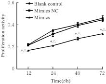
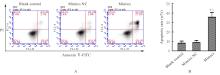
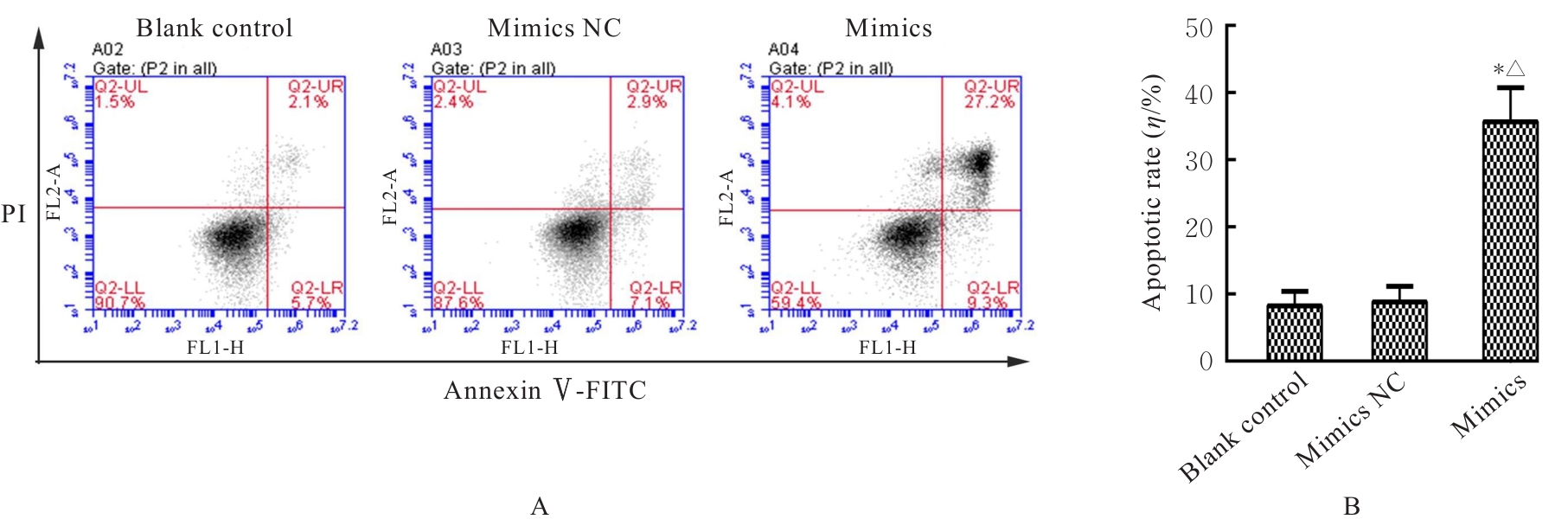
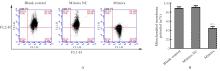
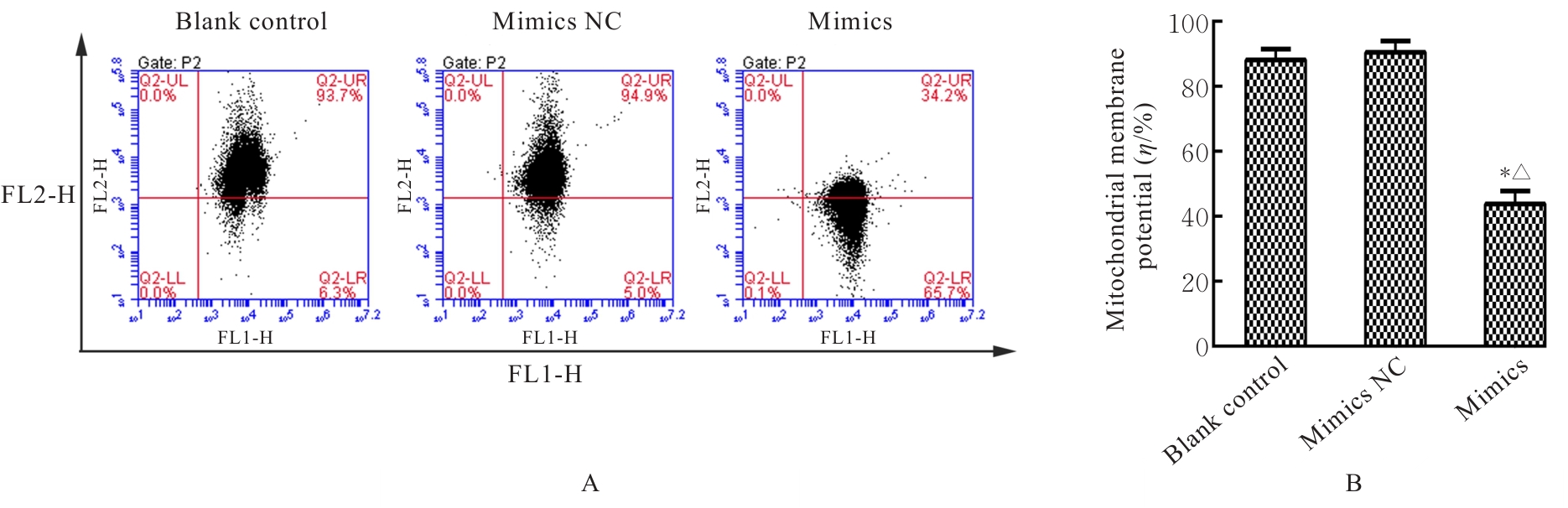
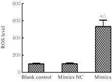
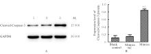
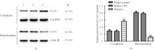
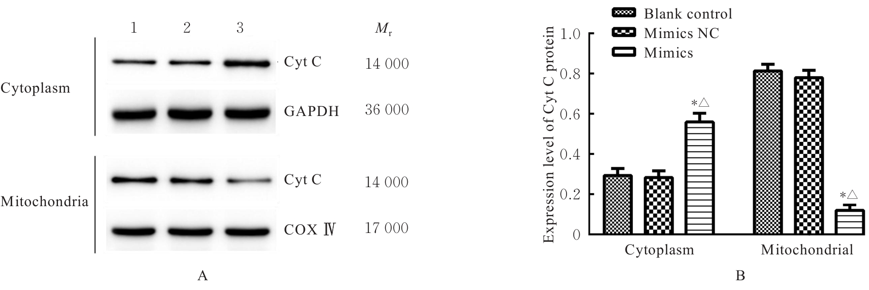
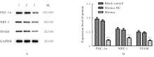
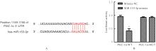
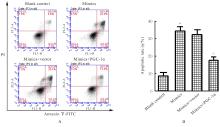
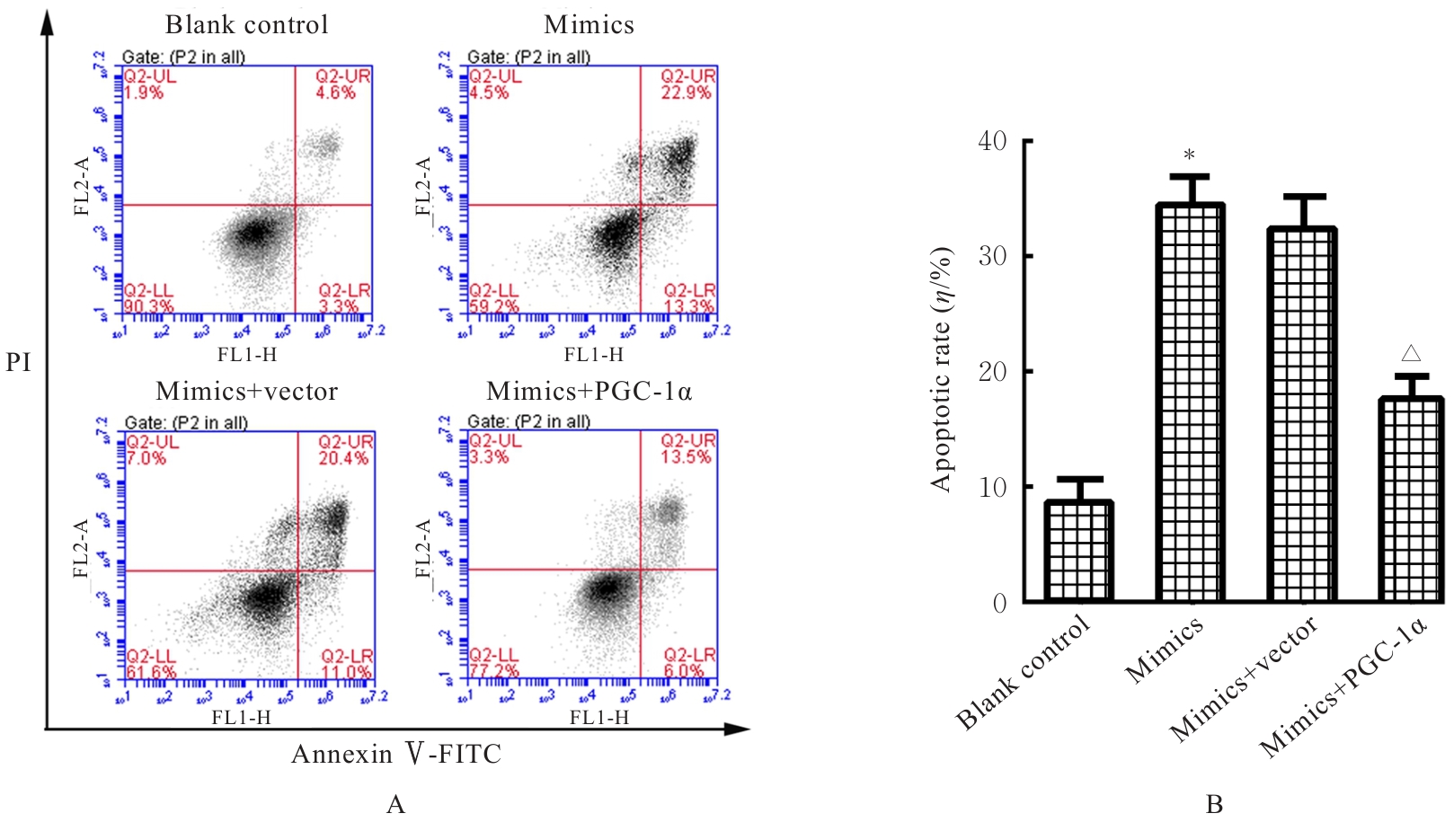
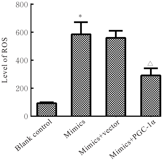
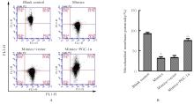
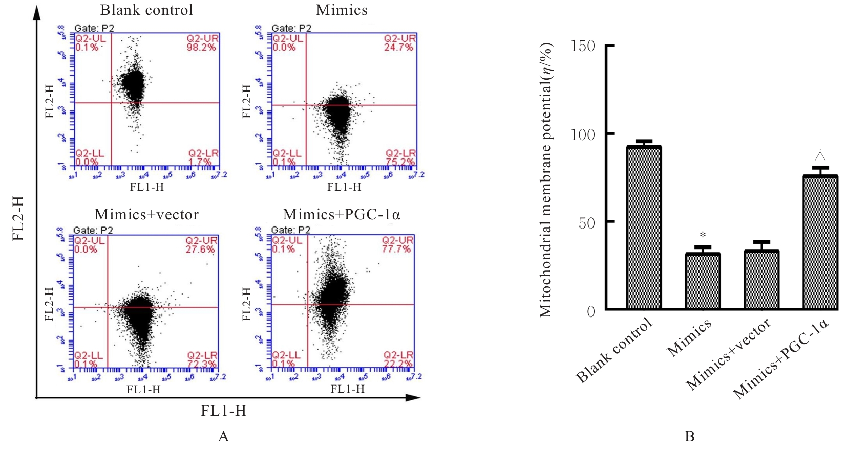
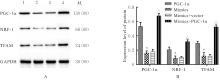
目的 探讨微小RNA-153-3p (miR-153-3p) 对口腔鳞状细胞癌(OSCC)细胞凋亡的影响,并阐明其作用机制。 方法 收集确诊为OSCC的84例患者的癌组织和癌旁组织,并培养5种OSCC细胞和正常人口腔黏膜HOK-16A细胞。采用实时荧光定量PCR(RT-qPCR)法检测OSCC患者癌组织和癌旁组织及5种OSCC细胞中miR-153-3p表达水平。将Tca8113细胞随机分为空白对照组、mimics NC组、mimics组、mimics+vector组和mimics+过氧化物酶体增殖物激活受体γ共激活因子1α(PGC-1α)组,分别转染mimics NC质粒、miR-153-3p mimic质粒、PGC-1α过表达质粒或共转染2种质粒。采用RT-qPCR法检测转染后各组细胞中miR-153-3p表达水平,MTT法检测各组细胞增殖活性,流式细胞术检测各组细胞凋亡率,2',7'-二氯二氢荧光素二乙酯(DCFH-DA)荧光染色法检测各组细胞中活性氧(ROS)水平,JC-1探针染色法检测各组细胞线粒体膜电位,Western blotting法检测各组细胞胞浆和线粒体中细胞色素C(Cyt C)蛋白表达水平及细胞中Cleaved-Caspase-3、PGC-1α、核呼吸因子1(NRF-1)及线粒体转录因子A(TFAM)蛋白表达水平,双荧光素酶报告基因检测实验验证miR-153-3pPGC-的靶向关系。 结果 与癌旁组织和HOK-16A细胞比较,OSCC癌组织及各种OSCC细胞中miR-153-3p表达水平均明显降低(P<0.05),其中Tca8113细胞中表达水平最低。转染后,与空白对照组和mimics NC组比较,mimics组Tca8113细胞中miR-153-3p表达水平明显升高(P<0.001);各时间点细胞增殖活性均明显降低(P<0.001);细胞凋亡率和ROS水平明显升高(P<0.001),细胞线粒体膜电位明显降低(P<0.001);细胞中Cleaved-Caspase-3蛋白表达水平和细胞浆中Cyt C蛋白表达水平均明显升高(P<0.001),线粒体中Cyt C蛋白和细胞中PGC-1α/TFAM通路相关蛋白PGC-1、NRF-1及TFAM表达水平均明显降低(P<0.05)。双荧光素酶报告基因实验证明miR-153-3p能够特异性靶向调控PGC-基因表达。共转染后,与空白对照组比较,mimics组细胞凋亡率和细胞中ROS水平明显升高(P<0.001),线粒体膜电位明显降低(P<0.001);与mimics组比较,mimics+vector组细胞凋亡率、细胞中ROS水平和线粒体膜电位差异无统计学意义(P>0.05);与mimics+vector组比较,mimics+PGC-1α组细胞凋亡率和细胞中ROS水平均明显降低(P<0.01),细胞线粒体膜电位明显升高(P<0.001)。与空白对照组比较,mimics组细胞中PGC-1、NRF-1和TFAM蛋白表达水平均明显降低(P<0.001);与mimics+vector组比较,mimics+PGC-1α组细胞中PGC-1、NRF-1和TFAM蛋白表达水平均明显升高(P<0.001)。 结论 MiR-153-3p过表达可通过靶向下调PGC-表达,阻断PGC-1α/TFAM信号通路,诱导线粒体功能障碍并激活细胞线粒体凋亡途径,诱导Tca8113细胞凋亡。

关键词: 口腔鳞状细胞癌, 微小RNA-153-3p, 过氧化物酶体增殖物激活受体γ共激活因子1α, 线粒体, 细胞凋亡

Abstract:

Objective To discuss the effect of microRNA-153-3p (miR-153-3p) on the apoptosis of oral squamous cell carcinoma (OSCC) cells, and to clarify its mechanism. Methods The Cancer tissue and adjacent normal tissue were collected from 84 patients diagnosed with OSCC, and five kinds of OSCC cells and normal human oral mucosa HOK-16A cells were cultured. Real-time fluorescence quantitative PCR (RT-qPCR) method was used to detect the expression levels of miR-153-3p in the OSCC cancer tissue and adjacent normal tissue, as well as in the five kinds of cells. The Tca8113 cells were randomly divided into blank control group, mimics NC group,mimics group, mimics+vector group, and mimics+peroxisome proliferator-activated receptor γ coactivator-1α (PGC-1α) group, and were transfected with mimics NC plasmid,miR-153-3p mimic plasmid, PGC-1α overexpression plasmid, or co-transfected with both plasmids. RT-qPCR method was used to detect the expression levels of miR-153-3p in the cells in various groups after transfection; MTT method was used to detect the proliferation activities of the cells in various groups; flow cytometry was used to detect the apoptotic rates of the cells in various groups; 2',7'- dichlorodihydrofluorescein diacetate (DCFH-DA) fluorescence staining was used to detect the levels of reactive oxygen species (ROS) in the cells in various groups; JC-1 probe staining was used to detect the mitochondrial membrane potential of the cells in various groups; Western blotting method was used to detect the expression levels of cytochrome C (Cyt C) protein in the cytoplasm and mitochondria, as well as the expression levels of Cleaved-Caspase-3, PGC-1α, nuclear respiratory factor 1 (NRF-1), and mitochondrial transcription factor A (TFAM) proteins in the cells in various groups; dual-luciferase reporter assay was used to verify the targeting relationship between miR-153-3p and PGC-. Results Compared with adjacent normal tissue and HOK-16A cells, the expression levels of miR-153-3p in OSCC cancer tissue and OSCC cells were significantly decreased (P<0.05), while the lowest expression level was observed in the Tca8113 cells. After transfection, compared with blank control group and mimics NC group, the expression level of miR-153-3p in the Tca8113 cells in mimics group was significantly increased (P<0.001); the proliferation activity of the cells at each time point was significantly decreased (P<0.001); the apoptotic rate and ROS level of the cells were significantly increased (P<0.001), while the mitochondrial membrane potential of the cells was significantly decreased (P<0.001); the expression level of Cleaved-Caspase-3 protein in the cells and the expression level of Cyt C protein in the cytoplasm were significantly increased (P<0.001), while the expression level of Cyt C protein in the mitochondria and the expression levels of PGC-1α/TFAM pathway-related proteins PGC-1α, NRF-1, and TFAM in the cells were significantly decreased (P<0.05). The dual-luciferase reporter assay results confirmed that miR-153-3p could specifically target and regulate the expression of the PGC- gene. After co-transfection, compared with blank control group, the apoptotic rate and ROS level in the cells in mimics group were significantly increased (P<0.001), while the mitochondrial membrane potential was significantly decreased (P<0.001); compared with mimics group, there were no significant differences in apoptotic rate, ROS level, and mitochondrial membrane potential in the cells in mimics+vector group (P>0.05); compared with mimics+vector group, the apoptotic rate and ROS level in the cells in mimics+PGC-1α group were significantly decreased (P<0.01), while the mitochondrial membrane potential was significantly increased (P<0.001). Compared with blank control group, the expression levels of PGC-1α, NRF-1, and TFAM proteins in the cells in mimics group were significantly decreased (P<0.001); compared with mimics+vector group, the expression levels of PGC-1α, NRF-1, and TFAM proteins in the cells in mimics+PGC-1α group were significantly increased (P<0.001). Conclusion ?Overexpression of miR-153-3p can induce apoptosis in the Tca8113 cells by targeting and down-regulating the expression of PGC-1α, thereby blocking the PGC-1α/TFAM signaling pathway, inducing mitochondrial dysfunction, and activating the mitochondrial apoptotic pathway.

Key words: Oral squamous cell carcinoma, MicroRNA-153-3p, Peroxisome proliferator-activated receptor γ coactivator-1α, Mitochondria, Apoptosis

中图分类号: 

  • R739.8