吉林大学学报(医学版) ›› 2026, Vol. 52 ›› Issue (1): 70-80.doi: 10.13481/j.1671-587X.20260108

• 基础研究 • 上一篇    下一篇

缓激肽B1受体拮抗剂ELN441958通过调节Akt/FoxO3a信号通路对肝癌HepG2细胞增殖的抑制作用

孙杉杉1,陆梅2,高新富2,吕光耀2,赵宝磊3,吕文文2()   

  1. 1.滨州医学院附属医院肿瘤科,山东 滨州 256600
    2.滨州医学院附属医院药学部,山东 滨州 256600
    3.滨州医学院附属医院肝胆外科,山东 滨州 256600
  • 收稿日期:2025-01-29 接受日期:2025-03-24 出版日期:2026-01-28 发布日期:2026-02-24
  • 通讯作者: 吕文文 E-mail:byyxlww@126.com
  • 作者简介:孙杉杉(1998-),女,山东省烟台市人,在读硕士研究生,主要从事抗肿瘤药物筛选及机制方面的研究。
  • 基金资助:
    山东省科技厅自然科学基金项目(ZR2021MH173);山东省卫健委齐鲁卫生与健康杰出青年人才项目(鲁卫人才字〔2020〕3号)

Inhibitory effect of Bradykinin 1 receptor antagonist ELN441958 on proliferation of HepG2 cells by regulating Akt/FoxO3a signaling pathway

Shanshan SUN1,Mei LU2,Xinfu GAO2,LYu Guangyao2,Baolei Zhao3,LYu Wenwen2()   

  1. 1.Department of Oncology,Affliated Hospital,Binzhou Medical University,Binzhou 256600,China
    2.Department of Pharmacy,Affliated Hospital,Binzhou Medical University,Binzhou 256600,China
    3.Department of Hepatobiliary Surgery,Affliated Hospital,Binzhou Medical University,Binzhou 256600,China
  • Received:2025-01-29 Accepted:2025-03-24 Online:2026-01-28 Published:2026-02-24
  • Contact: LYu Wenwen E-mail:byyxlww@126.com

摘要:

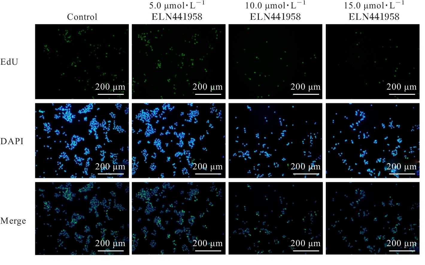
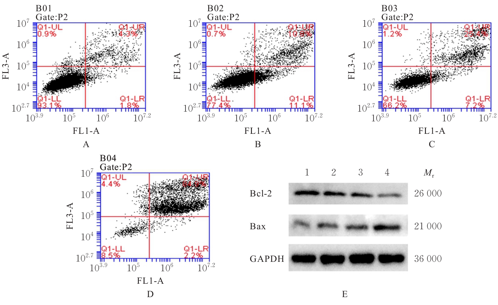
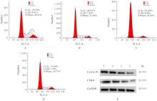
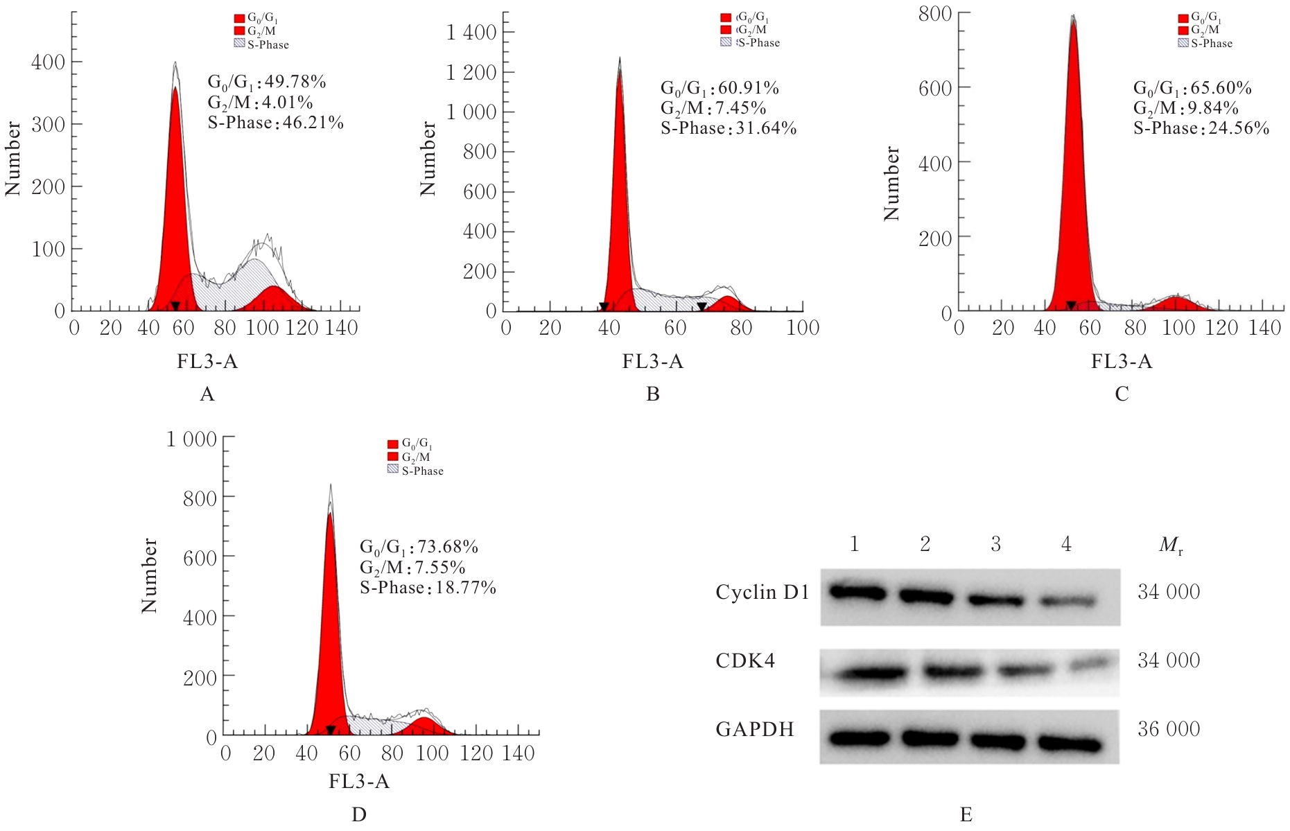
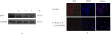
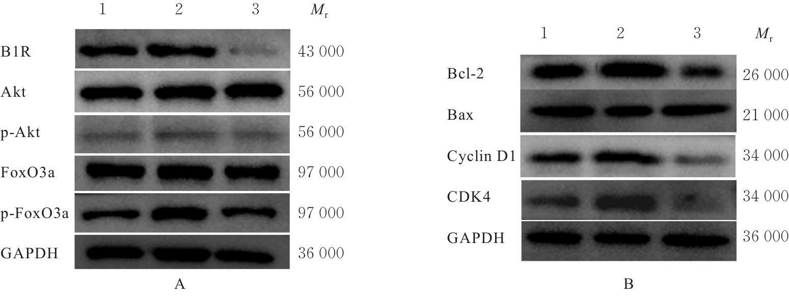
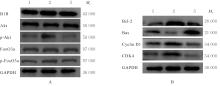
目的 探讨缓激肽B1受体(B1R)拮抗剂ELN441958对肝癌HepG2细胞的生长抑制作用及其对蛋白激酶B(Akt)/叉头框O 3a(FoxO3a)信号通路的调控机制,阐明B1R拮抗剂ELN441958在肝癌中的抗肿瘤作用。 方法 HepG2细胞经不同剂量(0、2.5、5.0、10.0和20.0 μmol·L-1)ELN441958处理24、48和72 h后,0 μmol·L-1 ELN441958作为对照组,采用细胞计数试剂盒8(CCK-8)法检测各组细胞增殖抑制率。 将HepG2细胞分为对照组、 5.0 μmol·L-1 ELN441958组、10.0 μmol·L-1 ELN441958组和15.0 μmol·L-1 ELN441958组,采用5-乙炔基-2'-脱氧尿嘧啶核苷(EdU)染色检测各组HepG2细胞的EdU阳性细胞率,流式细胞术检测各组细胞凋亡率及处于不同细胞周期的细胞比例,免疫荧光法检测各组HepG2细胞中B1R蛋白表达水平,Western blotting法检测各组HepG2细胞中B细胞淋巴瘤2(Bcl-2)、Bcl-2相关X蛋白(Bax)、细胞周期蛋白D1(Cyclin D1)、细胞周期蛋白依赖性激酶4(CDK4)、B1R、Akt、磷酸化Akt(p-Akt)、FoxO3a和磷酸化FoxO3a(p-FoxO3a)蛋白表达水平。将HepG2细胞分为对照组、B1R激动剂des-Arg9-BK(1.0 μmol·L-1 des-Arg9-BK)组和BK+ELN441958组,Western blotting法检测各组HepG2细胞中Bcl-2、Bax、Cyclin D1、CDK4、Akt、p-Akt、FoxO3a和p-FoxO3a蛋白表达水平。将HepG2细胞分为对照组、Akt激动剂SC79组和SC79+ELN441958组,Western blotting法检测各组HepG2细胞中Bcl-2、Bax、Cyclin D1、CDK4、Akt、p-Akt、FoxO3a和p-FoxO3a蛋白表达水平。 结果 CCK-8法,与对照组比较,当作用时间相同, HepG2细胞增殖抑制率随ELN441958剂量升高而明显升高(P<0.05或P<0.01);当ELN441958剂量相同时,HepG2细胞增殖抑制率随作用时间增加而明显升高(P<0.05或P<0.01)。ELN441958作用24、48和72 h的半数抑制浓度 (IC50) 分别为 (21.4±1.1)、 (10.5±0.3)和(3.2±0.3)μmol·L-1。与对照组比较,5.0、10.0和15.0 μmol·L-1 ELN441958组HepG2细胞的EdU阳性细胞率明显降低(P<0.05或P<0.01)。流式细胞术,与对照组比较,不同剂量ELN441958组HepG2细胞凋亡率明显升高(P<0.05或P<0.01),G0/G1期细胞百分率明显升高(P<0.05或P<0.01)。Western blotting法,与对照组比较,5.0、10.0和15.0 μmol·L-1 ELN441958组HepG2细胞中Bcl-2、CDK4和Cyclin D1蛋白表达水平明显降低(P<0.05或P<0.01),Bax蛋白表达水平明显升高(P<0.05或P<0.01)。免疫荧光法,与对照组比较,15.0 μmol·L-1 ELN441958组HepG2细胞中B1R荧光强度明显降低(P<0.01)。Western blotting法,与对照组比较,10.0和15.0 μmol·L-1 ELN441958组HepG2细胞中B1R蛋白表达水平明显降低(P<0.01),5、10和15 μmol·L-1 ELN441958组HepG2细胞中p-Akt和p-FoxO3a蛋白表达水平明显降低(P<0.05或P<0.01);与B1R激动剂des-Arg9-BK组比较,BK+ELN441958组HepG2细胞中Bcl-2、CDK4、Cyclin D1、B1R、p-Akt和p-FoxO3a蛋白表达水平明显降低(P<0.01),Bax蛋白表达水平明显升高(P<0.01);与SC79组比较,SC79+ELN441958组HepG2细胞中Bcl-2、CDK4、Cyclin D1、B1R、p-Akt和p-FoxO3a蛋白表达水平明显降低(P<0.01),Bax蛋白表达水平明显升高(P<0.01)。 结论 B1R拮抗剂ELN441958能够抑制HepG2细胞增殖,并诱导HepG2细胞凋亡和G0/G1期细胞周期阻滞,其作用机制可能与调节Akt/FoxO3a信号轴有关。

关键词: ELN441958, 肝肿瘤, 蛋白激酶B, 叉头框O 3a, 细胞凋亡, 细胞周期

Abstract:

Objective To discuss the inhibitory effect of the bradykinin B1 receptor (B1R) antagonist ELN441958 on the growth of hepatocellular carcinoma HepG2 cells and its regulatory mechanism on the protein kinase B (Akt)/forkhead box O3a (FoxO3a) signaling pathway, and to clarify the anti-tumor effect of the B1R antagonist ELN441958 in liver cancer. Methods After the HepG2 cells were treated with different doses (0, 2.5, 5.0, 10.0, and 20.0 μmol·L-1) of ELN441958 for 24, 48, and 72 h, 0 μmol·L-1 ELN441958 was regared as control group. CCK-8 method was used to detect the inhibitory rate of proliferation of the cells in various groups. The HepG2 cells were divided into control group, 5.0 μmol·L-1 ELN441958 group, 10.0 μmol·L-1 ELN441958 group, and 15.0 μmol·L-1 ELN441958 group; 5-ethynyl-2'-deoxyuridine (EdU) staining was used to detect the EdU positive cell rates of the HepG2 cells in various groups; flow cytometry was used to detect the apoptotic rates of the cells in various groups and the proportions of the cells at different cell cycles; immunofluorescence method was used to detect the expression level of B1R protein in the HepG2 cells in various groups; Western blotting method was used to detect the expression levels of B-cell lymphoma 2 (Bcl-2), Bcl-2-associated X protein (Bax), cyclin D1 (Cyclin D1), cyclin-dependent kinase 4 (CDK4), B1R, Akt, phosphorylated Akt (p-Akt), FoxO3a, and phosphorylated FoxO3a (p-FoxO3a) proteins in the HepG2 cells in various groups. The HepG2 cells were divided into control group, B1R agonist des-Arg9-BK (1.0 μmol·L-1 des-Arg9-BK) group, and BK+ELN441958 group; Western blotting method was used to detect the expression levels of Bcl-2, Bax, Cyclin D1, CDK4, Akt, p-Akt, FoxO3a, and p-FoxO3a proteins in the HepG2 cells in various groups. The HepG2 cells were divided into control group, Akt agonist SC79 group, and SC79+ELN441958 group; Western blotting method was used to detect the expression levels of Bcl-2, Bax, Cyclin D1, CDK4, Akt, p-Akt, FoxO3a, and p-FoxO3a proteins in the HepG2 cells in various groups. Results The CCK-8 assay results showed that compared with control group, when the action time was the same, the inhibitory rate of proliferation of the HepG2 cells was significantly increased with the increase of ELN441958 dose (P<0.05 or P<0.01); when the dose of ELN441958 was the same, the inhibitory rate of proliferation of the HepG2 cells was significantly increased with the extension of action time (P<0.05 or P<0.01). The half maximal inhibitory concentration (IC50) values of ELN441958 at 24, 48, and 72 h were (21.4±1.1), (10.5±0.3), and (3.2±0.3) μmol·L?1, respectively. Compared with control group, the EdU positive cell rates of the HepG2 cells in 5.0, 10.0, and 15.0 μmol·L?1 ELN441958 groups were significantly decreased (P<0.05 or P<0.01). The flow cytometry results showed that compared with control group, the apoptotic rates of the HepG2 cells in different doses of ELN441958 groups were significantly increased (P<0.05 or P<0.01), and the percentages of the cells at G0/G1 phase were significantly increased (P<0.05 or P<0.01). The Western blotting results showed that compared with control group, the; the expression levels of Bcl-2, CDK4, and Cyclin D1 proteins in the HepG2 cells in 5.0, 10.0 and 15.0 μmol·L-1 ELN441958 groups were significantly decreased (P<0.01), and the expression level of Bax protein was significantly increased (P<0.05 or P<0.01). The immunofluorescence results showed that compared with control group, the fluorescence intensity of B1R in the HepG2 cells in 15.0 μmol·L-1 ELN441958 group was significantly decreased (P<0.01). The Western blotting results showed that compared with control group, the expression level of B1R, p-Akt, and p-FoxO3a proteins in the HepG2 cells in 10.0 and 15.0 μmol·L-1 ELN441958 groups were significantly decreased (P<0.01); the expressions p-Akt, and p-FoxO3a proteins were decreased (P<0.05 or P<0.01) compared with B1R agonist des-Arg9-BK group, the expression levels of Bcl-2, CDK4, Cyclin D1, B1R, p-Akt, and p-FoxO3a proteins in the HepG2 cells in BK+ELN441958 group were significantly decreased (P<0.01), and the expression level of Bax protein was significantly increased (P<0.01); compared with SC79 group, the expression levels of Bcl-2, CDK4, Cyclin D1, B1R, p-Akt, and p-FoxO3a proteins in the HepG2 cells in SC79+ELN441958 group were significantly decreased (P<0.01), and the expression level of Bax protein was significantly increased (P<0.01). Conclusion The B1R antagonist ELN441958 can inhibit the proliferation of HepG2 cells, and induce the apoptosis and G0/G1 phase cell cycle arrest of HepG2 cells, and its mechanism may be related to the regulation of Akt/FoxO3a signaling axis.

Key words: ELN441958, Liver neoplasms, Protein kinase B, Forkhead box O3a, Apoptosis, Cell cycle

中图分类号: 

  • R965