吉林大学学报(医学版) ›› 2021, Vol. 47 ›› Issue (2): 352-359.doi: 10.13481/j.1671-587X.20210214

• 基础研究 • 上一篇    下一篇

MARCH1通过PI3K/AKT信号通路对人胃癌细胞迁移和侵袭的促进作用

王暖1,杨丽娟2(),代娟娟2,王爱丽1,武艳2,刘成霞1()   

  1. 1.滨州医学院附属医院消化内科,山东 滨州 256600
    2.滨州医学院附属医院肿瘤研究实验室,山东 滨州 256600
  • 收稿日期:2020-06-04 出版日期:2021-03-28 发布日期:2021-03-25
  • 通讯作者: 杨丽娟,刘成霞 E-mail:yljlw@163.com;phdlcx@163.com
  • 作者简介:王 暖(1995-),女,山东省淄博市人,在读医学硕士,主要从事胃肠道肿瘤基础和临床方面的研究。
  • 基金资助:
    山东省教育厅“临床医学+X”学科建设项目(200375);山东省卫健委中医药科技发展计划项目(2019-0517);山东省科技厅重点研发计划项目(2019GSF107099)

Promotion effects of MARCH1 on migration and invasion of human gastric cancer cells through PI3K/AKT signaling pathway

Nuan WANG1,Lijuan YANG2(),Juanjuan DAI2,Aili WANG1,Yan WU2,Chengxia LIU1()   

  1. 1.Department of Gastroenterology,Affiliated Hospital,Binzhou Medical College,Binzhou 256600,China
    2.Cancer Research Laboratory,Affiliated Hospital,Binzhou Medical College,Binzhou 256600,China
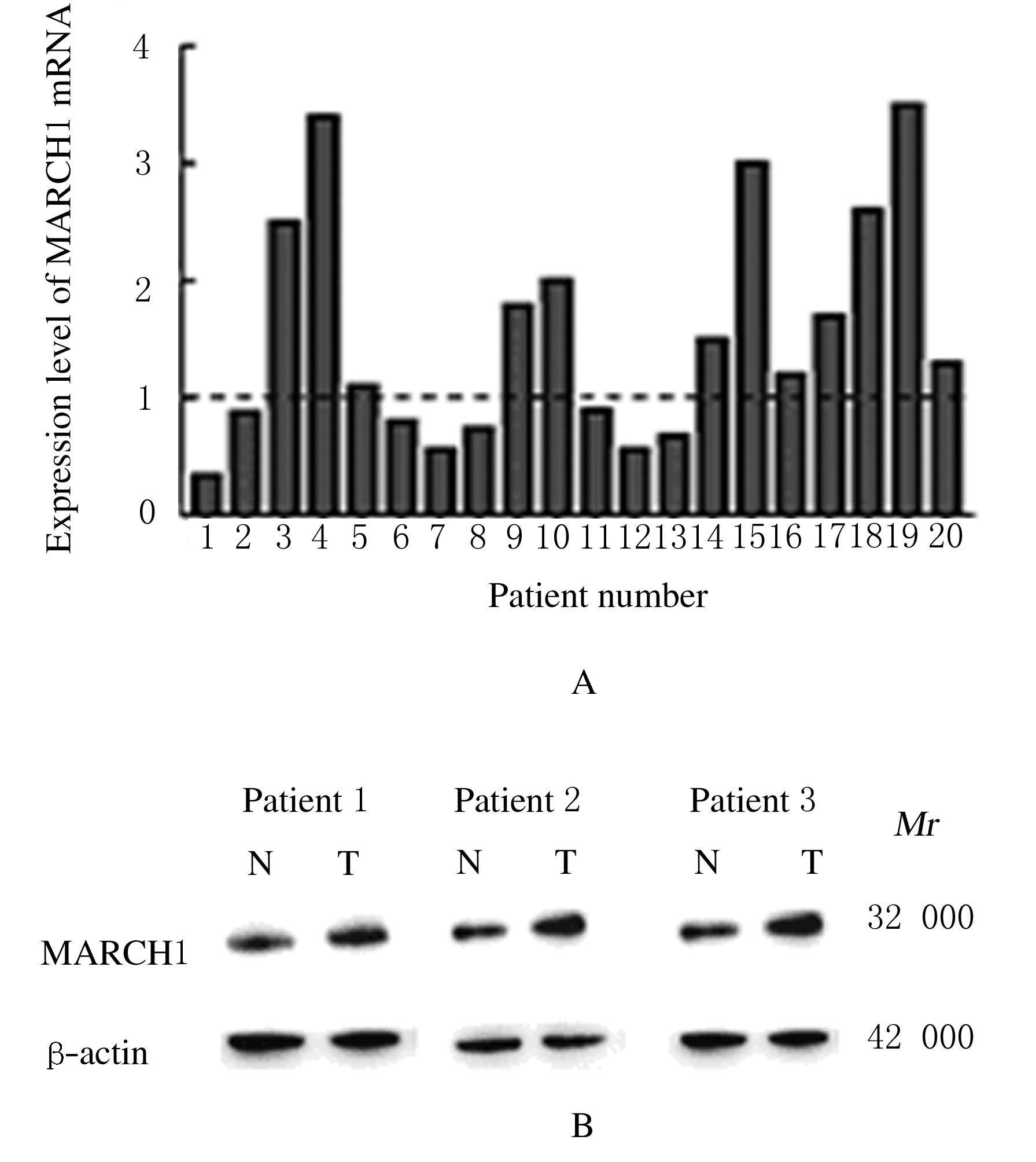
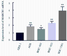
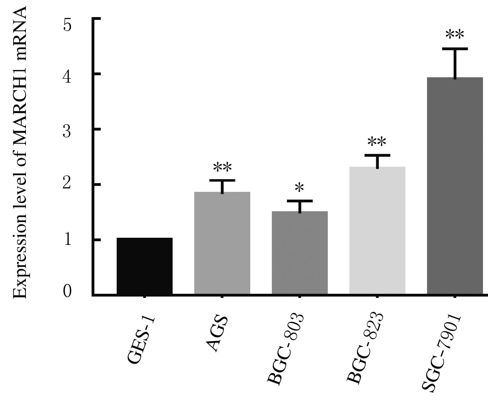
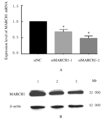
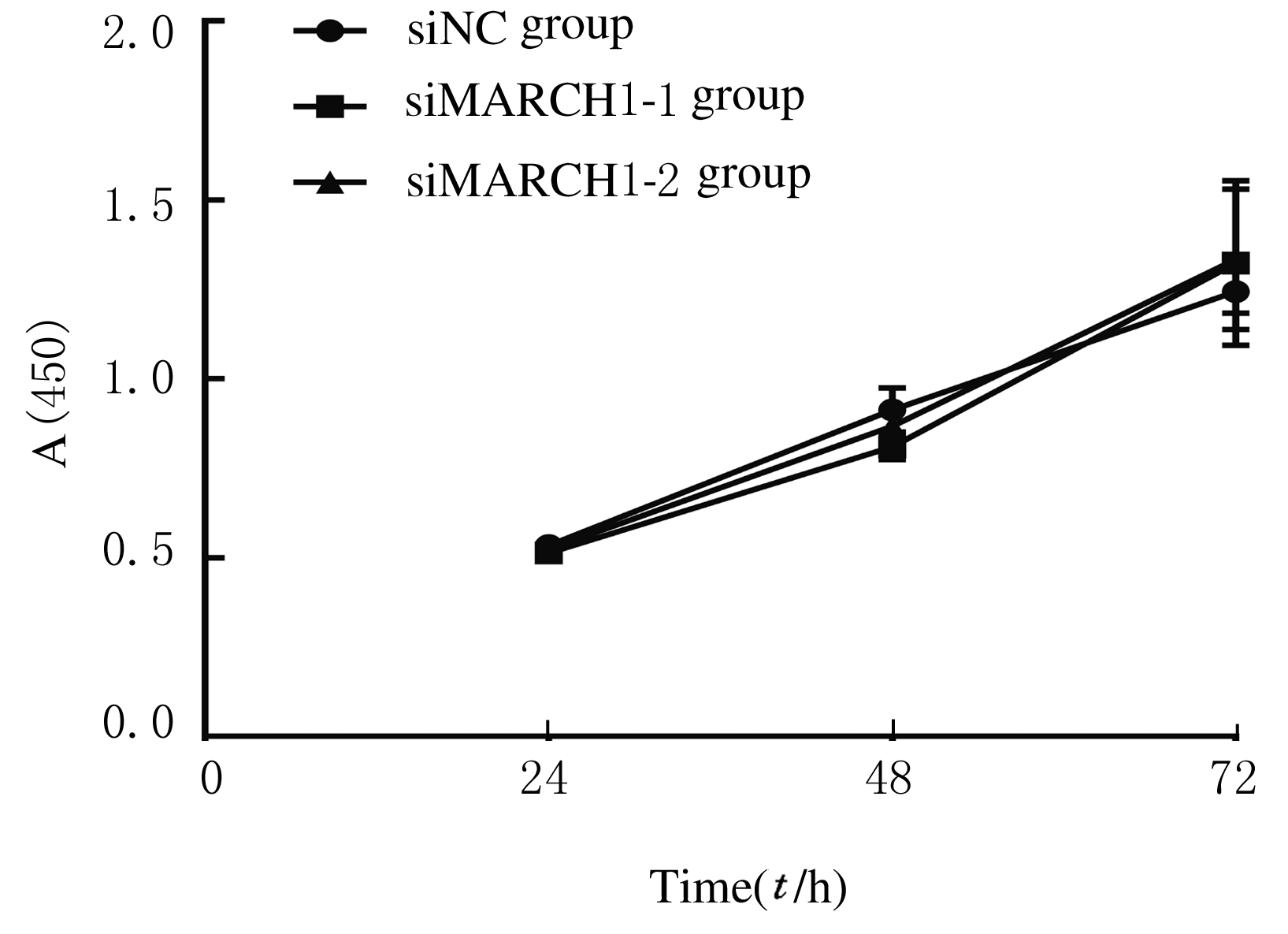
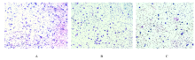
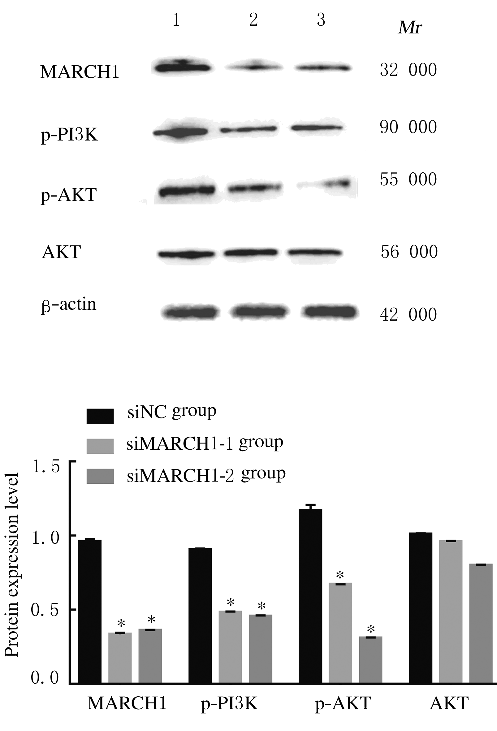
  • Received:2020-06-04 Online:2021-03-28 Published:2021-03-25
  • Contact: Lijuan YANG,Chengxia LIU E-mail:yljlw@163.com;phdlcx@163.com

摘要: 目的

探讨膜相关环状蛋白1(MARCH1)对胃癌细胞迁移和侵袭的影响,并阐明其可能的分子机制。

方法

收集胃癌手术切除的20例胃癌组织和癌旁组织,实时荧光定量聚合酶链式反应(RT-qPCR)法和Western blotting法检测不同组织中MARCH1 mRNA和蛋白表达水平。选择人胃黏膜正常上皮细胞GES-1和人胃癌细胞BGC-823、BGC-803、AGS及SGC-7901,RT-qPCR法检测不同细胞中MARCH1 mRNA表达水平。取对数生长期的SGC-7901细胞系分为siNC(转染siNC)、siMARCH1-1组(转染siMARCH1-1)和siMARCH1-2组(转染siMARCH1-2),RT-qPCR法和Western blotting法检测各组细胞中MARCH1 mRNA和蛋白表达水平,CCK-8法检测各组细胞增殖活性,细胞划痕实验检测各组细胞划痕愈合率,Transwell小室实验检测各组细胞侵袭能力,Western blotting法检测各组细胞中磷酸化磷脂酰肌醇3-激酶(p-PI3K)、磷酸化蛋白激酶B(p-AKT)和蛋白激酶B(AKT)蛋白表达水平。

结果

与癌旁组织比较,胃癌组织中MARCH1 mRNA和蛋白表达水平明显升高(P<0.05),与胃黏膜正常上皮细胞GES-1比较,人胃癌细胞BGC-823、BGC-803、AGS和SGC-7901中MARCH1 mRNA表达水平明显升高(P<0.05或P<0.01)。与siNC比较,siMARCH1-1组和siMARCH1-2组细胞中MARCH1 mRNA及蛋白表达水平均明显降低(P<0.01);与siNC比较,siMARCH1-1组和siMARCH1-2组细胞增殖活性差异无统计学意义(P>0.05),细胞划痕愈合率和侵袭数均明显降低(P<0.01),细胞中p-PI3K和p-AKT蛋白表达水平明显降低(P<0.01),AKT蛋白表达水平差异无统计学意义(P>0.05)。

结论

MARCH1能够促进胃癌细胞的迁移和侵袭,其机制可能与调控PI3K/AKT信号通路有关。

关键词: 胃肿瘤, 膜相关环状蛋白1, 细胞迁移, 细胞侵袭, 磷脂酰肌醇3-激酶, 蛋白激酶B

Abstract: Objective

To investigate the effects of membrane associated RING-CH 1 (MARCH1) on the migration and invasion of gastric cancer cells, and to clarify its possible molecular mechanism.

Methods

A total of 20 cases of gastric cancer tissue and adjacent tissue obtained from gastrectomy were collected, and the expression levels of MARCH1 mRNA and protein in the different tissues were detected by Real-time fluorescence quantitative polymerase chain reaction (RT-qPCR) method and Western blotting method. The expression levels of MARCH1 mRNA in the human gastric mucosal normal epithelial cells GES-1 and the human gastric cancer cells BGC-823, BGC-803, AGS and SGC-7901 were detected by RT-qPCR method. The SGC-7901 cells in logarithmic growth phase were divided into siNC group(transfected with siNC), siMARCH1-1 group(transfected with siMARCH1-1) and siMARCH1-2 group(transfected with siMARCH1-2). RT-qPCR method and Western blotting method were used to detect the expression levels of MARCH1 mRNA and protein in the cells in various groups. CCK-8 method was used to detect the cell proliferation activities in various groups; cell scratch test was used to detect the scratch healing rates of the cells in various groups; Transwell chamber test was used to detect the cell invasion abilities in various groups. The expression levels of phosphorylated phosphatidylinositol 3-kinase (p-PI3K), phosphorylated protein kinase B(p-AKT) and protein kinase B (AKT) proteins in various groups were detected by Western blotting method.

Results

Compared with the adjacent tissue, the expression levels of MARCH1 mRNA and protein in the gastric cancer tissue were increased (P<0.05). Compared with the human gastric mucosal normal epithelial cells GES-1, the expression levels of MARCH1 mRNA in human gastric cancer cells BGC-823, BGC-803, AGS, and SGC-7901 were all increased (P<0.05 or P<0.01). Compared with siNC group, the expression levels of MARCH1 mRNA and protein in siMARCH1-1 group and siMARCH1-2 group were significantly reduced (P<0.01). Compared with siNC group, the cell proliferation activities in siMARCH1-1 group and siMARCH1-2 group had no significant differences(P>0.05); the cell scratch healing rates and the number of invasion cells in siMARCH1-1 group and siMARCH1-2 group were significantly reduced (P<0.01 ). The expression levels of p-PI3K and p-AKT proteins in siMARCH1-1 group and siMARCH1-2 group were significantly reduced compared with siNC group (P<0.01), and the expression levels of AKT protein had no significant differences(P>0.05).

Conclusion

MARCH1 can promote the migration and invasion of gastric cancer cells, and its mechanism may be related to the regulation of PI3K/AKT signaling pathway.

Key words: stomach neoplasm, membrane associated RING-CH 1, cell migration, cell invasion, phosphatidylinositol 3-kinase, protein kinase B

中图分类号: 

  • R735.2