吉林大学学报(医学版) ›› 2021, Vol. 47 ›› Issue (1): 145-151.doi: 10.13481/j.1671-587x.20210120

• 基础研究 • 上一篇    下一篇

姜黄素对结直肠癌小鼠肿瘤生长的抑制作用及其PTEN/PI3K/Akt信号通路机制

裴永彬,王桂琦,李卫,姜霞,姜海波,赵增仁()   

  1. 河北医科大学第一医院普外三科,河北 石家庄 050031
  • 收稿日期:2020-05-06 出版日期:2021-01-28 发布日期:2021-01-27
  • 通讯作者: 赵增仁 E-mail:xiaoerbing112@sina.com
  • 作者简介:裴永彬(1977-)男,河北省石家庄市人,副教授,副主任医师,医学硕士,主要从事胃肠外科基础和临床 方面的研究。
  • 基金资助:
    河北省卫计委医学科学项目(172777164)

Inhibitory effect of curcumin on tumor growth in colorectal cancer mice and its mechanism of PTEN/PI3K/Akt signaling pathwayPEI Yongbin, WANG Guiqi, LI Wei, JIANG Xia, JIANG Haibo, ZHAO Zengren (Department of General Surgery,First Hospital,Hebei Medical University, Shijiazhuang 050031,China)
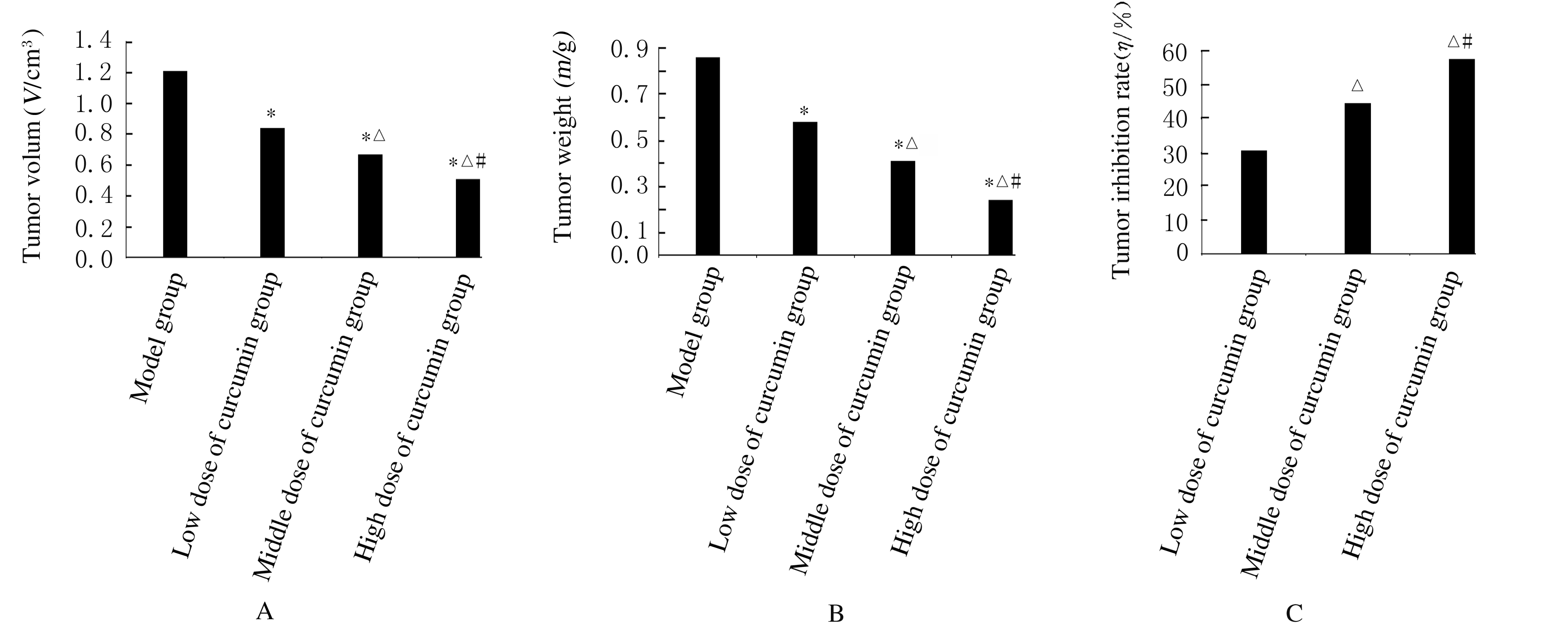
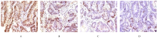
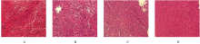
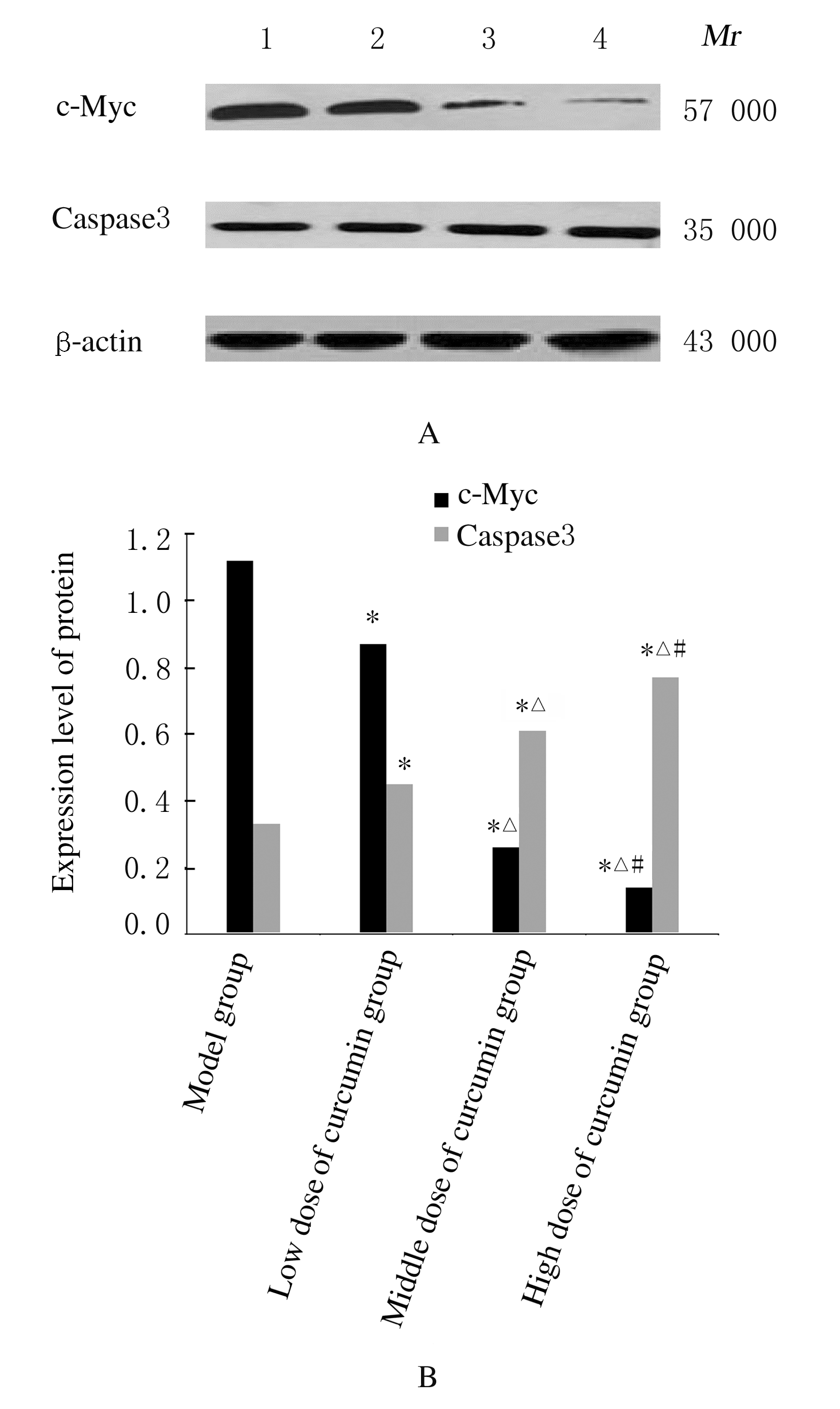
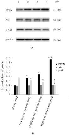
  • Received:2020-05-06 Online:2021-01-28 Published:2021-01-27

摘要: 目的

探讨姜黄素对结直肠癌小鼠肿瘤生长及第10号染色体缺失的磷酸酶和张力蛋白的同源基因/磷酯酰肌醇3-激酶/蛋白激酶B(PTEN/PI3K/Akt)信号通路的影响,阐明姜黄素对结直肠癌的可能作用机制。

方法

60只小鼠随机分为模型组、低剂量姜黄素组、中剂量姜黄素组和高剂量姜黄素组,每组15只。接种结直肠癌LOVO细胞建立小鼠结直肠癌模型,低、中和高剂量姜黄素组分别给予25、50和100 mg·kg-1姜黄素灌胃,共4周。末次灌胃后24 h,检测各组小鼠肿瘤体积、肿瘤质量和抑瘤率,免疫组织化学染色检测各组小鼠肿瘤组织中增殖细胞核抗原(PCNA)蛋白表达,并计算细胞增殖指数(PI),HE染色观察各组小鼠肿瘤组织病理形态表现,Western blotting法检测各组小鼠肿瘤组织中人髓细胞增生原癌基因c-Myc、半胱氨酸蛋白酶3(caspase3)、PTEN、Akt和磷酸化Akt(p-Akt)蛋白表达水平。

结果

各组小鼠肿瘤体积、肿瘤质量、抑瘤率、肿瘤组织PI值及肿瘤组织中c-Myc、caspase3、PTEN和p-Akt蛋白表达水平比较差异均有统计学意义(P<0.05)。与模型组比较,各剂量姜黄素组小鼠肿瘤体积和肿瘤质量均明显降低(P<0.05),抑瘤率增加(P<0.05),肿瘤组织PI值降低(P<0.05),肿瘤组织中c-Myc和p-Akt蛋白表达水平降低(P<0.05),caspase3和PTEN蛋白表达水平升高(P<0.05)。免疫组织化学染色,与模型组比较,各剂量姜黄素组小鼠肿瘤组织中PCNA阳性细胞数明显减少。HE染色,模型组小鼠肿瘤组织中细胞排列紊乱,核质比例增加,核染色深;与模型组比较,各剂量姜黄素组小鼠肿瘤组织中细胞排列相对整齐,核质比例下降,核染色浅。姜黄素对结直肠癌肿瘤体积、肿瘤质量、抑瘤率、肿瘤组织PI值及肿瘤组织中c-Myc、caspase3、PTEN和p-Akt蛋白表达的影响呈剂量依赖性,不同剂量姜黄素组间上述各指标比较差异均有统计学意义(P<0.05)。

结论

姜黄素可抑制结直肠癌小鼠肿瘤生长,其机制可能与姜黄素抑制PTEN/PI3K/Akt信号通路有关。

关键词: 姜黄素, 结直肠肿瘤, 细胞增殖, 细胞凋亡, 第10号染色体缺失的磷酸酶和张力蛋白的同源基因, 磷酯酰肌醇3-激酶, 蛋白激酶B, 信号通路

Abstract: Objective

To explore the effects of curcumin on the tumor growth and the phosphatase and tensin homologous genes deleted on chromosome ten/ phosphatidylinositol 3 kinase / protein kinase B (PTEN / PI3K / Akt) signaling pathway in the colorectal cancer mice,and to clarify the possible mechanism of curcumin in colorectal cancer.

Methods

A total of 60 mice were divided into model group, low dose of curcumin group, middle dose of curcumin group and high dose of curcumin group, with 15 mice in each group. The mice were inoculated with colorectal cancer LOVO cells to establish the mouse colorectal cancer models.The rats in low,middle and high doses of curcumin groups were given 25, 50 and 100 mg·kg-1 curcumin by gavage, respectively, for 4 weeks.Twenty four hours after the last administration,the tumor volumes, tumor weights and the inhibitory rates of tumor of the mice in various groups were detected. Immunohistochemical staining was used to detect the expressions of proliferating cell nuclear antigen (PCNA) in tumor tissue of the mice in various groups,and the proliferation index(PI) of cells was calculated. HE staining was used to observe the pathomorphology of tumor tissue of the mice in various groups, and Western blotting method was used to determine the expression levels of proto-oncogene c-Myc, caspase3, PTEN, Akt and phosphorylated Akt (p-Akt) proteins in tumor tissue of the mice in various groups.

Results

The tumor volumes, tumor weights, the inhibitory rates of tumor, PI values,and the expression levels of c-Myc, caspase3, PTEN and p-Akt proteins in tumor tissue of the mice in various groups had statistically significant differences (P<0.05).Compared with model group, the tumor volumes and tumor weights of the mice in different doses of curcumin groups were significantly decreased(P<0.05),the inhibitory rates of tumor were increased(P<0.05), the PI values were decreased(P<0.05),the expression levels of c-Myc and p-Akt proteins were decreased(P<0.05),and the expression levels of caspase3 and PTEN proteins were increased(P<0.05). The immunohistochemical results showed that the number of PCNA positive cells in tumor tissue of the mice in different doses of curcumin groups were significantly less than that in model group.The HE staining results showed that the tumor tissue cells in model group were disordered, the nucleus-cytoplasm ratio was increased, and the nuclear staining was deep; compared with model group, the tumor tissue cells of the mice in different doses of curcumin groups were relatively neatly arranged, the nucleus-cytoplasm ratios were decreased, and the nuclear staining was light.The effects of curcumin on tumor volume, tumor weight,inhibiory rate of tumor, PI value of tumor tissue and the expressions of c-Myc,caspase3,PTEN and p-Akt proteins in tumor tissue were dose-dependent, and there were statistically significant differences in the indicators among different doses of curcumin groups (P<0.05).

Conclusion

Curcumin can inhibit the tumor growth in the colorectal cancer mice,and its mechanism may be related to that curcumin can inhibit PTEN/PI3K/Akt signaling pathway.

Key words: curcumin, colorectal tumor, cell proliferation, apoptosis, phosphatase and tensin homologous genes deleted on chromosome ten, phosphatidylinositol 3 kinase, protein kinase B, signaling pathway

中图分类号: 

  • R735.3