吉林大学学报(医学版) ›› 2021, Vol. 47 ›› Issue (6): 1371-1379.doi: 10.13481/j.1671-587X.20210605

• 基础研究 • 上一篇    下一篇

黄芪多糖对野百合碱诱导的肺动脉高压大鼠肺动脉血管的保护作用及其机制

李聪1,2,赵坤2,张景良2,张英杰2(),王洪新1()   

  1. 1.锦州医科大学辽宁省心脑血管药物研究重点实验室,辽宁 锦州 121001
    2.锦州医科大学附属第一医院心血管内科,辽宁 锦州 121000
  • 收稿日期:2021-03-17 出版日期:2021-11-28 发布日期:2021-12-14
  • 通讯作者: 张英杰,王洪新 E-mail:zhangyingjie@jzmu.edu.cn;hongxinwang@jzmu.edu.cn
  • 作者简介:李 聪(1994-),男,重庆市人,在读硕士研究生,主要从事心血管疾病方面的研究。
  • 基金资助:
    国家自然科学基金项目(81973553)

Protective effect of astragalus polysaccharides on pulmonary artery vessels in rats with MCT-induced pulmonary hypertension and its mechanism

Cong LI1,2,Kun ZHAO2,Jingliang ZHANG2,Yingjie ZHANG2(),Hongxin WNAG1()   

  1. 1.Key Laboratory of Cardiovascular and Cerebrovascular Drug Research,Liaoning Province,Jinzhou Medical University,Jinzhou 121001,China
    2.Department of Cardiology,First Affiliated Hospital,Jinzhou Medical University,Jinzhou 121000,China
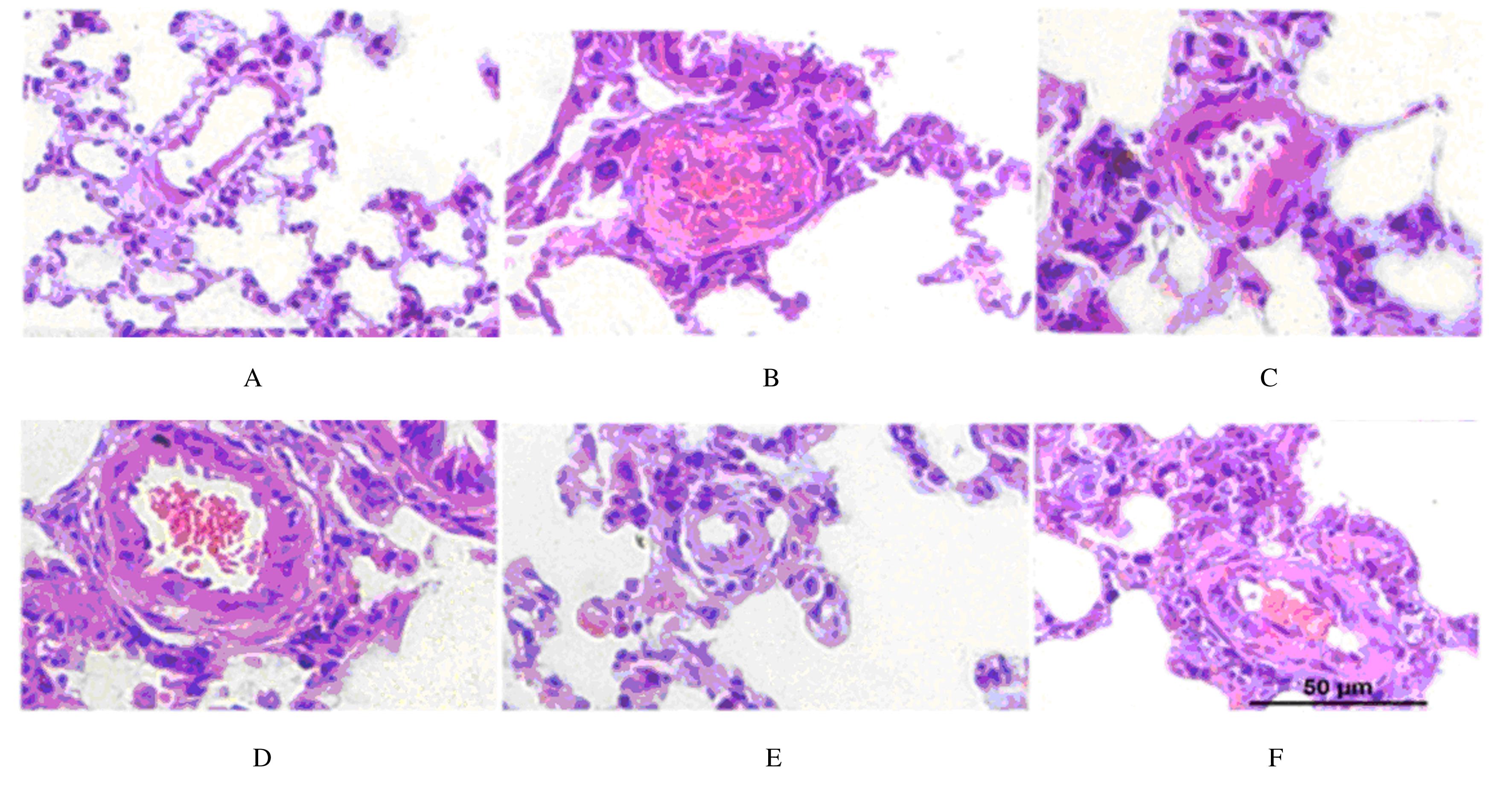
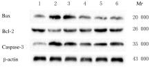
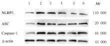
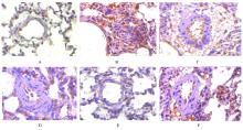
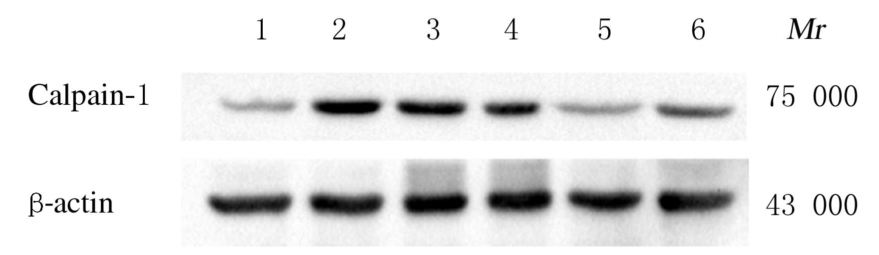
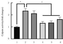
  • Received:2021-03-17 Online:2021-11-28 Published:2021-12-14
  • Contact: Yingjie ZHANG,Hongxin WNAG E-mail:zhangyingjie@jzmu.edu.cn;hongxinwang@jzmu.edu.cn

摘要: 目的

探讨黄芪多糖(APS)对野百合碱(MCT)诱导的肺动脉高压(PAH)大鼠肺动脉血管炎症的干预作用,并阐明其作用机制。

方法

60只雄性SD大鼠随机分为对照组、PAH组、钙蛋白酶1(Calpain-1)抑制剂组、NOD样受体家族蛋白3(NLRP3)抑制剂组、低剂量APS组(400 mg·kg-1 APS)和高剂量APS组(800 mg·kg-1 APS),每组10只。采用60 mg·kg-1 MCT腹腔注射的方法建立PAH模型。ELISA法检测各组大鼠血清白细胞介素18(IL-18)和白细胞介素1β(IL-1β)水平,HE染色观察各组大鼠肺组织病理形态表现,检测各组大鼠右心室收缩压(RVSP)、右心室肥厚指数(RVHI)、肺小动脉管壁厚度占血管总直径的百分率(WT%)和管壁面积占血管总面积的百分率(WA%),免疫组织化学染色检测各组大鼠肺组织中Calpain-1表达量,Western blotting法检测各组大鼠肺组织中B细胞淋巴瘤2(Bcl-2)、Bcl-2相关X蛋白(Bax)、半胱氨酸天冬氨酸蛋白酶3(Caspase-3)、NLRP3、凋亡相关斑点样蛋白(ASC)、半胱氨酸天冬氨酸蛋白酶1(Caspase-1)和Calpain-1蛋白表达水平。

结果

与PAH组比较,低和高剂量APS组及Calpain-1抑制剂组大鼠血清IL-1β和IL-18水平降低(P<0.05)。HE染色,对照组大鼠肺组织形态正常,细胞无脱落、增生,未见炎性细胞浸润;PAH组大鼠肺小动脉内皮脱落,平滑肌细胞严重增生,肺泡腔大量炎性细胞浸润;低和高剂量APS组、Calpain-1抑制剂组和NLRP3抑制剂组大鼠肺小动脉内皮轻度脱落,平滑肌细胞增生减少,炎性细胞浸润减少。与PAH组比较,低和高剂量APS组、Calpain-1抑制剂组及NLRP3抑制剂组大鼠RVSP和RVHI明显降低(P<0.05)。免疫组织化学染色,与PAH组比较,低和高剂量APS组及Calpain-1抑制剂组大鼠肺组织中Calpain-1表达量降低;与PAH组比较,低和高剂量APS组及Calpain-1抑制剂组大鼠肺组织中Calpain活性降低(P<0.05)。Western blotting法检测,与PAH组比较,低和高剂量APS组、Calpain-1抑制剂组及NLRP3抑制剂组大鼠肺组织中Bax、NLRP3、Caspase-3、ASC、Caspase-1和Calpain-1蛋白表达水平降低(P<0.05),Bcl-2蛋白表达水平升高(P<0.05)。

结论

APS对MCT诱导的PAH大鼠肺动脉血管有保护作用,其机制可能与APS影响PAH大鼠肺组织中Calpain-1蛋白表达有关。

关键词: 黄芪多糖, 肺动脉高压, 野百合碱, 钙蛋白酶1, 核苷酸结合寡聚结构域样受体蛋白3, 炎症小体

Abstract: Objective

To investigate the intervention effect of astragalus polysaccharide (APS) on the pulmonary vascular inflammation of the rats with pulmonary hypertension (PAH) induced by monocrotaline (MCT), and to clarify its mechanism.

Methods

Sixty male SD rats were randomly divided into control group, PAH group, Calpain-1 inhibitor group, NOD like receptor family protein 3 (NLRP3) inhibitor group, low dose of APS group(400 mg·kg-1 APS),and high dose of APS group(800 mg·kg-1 APS).The PAH rat model was established by intraperitoneal injection of 60 mg·kg-1MCT.The serum levels of interleukin-18 (IL-18) and interleukin-1 β(IL-1 β)of the rats in various groups were detected by ELISA method. HE staining was used to observe the pathomorphology of lung tissue of the rats in various groups.The right ventricular systolic pressure (RVSP), right ventricular hypertrophy index (RVHI),the percentage of pulmonary arteriole wall thickness in total vessel diameter (WT%) and the percentage of pulmonary arteriole wall area in total vessel area (WA%)of the rats in various groups were measured.Immunohistochemistry staining was used to detect the expressions of Calpain-1 in lung tissue of the rats in various groups.The expression levels of B-cell lymphoma-2(Bcl-2),Bcl-2 assaciated X protein(Bax), Caspase-3, NLRP3, apoptosis related particulate protein (ASC),Caspase-1, and Calpain-1 in lung tissue of the rats in various groups were detected by Western blotting method.

Results

Compared with PAH group, the serum levels of IL-18 and IL-1β in low and high doses of APS groups and Calpain-1 inhibitor group were decreased (P<0.05). The HE staining results showed that the morphology of lung tissue of the rats in control group was normal, and the cells did not fall off and proliferate, and there was no inflammatory cell infiltration; in PAH group, the endothelium of pulmonary arterioles fell off, the smooth muscle cells proliferated seriously, and a large number of inflammatory cells infiltrated in the alveolar cavity;the endothelium of pulmonary arterioles in low and high doses of APS groups, Calpain-1 inhibitor group and NLRP3 inhibitor group fell off slightly, the proliferation of smooth muscle cells and inflammatory cell infiltration were decreased. Compared with PAH group, the RVSP and RVHI of the rats in low and high doses of APS groups, Calpain-1 inhibitor group and NLRP3 inhibitor group were decreased significantly (P<0.05).The immunohistochemistry staining results showed that compared with PAH group, the expression amounts of Calpain-1 in lung tissue of the rats in low and high doses of APS groups and Calpain-1 inhibitor group were decreased.Compared with PAH group,the Calpain-1 activities in lung tissue of the rats in low and high doses of APS groups and Calpain-1 inhibitor group were decreased(P<0.05). The Western blotting results showed that compared with PAH group, the expression levels of Bax,Caspase-3,NLRP3, ASC,Caspase-1,and Calpain-1 proteins in lung tissue of the rats in low and high doses of APS groups, Calpain-1 inhibitor group, and NLRP3 inhibitor group were decreased (P<0.05), and the expression levels of Bcl-2 protein were increased (P<0.05).

Conclusion

APS has a protective effect on the pulmonary arteries of the PAH rats induced by MCT, and its mechanism may be related to the effect of APS on the expression of Calpain-1 protein in lung tissue of the PAH rats.

Key words: astragalus polysaccharides, pulmonary arteries hypertension, monocrotaline, Calpain-1, NOD like receptor family protein 3, inflammasome

中图分类号: 

  • R543.2