吉林大学学报(医学版) ›› 2022, Vol. 48 ›› Issue (3): 702-710.doi: 10.13481/j.1671-587X.20220319

• 基础研究 • 上一篇    

脂联素受体激动剂AdiopRon对胶质瘤细胞生物学行为的影响及其机制

刘翠兰,胡凤爱,刘晶,王丹,邱长云,柳敦江,赵娣()   

  1. 滨州医学院附属医院医学研究中心,山东 滨州 256603
  • 收稿日期:2021-08-19 出版日期:2022-05-28 发布日期:2022-06-21
  • 通讯作者: 赵娣 E-mail:zhaodi914@126.com
  • 作者简介:刘翠兰(1987-),女,山东省滨州市人,主管技师,理学硕士,主要从事胶质瘤发病机制方面的研究。
  • 基金资助:
    山东省卫健委医药卫生科技发展计划项目(202003090720);山东省卫健委中医药科技发展计划项目(2019-0523);滨州医学院科研计划与科研启动基金项目(BY2019KJ02)

Effect of adiponectin receptor agonist AdiopRon on biological behaviors of glioma cells and its mechanism

Cuilan LIU,Fengai HU,Jing LIU,Dan WANG,Changyun QIU,Dunjiang LIU,Di ZHAO()   

  1. Medical Research Center,Affiliated Hospital,Binzhou Medical University Hospital,Binzhou 256603,China
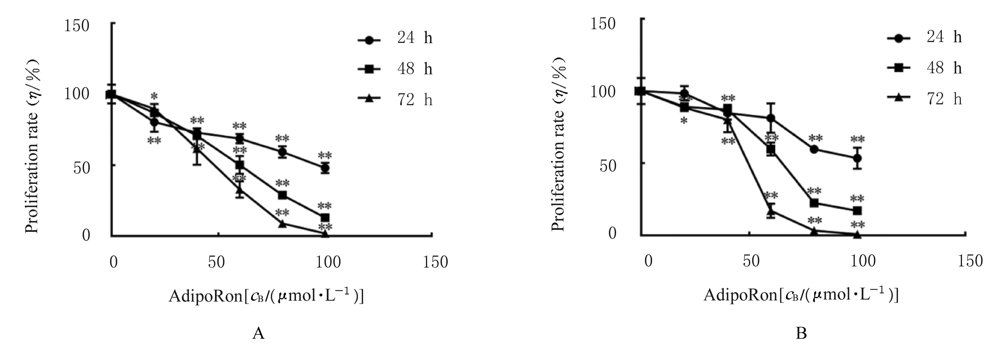
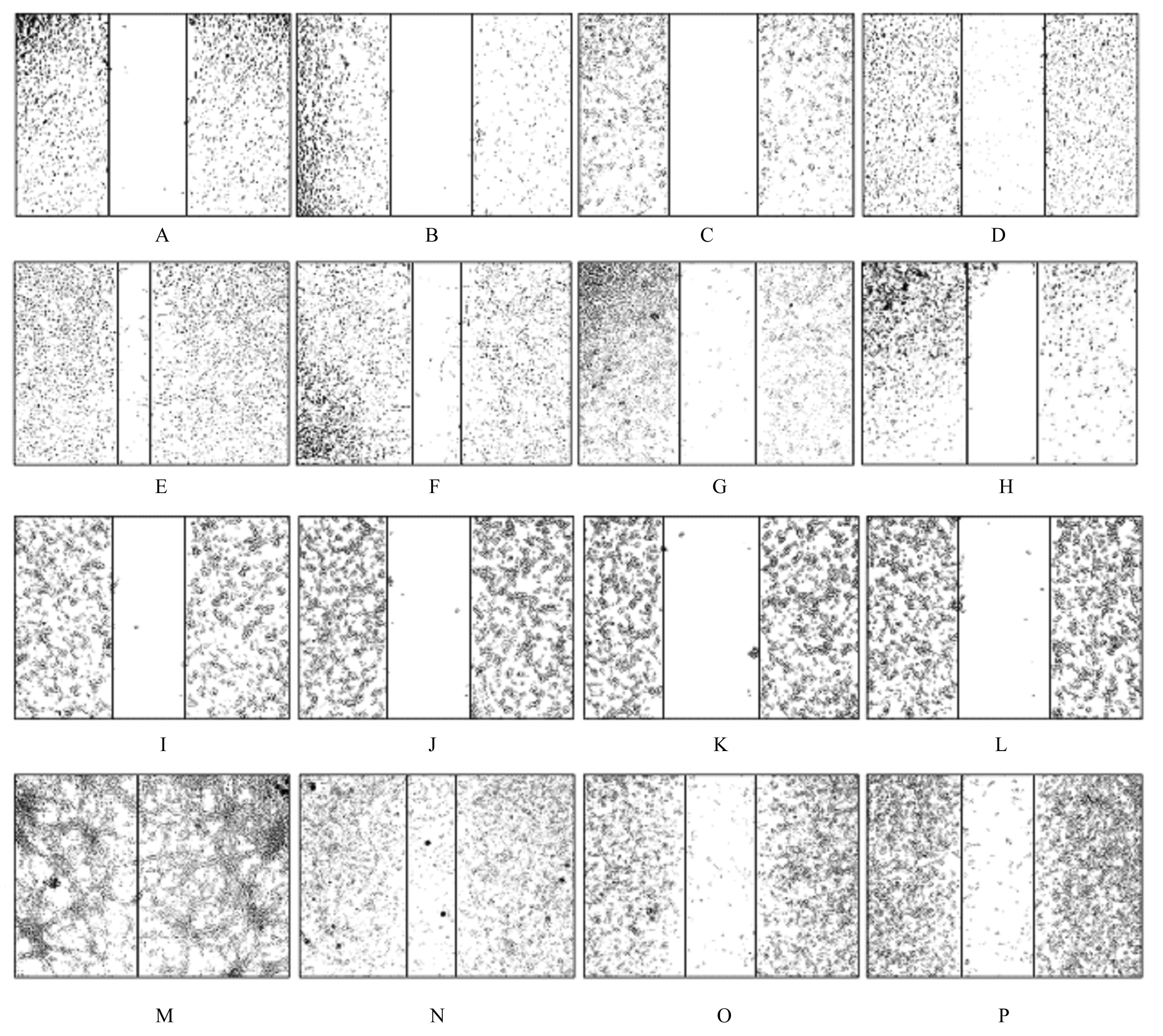
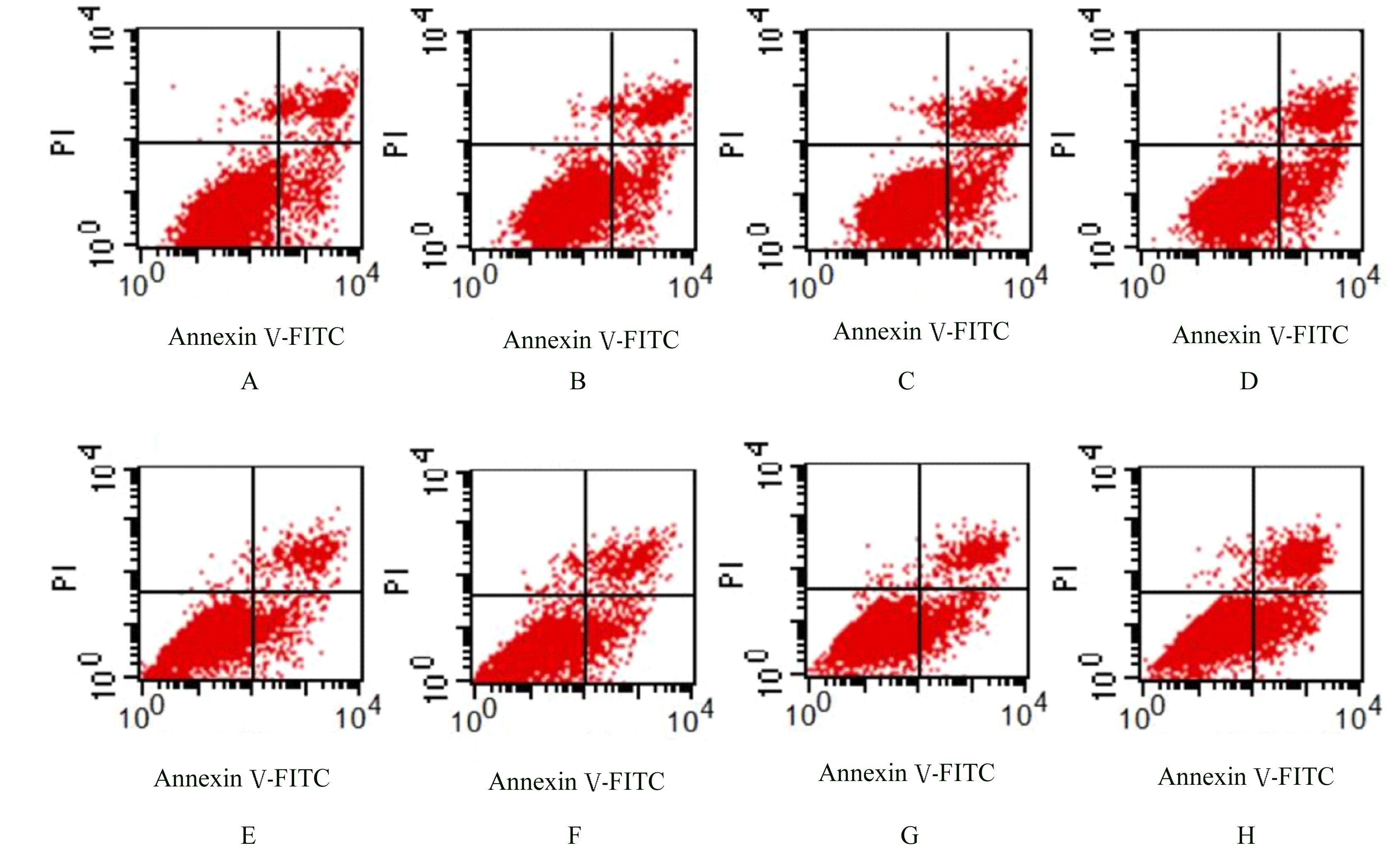
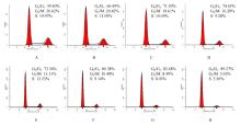
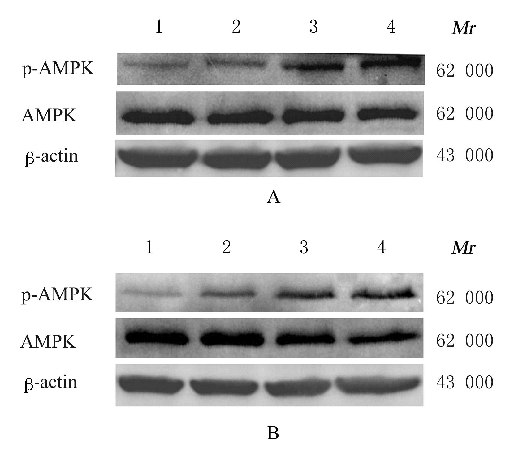
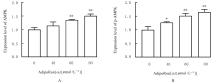
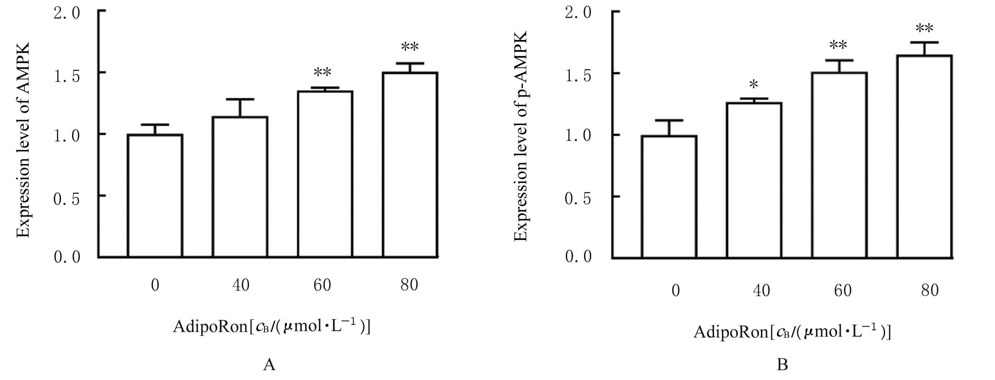
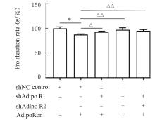
  • Received:2021-08-19 Online:2022-05-28 Published:2022-06-21
  • Contact: Di ZHAO E-mail:zhaodi914@126.com

摘要: 目的

探讨脂联素受体激动剂AdipoRon对胶质瘤细胞生物学行为的影响,阐明其可能的作用机制。

方法

取对数生长期的神经胶质瘤U251和U87 MG细胞分为对照组(0 μmol·L-1 AdipoRon)和20、40、60、80及100 μmol·L-1 AdipoRon组,CCK-8法检测24、48和72 h时各组细胞增殖率。U251和U87 MG细胞分为对照组(0 μmol·L-1 )和40、60及80 μmol·L-1 AdipoRon组,细胞克隆形成实验检测各组细胞克隆形成率,划痕愈合实验检测各组细胞划痕愈合率,流式细胞术检测各组细胞凋亡率及不同细胞周期细胞百分率,Western blotting法检测各组细胞中AMP依赖蛋白激酶(AMPK)和磷酸化AMPK(p-AMPK)蛋白表达水平。将U251细胞分为shNC对照组(未处理)、shNC组(经40 μmol·L-1 AdipoRon处理,未敲低)、AdipoR1敲低组(经40 μmol·L-1 AdipoRon处理,敲低AdipoR1)、AdipoR2敲低组(经40 μmol·L-1 AdipoRon处理,敲低AdipoR2)和AdipoR1+AdipoR2共同敲低组(经40 μmol·L-1 AdipoRon处理,敲低AdipoR1和AdipoR2)。CCK-8法检测各组U251细胞增殖率,实时荧光定量PCR(RT-qPCR)法检测各组U251细胞中AdiopR1和 AdiopR2 mRNA表达水平。

结果

与对照组比较,24、48和72 h时,不同浓度AdipoRon组U251和U87 MG细胞增殖率明显降低(P<0.05或P<0.01);与对照组比较,40、60和80 μmol·L-1 AdipoRon组U251和U87 MG细胞克隆形成率明显降低(P<0.01);与对照组比较,作用48 h时,40、60和80 μmol·L-1 AdipoRon组U251和U87 MG细胞划痕愈合率降低(P<0.01);与对照组比较,60和80 μmol·L-1 AdipoRon组U251细胞及80 μmol·L-1 AdipoRon组U87 MG细胞凋亡率升高(P<0.01),40、60和80 μmol·L-1 AdipoRon组U251和U87 MG细胞中G0/G1期细胞百分率升高(P<0.01);与对照组比较,60和80 μmol·L-1 AdipoRon组U251细胞中p-AMPK蛋白表达水平升高(P<0.01),40、60和80 μmol·L-1 AdipoRon组U87 MG细胞中p-AMPK蛋白表达水平升高(P<0.05或P<0.01)。与shNC对照组比较,AdipoR1敲低组细胞中AdipoR1 mRNA表达水平降低(P<0.01),AdipoR2敲低组细胞中AdipoR2 mRNA表达水平降低(P<0.01)。与shNC对照组比较,经40 μmol·L-1 AdipoRon作用后,敲低AdipoR1组、敲低AdipoR2组和AdipoR1+AdipoR2共同敲低组U251细胞增殖率均升高(P<0.05或P<0.01)。

结论

AdipoRon可抑制神经胶质瘤细胞增殖、迁移和凋亡,并将细胞周期阻滞在G0/G1期,其作用机制可能是AdipoRon与脂联素受体AdipoR1和AdipoR2相互作用后促进AMPK磷酸化,从而影响胶质瘤细胞的生物学行为。

关键词: 胶质瘤, AdipoRon, 细胞增殖, 细胞迁移, 细胞凋亡, 细胞周期, AMP依赖蛋白激酶

Abstract:

Objective: To investigate the effect of adiponectin receptor agonist AdipoRon on the biological behavior of the glioma cells, and to clarify its possible mechanism.

Methods

The glioma cells U251 and U87 MG at the logarithmic phase were divided into control group (0 μmol·L-1 AdipoRon) and 20, 40, 60, 80,and 100 μmol·L-1 AdipoRon groups. The proliferation rates of the cells in various groups at 24, 48,and 72 h were detected by CCK-8 assay.The U251 and U87 MG cells were divided into control group (0 μmol·L-1 AdipoRon) and 40, 60, and 80 μmol·L-1 AdipoRon groups.The clone formation rates of the cells in various groups were detected by clone formation assay, the scratch healing rates of the cells in various groups were detected by scratch healing experiment, the apoptotic rates and the percentages of the cells at different cell cycles were detected by flow cytometry, and the expression levels of AMP-dependent protein kinase (AMPK) and phosphorylated AMPK (p-AMPK) proteins in the cells in various groups were detected by Western blotting method.The U251 cells were divided into shNC control group(given no treatment) and shNC group(given 40 μmol·L-1 Adiporon), AdipoR1 knockdown group (given 40 μmol·L-1 Adiporon, knockdown AdipoR1), AdipoR2 knockdown group (given 40 μmol·L-1 Adiporon, knockdown AdipoR2) and AdipoR1+AdipoR2 co-knockdown group (given 40 μmol·L-1 Adiporon, knockdown AdipoR1 and AdipoR2). The proliferation rates of the U251 cells in various groups were detected by CCK-8 assay, and the expression levels of AdiopR1 and AdiopR2 mRNA in the U251 cells were detected by real-time fluorescence quantitative PCR (RT-qPCR)method.

Results

Compared with control group, the proliferation rates of the U251 and U87 MG cells in different concentrations of AdipoRon groups were decreased at 24, 48 and 72 h (P<0.05 or P<0.01). Compared with control group, the clone formation rates of the U251 and U87 MG cells in 40, 60,and 80 μmol·L-1 AdipoRon groups were decreased (P<0.01). Compared with control group, the scratch healing rates of the U251 and U87 MG cells in 40, 60,and 80 μmol·L-1 AdipoRon groups were decreased after 48 h treatment (P<0.01). Compared with control group, the apoptotic rates of the U251 cells in 60 and 80 μmol·L-1 AdipoRon groups and the U87 MG cells in 80 μmol·L-1 AdipoRon group were increased (P<0.01); the percentages of the U251 and U87 MG cells at G0/G1 phase in 40, 60, and 80 μmol·L-1 AdipoRon groups were increased (P<0.01). Compared with control group, the expression levels of p-AMPK protein in the U251 cells in 60 and 80 μmol·L-1 Adiporon groups were increased (P<0.01),and the expression levels of p-AMPK protein in the U87 MG cells in 40, 60, and 80 μmol·L-1 Adiporon group were increased (P<0.05 or P<0.01). Compared with shNC control group, the expression level of AdipoR1 mRNA in the U251 cells in AdipoR1 knockdown group was decreased (P<0.01), and the expression level of AdipoR2 mRNA in the U251 cells in AdipoR2 knockdown group was decreased (P<0.01). Compared with shNC control group, after treated with of 40 μmol·L-1 Adiporon, the proliferation rates of the U251 cells in AdipoR1 knockdown group, AdipoR2 knockdown group, and AdipoR1+AdipoR2 co-knockdown group were increased (P<0.05 or P<0.01).

Conclusion

Adiporon can inhibit the proliferation, migration and apoptosis of the glioma cells and block the cell cycle at G0/G1 phase, and its mechanism may be that adiporon interacts with the adiponectin receptors AdipoR1 and AdipoR2 to promote the AMPK phosphorylation, thus affecting the biological behaviors of the glioma cells.

Key words: Glioma, AdipoRon, Cell proliferation, Cell migration, Apoptosis, Cell cycle, AMP-dependent protein kinase

中图分类号: 

  • R739.41