吉林大学学报(医学版) ›› 2025, Vol. 51 ›› Issue (5): 1155-1164.doi: 10.13481/j.1671-587X.20250501

• 基础研究 •    

洛伐他汀对高脂血症诱导大鼠肝损伤的改善作用及其机制

赵艺1,周冰2,邱惠蕊3,李轩3,崔向丽1()   

  1. 1.首都医科大学附属北京友谊医院药剂科,北京 100050
    2.吉林大学第一医院麻醉科,吉林 长春 130021
    3.天津科技大学生物工程学院生物与医学专业,天津 300457
  • 收稿日期:2025-04-08 接受日期:2025-05-27 出版日期:2025-09-28 发布日期:2025-11-05
  • 通讯作者: 崔向丽 E-mail:cui10@163.com
  • 作者简介:赵 艺(1989-),女,吉林省长春市人,初级药师,理学硕士,主要从事高脂血症基础和临床方面的研究。
  • 基金资助:
    国家卫健委医院管理研究所医院药学高质量发展研究项目(NIHAYS2302)

Improvement effect of lovastatin on hyperlipidemia-induced liver injury in rats and its mechanism

Yi ZHAO1,Bing ZHOU2,Huirui QIU3,Xuan LI3,Xiangli CUI1()   

  1. 1.Department of Pharmacy,Beijing Friendship Hospital,Capital Medical University,Beijing 100050,China
    2.Department of Anesthesiology,First Hospital,Jilin University,Changchun 130021,China
    3.Department of Biology and Medicine,School of Biological Engineering,Tianjin University of Science and Technology,Tianjin 300457,China
  • Received:2025-04-08 Accepted:2025-05-27 Online:2025-09-28 Published:2025-11-05
  • Contact: Xiangli CUI E-mail:cui10@163.com

摘要:

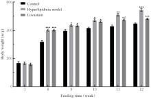
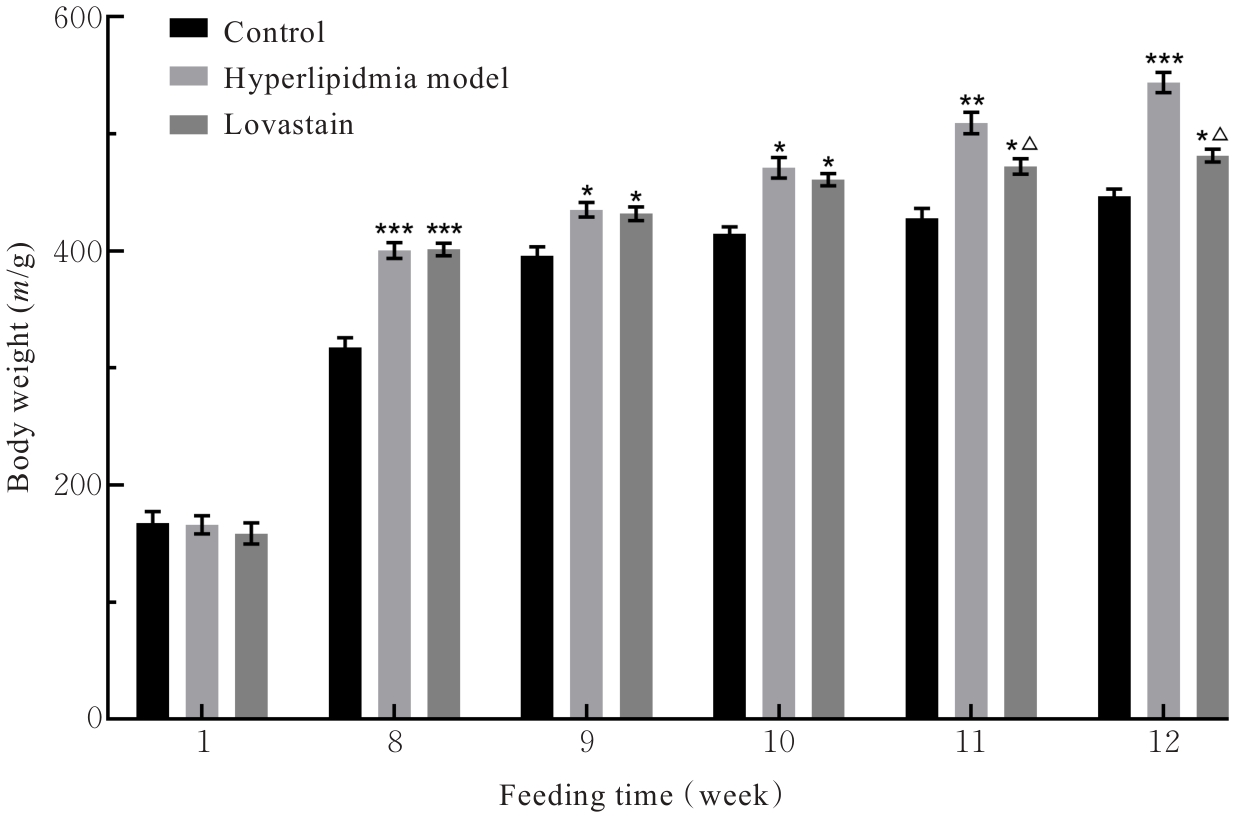
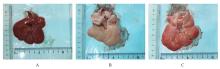
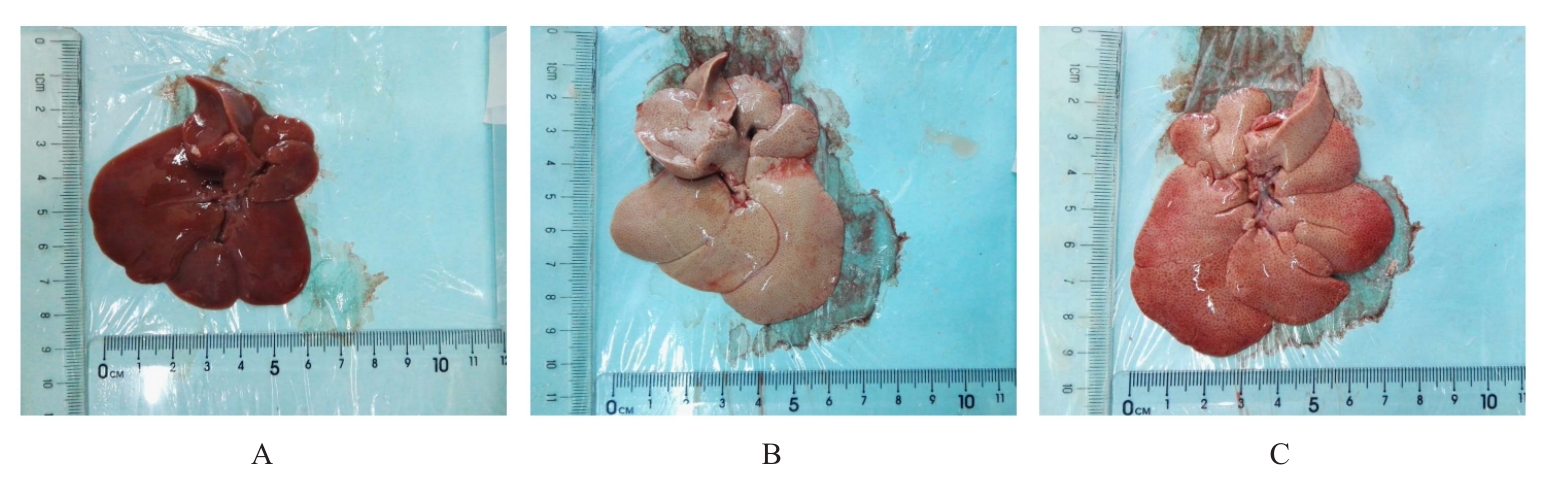
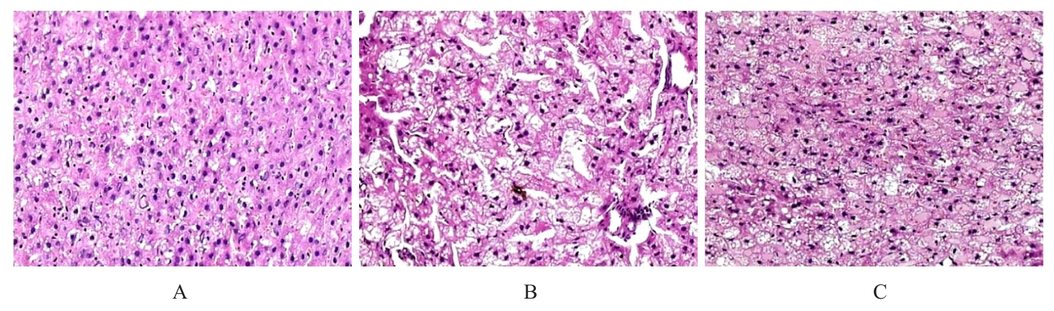
目的 探讨洛伐他汀对高脂血症诱导的大鼠肝损伤的保护作用,并阐明其可能的作用机制。 方法 将15只SD大鼠随机分为对照组、高脂血症模型组和洛伐他汀组,每组5只。对照组大鼠常规饲养,高脂血症模型组和洛伐他汀组大鼠采用高脂饲料饲养12周;第8周开始灌胃给药,洛伐他汀组大鼠给予2 mg?kg-1洛伐他汀,对照组和高脂血症模型组大鼠给予等体积生理盐水,每天1次,持续给药4周。检测各组大鼠实验开始后第1、8、9、10、11和12周时体质量,采用HE染色检测各组大鼠肝组织病理形态表现,采用试剂盒检测各组大鼠血清中总胆固醇(TC)、甘油三酯(TG)、高密度脂蛋白胆固醇(HDL-C)、低密度脂蛋白胆固醇(LDL-C)和丙二醛(MDA)水平及超氧化物歧化酶(SOD)、谷胱甘肽过氧化物酶(GSH-Px)、天门冬氨酸氨基转移酶(AST)和谷氨酸氨基转移酶(ALT)活性以及白细胞介素2(IL-2)、白细胞介素6(IL-6)、白细胞介素12(IL-12)和肿瘤坏死因子α(TNF-α)水平,采用16S rRNA测序方法检测各组大鼠肠道菌群组成。 结果 与对照组比较,高脂血症模型组大鼠体质量从高脂饲料饲养第8周开始明显升高(P<0.05或P<0.01或P<0.001);与高脂血症模型组比较,第11和12周洛伐他汀组大鼠体质量明显降低(P<0.05)。与对照组比较,高脂血症模型组大鼠肝脏表面较为粗糙并呈苍白色,边缘厚钝,形态肿大,有颗粒感和油腻感;与高脂血症模型组比较,洛伐他汀组大鼠肝脏呈淡褐红色,质软,边缘稍钝,体积减小,颗粒感和油腻感较轻。与对照组比较,高脂血症模型组大鼠肝脏细胞肿胀且排列紊乱,细胞核固缩,分布大量炎性细胞,且胞内有大量空泡样变性;与高脂血症模型组比较,洛伐他汀组大鼠肝脏细胞肿胀程度和变性程度明显减轻,肝细胞排列较整齐、结构较完整,炎症细胞浸润减少,空泡样变性减少。与对照组比较,高脂血症模型组血清中TC、TG和LDL-C水平明显升高(P<0.05),HDL-C水平明显降低(P<0.05);与高脂血症模型组比较,洛伐他汀组大鼠血清中TC、TG和LDL-C水平明显降低(P<0.05),HDL-C水平明显升高(P<0.05)。与对照组比较,高脂血症模型组大鼠血清中MDA水平和ALT及AST活性明显升高(P<0.05),SOD和GSH-Px活性明显降低(P<0.05);与高脂血症模型组比较,洛伐他汀组大鼠血清中MDA水平和ALT及AST活性明显降低(P<0.05),SOD和GSH-Px活性明显升高(P<0.05)。与对照组比较,高脂血症模型组大鼠血清中IL-2、IL-6、IL-12和TNF-α水平明显升高(P<0.05);与高脂血症模型组比较,洛伐他汀组大鼠血清中IL-2、IL-6、IL-12和TNF-α水平明显降低(P<0.05)。与对照组比较,高脂血症模型组大鼠ACE指数和Chao1指数明显降低(P<0.05);与高脂血症模型组比较,洛伐他汀组大鼠ACE指数和Chao1指数明显升高(P<0.05或P<0.01)。与对照组比较,高脂血症模型组大鼠肠道内厚壁菌门(Firmicutes)和变形菌门(Proteobacteria)相对丰度明显升高(P<0.001),拟杆菌门(Bacteroidetes)和放线菌门(Actinobacteria)相对丰度明显降低(P<0.001);与高脂血症模型组比较,洛伐他汀组大鼠肠道内FirmicutesProteobacteria相对丰度明显降低(P<0.05或P<0.01),BacteroidetesActinobacteria相对丰度未见明显变化。与对照组比较,高脂血症模型组大鼠肠道内乳杆菌属(Lactobacillus)相对丰度明显降低(P<0.001),拟杆菌属(Bacteroides脱硫弧菌属(Desulfovibrio)和梭菌属(Clostridium)相对丰度明显升高(P<0.01或P<0.001);与高脂血症模型组比较,洛伐他汀组大鼠肠道内Lactobacillus相对丰度未见明显变化,Bacteroides、DesulfovibrioClostridium相对丰度明显降低(P<0.05或P<0.01或P<0.001)。 结论 洛伐他汀对高脂血症诱导的大鼠肝损伤具有改善作用,其机制可能与洛伐他汀改善肠道菌群组成和抑制氧化应激及炎症损伤有关。

关键词: 洛伐他汀, 高脂血症, 肝损伤, 抗炎, 肠道菌群, 氧化应激

Abstract:

Objective To investigate the protective effect of lovastatin on liver injury in the rats induced by hyperlipidemia, and to elucidate its possible mechanism. Methods Fifteen SD rats were randomly divided into control group, hyperlipidemia model group, and lovastatin group, with 5 rats in each group. The rats in control group were fed with standard diet, while the rats in hyperlipidemia model group and lovastatin group were fed high-fat diet for 12 weeks. Starting from the 8th week, the rats were administered treatments via gavage once a day for 4 weeks: the rats in lovastatin group received 2 mg?kg?1 lovastatin, while the rats in control group and hyperlipidemia model group received an equal volume of normal saline. The body weights of the rats in various groups were measured at weeks 1, 8, 9, 10, 11, and 12 after the experiment began; the histopathology of liver tissue of the rats in various groups was observed using HE staining; the serum levels of total cholesterol (TC), triglycerides (TG), high-density lipoprotein cholesterol (HDL-C), low-density lipoprotein cholesterol (LDL-C), malondialdehyde (MDA), as well as the activities of superoxide dismutase (SOD), glutathione peroxidase (GSH-Px), aspartate aminotransferase (AST), and alanine aminotransferase (ALT), and the levels of interleukin-2 (IL-2), interleukin-6 (IL-6), interleukin-12 (IL-12), and tumor necrosis factor-α (TNF-α) of the rats in various groups were detected using commercial kits; the composition of the gut microbiota of the rats in various groups was analyzed by 16S rRNA sequencing. Results Compared with control group, the body weight of the rats in hyperlipidemia model group was significantly increased from the 8th week of high-fat diet feeding (P<0.05 or P<0.01 or P<0.001). Compared with hyperlipidemia model group, the body weight of the rats in lovastatin group was significantly decreased at weeks 11 and 12 (P<0.05). Compared with control group, the livers of the rats in hyperlipidemia model group appeared rough, pale, enlarged, with blunt edges, and had a granular and greasy texture. Compared with hyperlipidemia model group, the livers of the rats in lovastatin group were light brownish-red, soft, with slightly blunt edges, reduced volume, and less granularity and greasiness. Compared with control group, the liver cells of the rats in hyperlipidemia model group were swollen and disorganized, with pyknotic nuclei, extensive inflammatory cell infiltration, and numerous vacuolar degenerations. Compared with hyperlipidemia model group, the rats in lovastatin group showed significantly reduced hepatocyte swelling and degeneration, more orderly and intact liver cell arrangement, decreased inflammatory cell infiltration, and reduced vacuolar degeneration. Compared with control group, the serum levels of TC, TG, and LDL-C of the rats in hyperlipidemia model group were significantly increased (P<0.05), and the serum HDL-C level was decreased (P<0.05). Compared with hyperlipidemia model group, the serum levels of TC, TG, and LDL-C of the rats in lovastation group were significantly decreased (P<0.05), and the serum HDL-C level was increased (P<0.05). Compared with control group, the serum MDA levels and the ALT and AST activities of the rats in hyperlipidemia model group were significantly increased (P<0.05), and the SOD and GSH-Px activities were significantly decreased (P<0.05). Compared with hyperlipidemia model group, the serum MDA levels and ALT and AST activities of the rats in lovastatin group were decreased (P<0.05), and the SOD and GSH-Px activities were increased (P<0.05). Compared with control group, the serum levels of IL-2, IL-6, IL-12, and TNF-α of the rats in hyperlipidemia model group were significantly increased (P<0.05). Compared with hyperlipidemia model group, the serum levels of IL-2, IL-6, IL-12, and TNF-α of the rats in lovastatin group were significantly decreased (P<0.05). Compared with control group, the ACE and Chao1 indexes of the rats in hyperlipidemia model group were significantly decreased (P<0.05). Compared with hyperlipidemia model group, the ACE and Chao1 indexes of the rats in lovastatin group were significantly increased (P<0.05 or P<0.01). Compared with control group, the relative abundances of Firmicutes and Proteobacteria of the rats in hyperlipidemia model group were significantly increased (P<0.001), and the relative abundances of Bacteroidetes and Actinobacteria were decreased (P<0.001). Compared with hyperlipidemia model group, the relative abundances of Firmicutes and Proteobacteria of the rats in lovastatin group were significantly decreased (P<0.05 or P<0.01), while the relative abundances of Bacteroidetes and Actinobacteria showed no significant changes. Compared with control group, the relative abundance of Lactobacillus of the rats in hyperlipidemia model group was significantly decreased (P<0.001), and the relative abundances of BacteroidesDesulfovibrio, and Clostridium were significantly increased (P<0.01 or P<0.001). Compared with hyperlipidemia model group, the relative abundance of Lactobacillus of the rats in lovastatin group showed no significant change but the relative abundances of BacteroidesDesulfovibrio, and Clostridium were significantly decreased (P<0.05 or P<0.01 or P<0.001). Conclusion Lovastatin ameliorates liver injury induced by hyperlipidemia, and the mechanism may be related to its ability to improve gut microbiota composition and inhibit oxidative stress and inflammatory damage.

Key words: Lovastatin, Hyperlipidemia, Liver injury, Anti-inflammatory, Intestinal microorganism, Oxidative stress

中图分类号: 

  • R589.2