吉林大学学报(医学版) ›› 2025, Vol. 51 ›› Issue (1): 105-114.doi: 10.13481/j.1671-587X.20250113

• 基础研究 • 上一篇    

片仔癀在对乙酰氨基酚所致肝损伤中的保护作用及其机制

张潮鹤1,张昕玮,王相峰2()   

  1. 1.吉林大学第二医院肿瘤血液科, 吉林 长春 130021
    2.吉林大学第一医院乐群院区药学部, 吉林 长春 130031
  • 出版日期:2025-01-28 发布日期:2025-03-06
  • 通讯作者: 王相峰 E-mail:xiangfeng1012@163.com
  • 作者简介:吉林大学药学院2019级临床药学专业

Protective effect of Pien-Tze-Huang on acetaminophen-induced liver injury and its mechanism

Chaohe ZHANG1,Xinwei ZHANG,Xiangfeng WANG2()   

  1. 1.Department of Hematology,Second Hospital,Jilin University,Changchun 130031,China
    2.Department of Pharmacy,Lequn Branch,First Hospital,Jilin University,Changchun 130021,China
  • Online:2025-01-28 Published:2025-03-06
  • Contact: Xiangfeng WANG E-mail:xiangfeng1012@163.com

摘要:

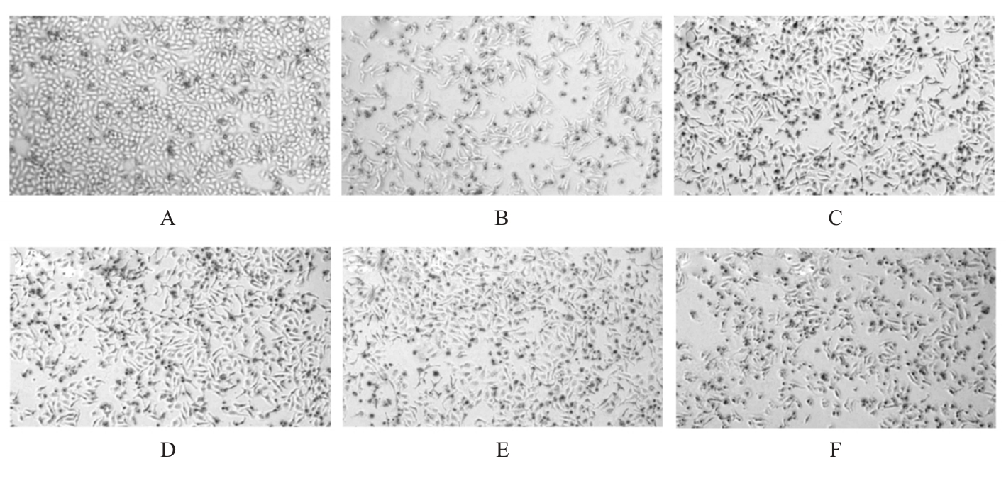
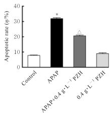
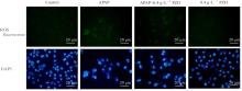
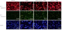
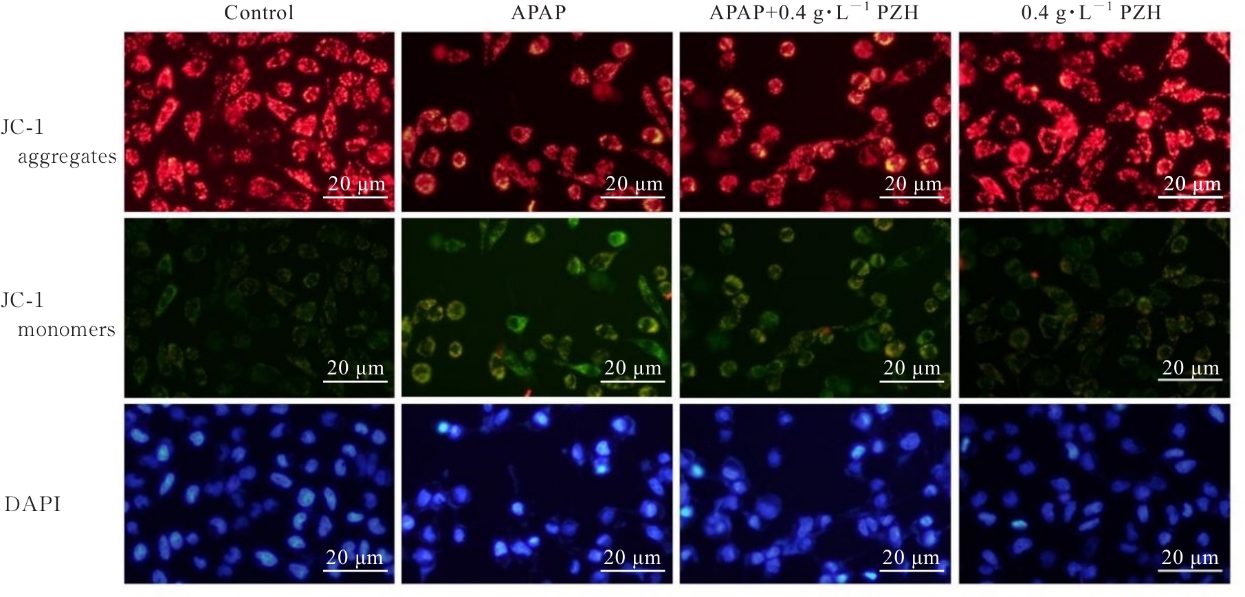
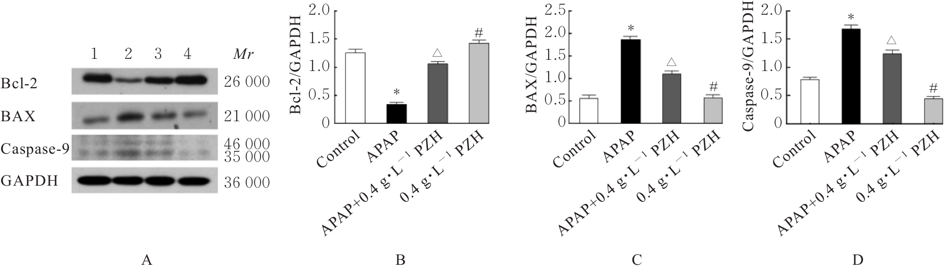
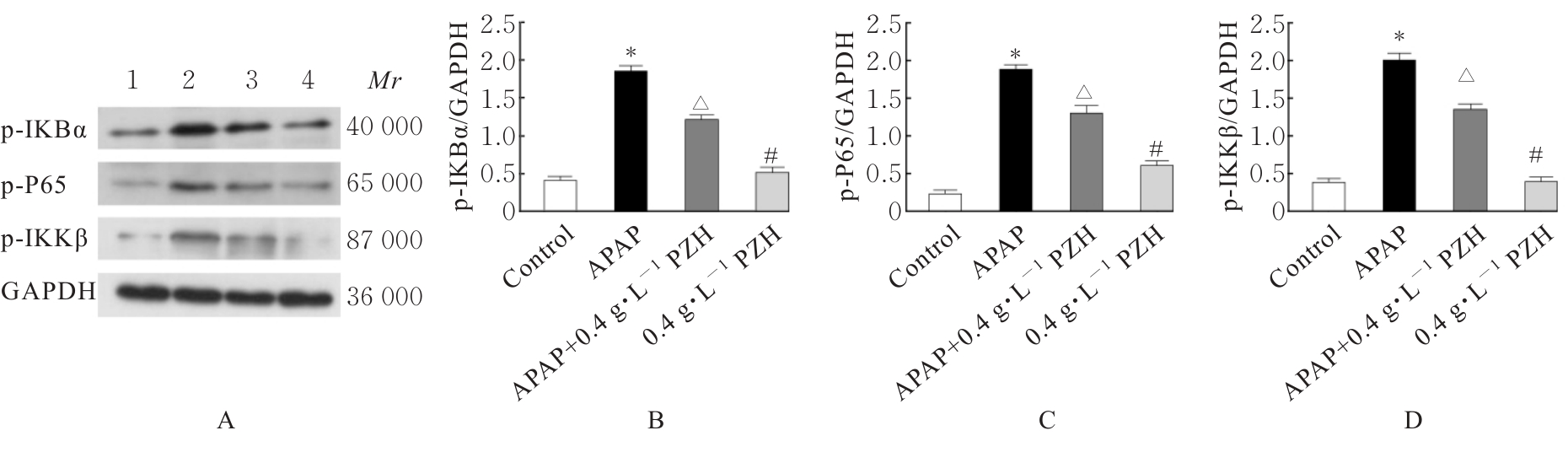
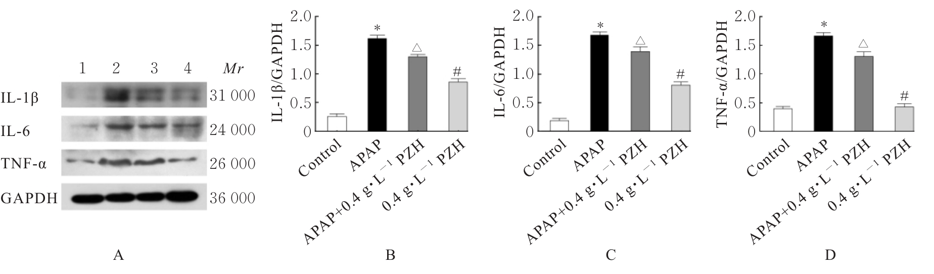
目的 探讨片仔癀(PZH)对对乙酰氨基酚(APAP)引起的肝损伤的保护作用,并阐明其可能的作用机制。 方法 将人正常肝细胞(L02细胞)分为对照组、APAP组(10 mmol·L-1 APAP)、APAP+PZH组(10 mmol·L-1APAP和0.4 g·L-1 PZH)和PZH组(0.4 g·L-1 PZH)。采用噻唑蓝(MTT)法检测各组细胞存活率,倒置显微镜观察各组肝细胞形态表现,流式细胞术检测各组肝细胞凋亡率,生化试剂盒检测各组细胞上清液中超氧化物歧化酶(SOD)和乳酸脱氢酶(LDH)活性及脂质过氧化物(MDA)水平,荧光探针技术检测各组肝细胞中活性氧(ROS)水平和线粒体膜电位(MMP),Western blotting法检测各组细胞中凋亡相关蛋白、磷脂酰肌醇3-激酶(PI3K)/蛋白激酶B(AKT)信号通路蛋白、核因子κB(NF-κB)信号通路蛋白和炎症因子的蛋白表达水平。 结果 MTT法,与对照组比较,APAP组细胞存活率明显降低(P<0.05);与APAP组比较,APAP+PZH组细胞存活率明显升高(P<0.05)。倒置显微镜观察,与对照组比较,APAP组细胞呈现数量减少且排列疏松的趋势;与APAP组比较,APAP+PZH组细胞数量及排列得到显著改善。流式细胞术检测,与对照组比较,APAP组细胞凋亡率明显升高(P<0.05);与APAP组比较,APAP+PZH组细胞凋亡率明显降低(P<0.05)。试剂盒检测,与对照组比较,APAP组细胞中LDH活性和MDA水平明显升高(P<0.05),SOD活性明显降低(P<0.05);与APAP组比较,APAP+PZH组细胞中LDH活性和MDA水平明显降低(P<0.05),SOD活性明显升高(P<0.05)。与对照组比较,APAP组肝细胞中ROS荧光强度明显升高;与APAP组比较,APAP+PZH组肝细胞中ROS荧光强度明显降低。与对照组比较,APAP组肝细胞中MMP明显降低;与APAP组比较,APAP+PZH组肝细胞中MMP明显升高。Western blotting法,与对照组比较,APAP组细胞中含半胱氨酸的天冬氨酸蛋白酶(caspase)-9、B细胞淋巴瘤2(Bcl-2)相关X蛋白(BAX)、磷酸化PI3K(p-PI3K)、磷酸化AKT(p-AKT)、磷酸化NF-κB抑制蛋白α(p-IKBα)、p-P65、磷酸化kappa B抑制因子激酶β(p-IKKβ)、白细胞介素1β(IL-1β)、白细胞介素6(IL-6)和肿瘤坏死因子α(TNF-α)蛋白表达水平明显升高(P<0.05),Bcl-2蛋白表达水平明显降低(P<0.05);与APAP组比较,APAP+PZH组细胞中caspase-9、BAX、p-PI3K、p-AKT、p-IKBα、p-P65、p-IKKβ、IL-1β、IL-6和TNF-α蛋白表达水平明显降低(P<0.05),Bcl-2蛋白表达水平明显升高(P<0.05)。 结论 PZH能够降低APAP引起的细胞内氧化应激和炎症反应,缓解L02细胞损伤,其作用机制可能与调控PI3K/AKT和NF-κB信号通路有关。

关键词: 片仔癀, 药物性肝损伤, 氧化应激, 磷脂酰肌醇3-激酶, 蛋白激酶B, 核因子κB

Abstract:

Objective To study the protective effect of Pien-Tze-Huang on acetaminophen-induced liver injury,and to clarify the possible mechanism. Methods The human normal hepatocytes(L02 cells) were divided into control group, APAP group (10 mmol·L-1 APAP),APAP+PZH group (10 mmol·L-1 APAP and 0.4 mg·mL-1 PZH), and PZH group (0.4 mg·mL-1 PZH). The survival rates of the cells in various groups were determined by MTT method, the morphology was observed by inverted microscope, and the apoptotic rates were detected by flow cytometry. The activities of superoxide dismutase (SOD) and lactate dehydrogenase (LDH) and the levels of malondialdehyde (MDA) in the cell supernant in various groups were detected by the kits, the reactive oxygen species(ROS) levels and the mitochondrial membrane potential (MMP) in the hepatocytes in various groups were detected by fluorescence probe. Western blotting method was used to examine the expression levels of apoptosis-related proteins, phosphatidylinositol 3 kinase (PI3K)/protein kinase B(AKT) signaling pathway proteins, nuclear factor-κB(NF-κB) signaling pathway proteins and inflammatory factors in the cells in various groups. Results The results of MTT showed that compared with control group, the survival rate of the cells in APAP group was markedly decreased (P<0.05); compared with APAP group, the survival rate of the cells in APAP+PZH group was significantly increased (P<0.05). Compared with control group, the number of the L02 cells in APAP group showed a decreasing and loosely arranged trend; compared with APAP group, the number and arrangement of the L02 cells in APAP+PZH group were notably improved. The results of flow cytometry showed that compared with control group, the apoptotic rate of the L02 cells in APAP group was significantly increased(P<0.05); compared with APAP group, the apoptotic rate of the cells in APAP+PZH group was significantly decreased(P<0.05). Compared with control group,the activity of LDH and level of MDA in the cells in APAP group were significantly increased (P<0.05), while the activity of SOD was significantly decreased (P<0.05); compared with APAP group,the activity of LDH and level of MDA in the cells in APAP+PZH group were significantly decreased(P<0.05), while the activity of SOD was significantly increased(P<0.05). Compared with control group, the fluorescence intensity of ROS in the cells in APAP group was significantly increased;compared with APAP group,the fluorescence intensity of ROS in APAP+PZH group was significantly decreased. Compared with control group, the MMP of the L02 cells in APAP group was significantly decreased; compared with APAP group,the MMP of the L02 cells in APAP+PZH group was significantly increased. The results of Western blotting showed that compared with control group, the expression levels of caspase-9, B-cell lymphoma 2(Bcl-2) associated X protein(BAX), phosphorylated PI3K (p-PI3K), phosphorylated AKT (p-AKT), phosphorylated NF-κB inhibitor alpha (p-IKBα), p-P65, phosphorylated inhibitor of kappaB kinaseβ (p-IKKβ), interleukin-1β (IL-1β), interleukin-6 (IL-6), and tumor necrosis factor-α(TNF-α) proteins in the cells in APAP group were significantly increased (P<0.05), while the level of Bcl-2 proteins in the cells in APAP group was significantly decreased (P<0.05); compared with APAP group, the levels of caspase-3, BAX, p-PI3K, p-AKT, p-IKBα, p-P65, p-IKKβ, IL-1β, IL-6, and TNF-α proteins in the cells in APAP+PZH group were significantly decreased, while the level of Bcl-2 protein was significantly increased(P<0.05). Conclusion PZH may reduce the oxidative stress and inflammatory response in the cells by regulating the PI3K/AKT and NF-κB signaling pathway, therefore attenuate the L02 cell injury induced by APAP.

Key words: Pien-Tze-Huang, Drug-induced liver injury, Oxidative stress, Phosphatidylinositol 3 kinase, Protein kinase B, Nuclear factor-κB

中图分类号: 

  • R285.5