吉林大学学报(医学版) ›› 2025, Vol. 51 ›› Issue (5): 1177-1184.doi: 10.13481/j.1671-587X.20250503

• 基础研究 • 上一篇    

胎盘表达转录因子1在多囊卵巢综合征大鼠卵巢组织中的表达及其对大鼠卵巢颗粒细胞增殖的影响

付璐璐1,邹颖刚1,郑晓宇2,张雪莹1,张京顺1,王敏1,张嫱1,郑连文1()   

  1. 1.吉林大学第二医院生殖中心,吉林 长春 130022
    2.东莞松山湖东华医院妇科,广东 东莞 523000
  • 收稿日期:2024-10-15 接受日期:2024-12-18 出版日期:2025-09-28 发布日期:2025-11-05
  • 通讯作者: 郑连文 E-mail:zhenglw@jlu.edu.cn
  • 作者简介:付璐璐(1988-),女,山东省聊城市人,主治医师,医学博士,主要从事生殖内分泌方面的研究。
  • 基金资助:
    吉林省科技厅科技发展计划项目(20210101462JC)

Expression of placenta expressed transcription factor 1 in ovarian tissue of polycystic ovary syndrome rats and its effect on proliferation of rat ovarian granulosa cells

Lulu FU1,Yinggang ZOU1,Xiaoyu ZHENG2,Xueying ZHANG1,Jingshun ZHANG1,Min WANG1,Qiang ZHANG1,Lianwen ZHENG1()   

  1. 1.Reproductive Center,Second Hospital,Jilin University,Changchun 130022,China
    2.Department of Gynecology,Dongguan Songshan Lake Tungwah Hospital,Dongguan 523000,China
  • Received:2024-10-15 Accepted:2024-12-18 Online:2025-09-28 Published:2025-11-05
  • Contact: Lianwen ZHENG E-mail:zhenglw@jlu.edu.cn

摘要:

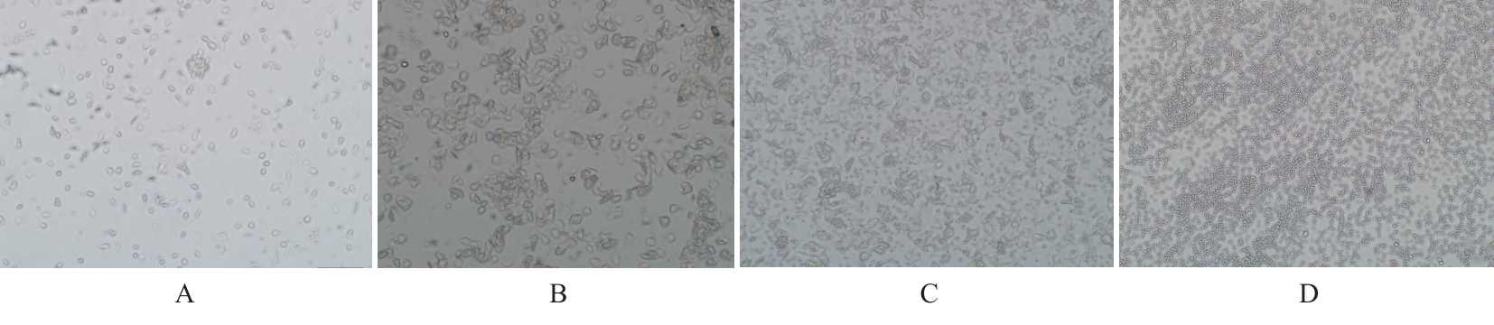
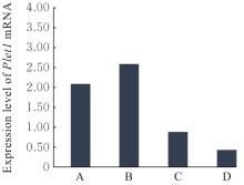
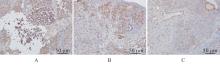
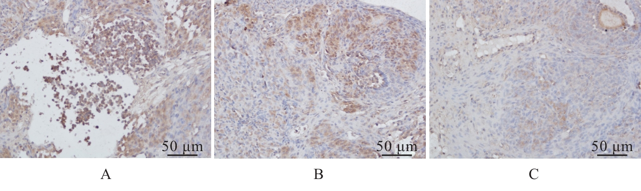
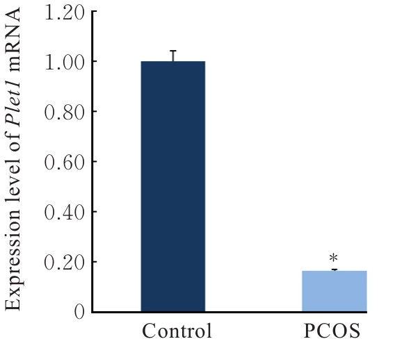
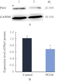
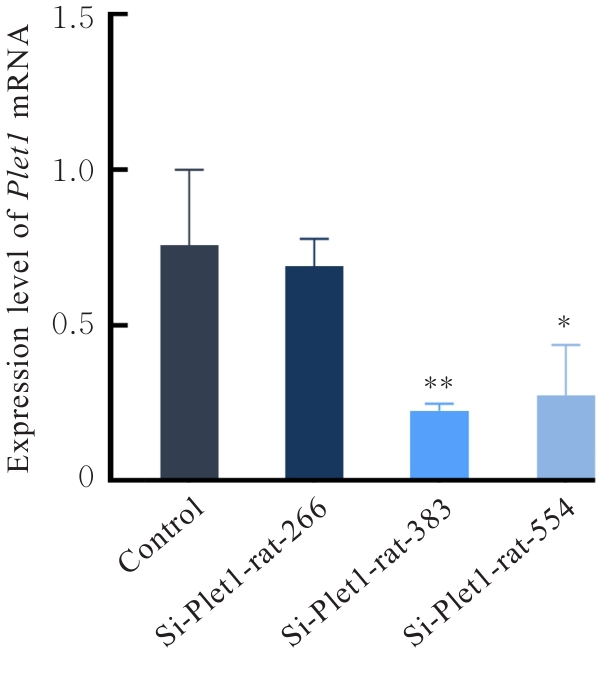
目的 探讨胎盘表达转录因子1(Plet1)在来曲唑构建的多囊卵巢综合征(PCOS)模型大鼠卵巢组织中的表达及其对大鼠卵巢颗粒细胞增殖调控作用,并阐明Plet1参与PCOS发病的可能机制。 方法 取课题组前期研究采集的大鼠卵巢样本,分为对照组和PCOS组,采用实时荧光定量PCR(RT-qPCR)和Western blotting法检测2组大鼠卵巢组织中Plet1 mRNA和蛋白表达水平。另取24只大鼠,采用阴道涂片鉴定将大鼠分为发情前期、发情期、发情后期及发情间期4组,RT-qPCR法检测不同发情周期大鼠卵巢组织中Plet1 mRNA表达水平,免疫组织化学法检测大鼠卵巢组织中Plet1表达定位。转染大鼠卵巢颗粒细胞,分为对照组、si-Plet1-rat-266组、si-Plet1-rat-383组和si-Plet1-rat-554组。采用细胞计数试剂盒8(CCK-8)法检测各组大鼠卵巢颗粒细胞增殖活性,采用RT-qPCR法检测各组大鼠卵巢颗粒细胞中细胞分裂蛋白激酶6(CDK6)和P53 mRNA表达水平。 结果 RT-qPCR法检测,Plet1 mRNA在正常大鼠卵巢中表达,Plet1 mRNA表达水平在各发情周期组间比较差异无统计学意义(P>0.05)。免疫组织化学检测,Plet1蛋白在大鼠卵巢组织中表达定位主要在卵巢颗粒细胞和黄体细胞。与对照组比较,PCOS组大鼠卵巢中Plet1 mRNA和蛋白表达水平均明显降低 (P<0.05)。RT-qPCR法检测,与对照组比较,si-Plet1-rat-383组大鼠卵巢颗粒细胞中Plet1 mRNA表达水平明显降低(P<0.01),且下降幅度最大。CCK-8法检测,与对照组比较,si-Plet1-rat-383组大鼠卵巢颗粒细胞增殖活性明显降低(P<0.05)。与对照组比较,si-Plet1-rat-383组CDK6P53 mRNA表达水平明显降低(P<0.05或P<0.01)。 结论 Plet1蛋白在正常大鼠卵巢组织中表达分布主要集中于卵巢颗粒细胞和黄体细胞。PCOS模型大鼠卵巢组织内Plet1基因表达下调,干扰Plet1基因表达可抑制大鼠卵巢颗粒细胞增殖。

关键词: 多囊卵巢综合征, 胎盘表达转录因子1, 发情周期, 卵巢颗粒细胞, 细胞增殖

Abstract:

Objective To investigate the expression of placenta expressed transcript 1(Plet1)in ovarian tissue of the letrozole-induced model rats of polycystic ovary syndrome (PCOS) and its effect on the proliferation of rat ovarian granulosa cells,and to clarify the possible mechanism by which Plet1 may contribute to the pathogenesis of PCOS. Methods The ovarian tissue samples from the rats collected in previous studies were used and divided into control and PCOS groups. Real-time fluorescence quantitative PCR (RT-qPCR) and Western blotting methods were used to detect the expression levels of Plet1 mRNA and protein in ovarian tissue of the rat in two groups. Additionally, twenty-four rats underwent vaginal smear cytology were divided into four groups by estrous cycle phase: proestrus,estrus,metestrus and diestrus. RT-qPCR was used to detect the expression level of Plet1 mRNA in ovarian tissue of the rats in various groups,and immunohistochemistry (IHC) method was used to detect the location of Plet1 expression in the rat ovarian tissue in various groups. The rat ovarian granulosa cells were transfected and divided into control group, si-Plet1-rat-266 group, si-Plet1-rat-383 group, and si-Plet1-rat-554 group. Cell counting kit-8(CCK-8)method was used to assess the cell proliferation activities of rat ovarian granulosa cells in various groups, and RT-qPCR method was used to detect the expression levels of cyclin-dependent kinase 6 (CDK6) and P53 mRNA in rat ovarian granulosa cells in various groups. Results The RT-qPCR results revealed that Plet1 mRNA was expressed in the ovaries of normal rats, while no statistically significant difference was observed across estrous cycle phases (P>0.05). The immunohistochemistry results showed that the expression of Plet1 protein was mainly localized in ovarian granulosa cells and luteal cells in the rat ovarian tissue. Compared with control group,the expression levels of Plet1 mRNA and protein in ovarian tissue of the rats in PCOS group were significantly decreased (P<0.05). The RT-qPCR results showed that compared with control group, the expression level of Plet1 mRNA in ovarian granulosa cells in si-Plet1-rat-383 group was decreased (P<0.01), exhibiting the most pronounced reduction. Compared with control group, the proliferation activity of rat ovarian granulosa cells in si-Plet1-rat-383 group was decreased (P<0.05). Compared with control group, the expression levels of CDK6 and P53 mRNA in rat ovarian granulosa cells in si-Plet1-rat-383 group were significantly decreased (P<0.05 or P<0.01). Conclusion Plet1 proteinis predominantly expressed and localized in granulosa cells and luteal cells in normal rat ovarian tissue. Its expression is downregulated in the ovarian tissue of PCOS model rats, and interference with Plet1 gene expression may inhibit the proliferation of rat ovarian granulosa cells.

Key words: Polycystic ovary syndrome, Placenta expressed transcription factor 1, Estrous cycle, Ovarian granulosa cells, Cell proliferation

中图分类号: 

  • R711.7