吉林大学学报(医学版)

• •    

lncRNA尿路上皮癌胚抗原1对人胃肠道间质瘤细胞失巢凋亡的调控作用及其机制

赵宇1,何小双2,董晓寅1,高丰毅1,何家赓1   

  1. 1.石河子大学第一附属医院胃肠外科,新疆 石河子 832000
    2.石河子大学第一附属医院呼吸与 危重症医学科,新疆 石河子 832000

Regulatory effect of lncRNA urothelial carcinoembryonic antigen 1 on anoikis in human gastrointestinal stromal tumor cells and its mechanism

Yu ZHAO1,Xiaoshuang HE2,Xiaoyin DONG1,Fengyi GAO1,Jiageng HE1   

  1. 1.Department of Gastroenterology,First Affiliated Hospital,Shihezi University,Shihezi 832000,China
    2.Department of Respiratory and Critical Care Medicine,First Affiliated Hospital,Shihezi University,Shihezi 832000,China

摘要:

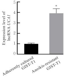
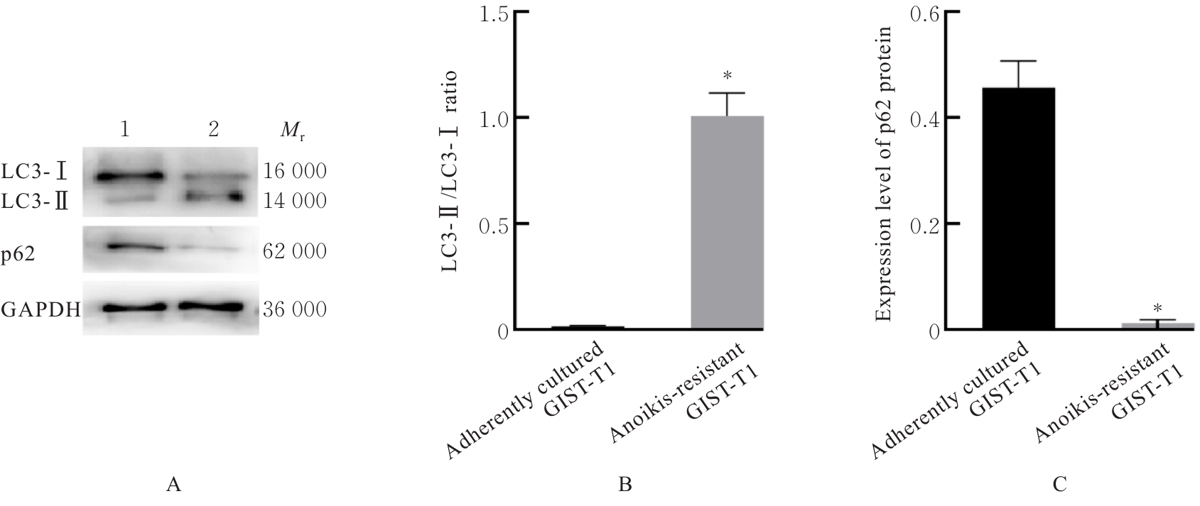
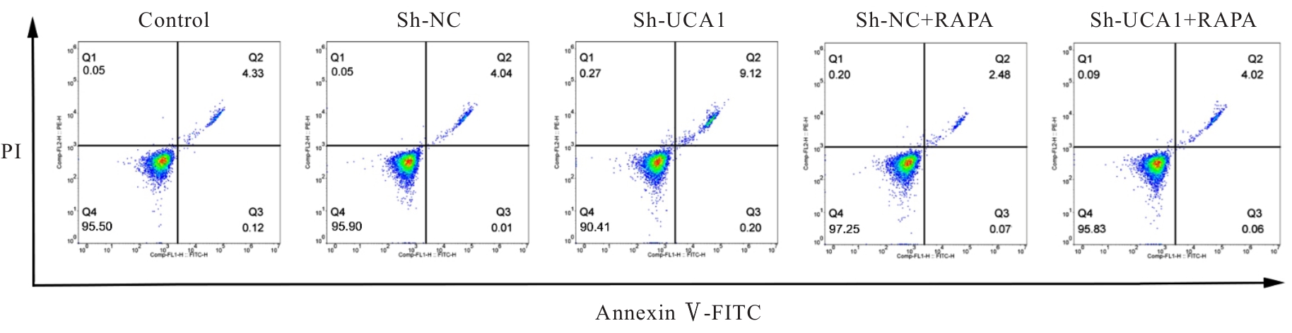
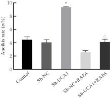
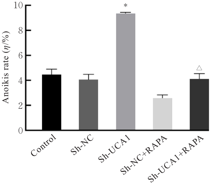
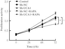
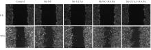
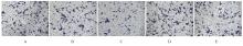
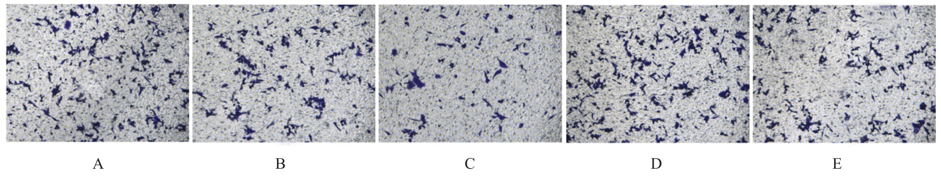
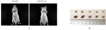
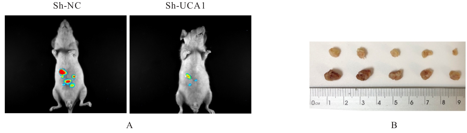
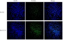
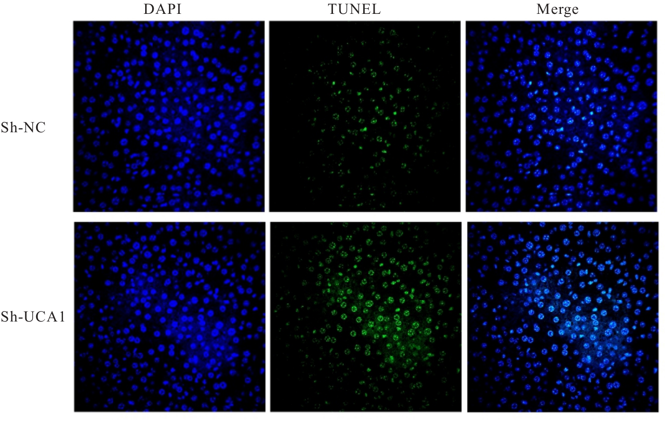
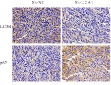
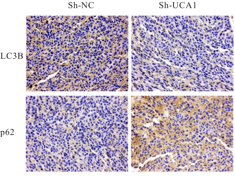
目的 探讨长链非编码RNA(lncRNA)尿路上皮癌胚抗原1(UCA1)对人胃肠道间质瘤(GIST)细胞失巢凋亡的影响,并阐明其作用机制。 方法 分别于贴壁和失巢状态下培养G1ST细胞株GIST-T1并诱导失巢凋亡抵抗的GIST-T1细胞,采用实时荧光定量PCR(RT-qPCR)法检测2种细胞中lncRNA UCA1表达,Western blotting法检测细胞中自噬标志物微管相关蛋白轻链3(LC3)-Ⅰ、LC3-Ⅱ和p62表达水平,并计算LC3-Ⅱ/LC3-Ⅰ比值。采用敲减lncRNA UCA1或阴性对照慢病毒转染GIST-T1细胞并给予自噬激活剂雷帕霉素(RAPA)干预,将GIST-T1细胞分为对照组、sh-NC组、sh-UCA1组、sh-NC+RAPA组和sh-UCA1+RAPA组,并对细胞进行失巢诱导。采用流式细胞术检测各组细胞失巢凋亡率,Western blotting法检测各组细胞中LC3-Ⅰ、LC3-Ⅱ和p62蛋白表达水平,细胞计数试剂盒8(CCK-8)法检测各组细胞增殖活性,细胞划痕实验检测各组细胞划痕愈合率,Transwell小室实验检测各组细胞的侵袭细胞数。收集sh-NC组和sh-UCA1组GIST-T1细胞,分别通过尾静脉注射建立2组裸鼠移植瘤模型,4周后采用荧光活体成像仪检测2组裸鼠体内肿瘤生长和肝脏转移情况,测量2组裸鼠肿瘤组织质量。采用TUNEL染色法观察2组裸鼠肿瘤组织中细胞凋亡情况,免疫组织化学染色法检测2组裸鼠肿瘤组织中LC3B和p62蛋白表达水平。 结果 与贴壁培养的GIST-T1细胞比较,失巢凋亡抵抗的GIST-T1细胞中lncRNA UCA1表达水平明显升高(P<0.05),LC3-Ⅱ/LC3-Ⅰ比值明显升高(P<0.05),p62蛋白表达水平明显降低(P<0.05)。与sh-NC组比较,sh-UCA1组GIST-T1细胞凋亡率升高(P<0.05),LC3-Ⅱ/LC3-Ⅰ比值明显降低(P<0.05),p62蛋白表达水平明显升高(P<0.05),细胞增殖活性和划痕愈合率明显降低(P<0.05),侵袭细胞数明显减少(P<0.05);与sh-UCA1组比较,sh-UCA1+RAPA组GIST-T1细胞失巢凋亡率明显降低(P<0.05),LC3-Ⅱ/LC3-Ⅰ比值明显升高(P<0.05),p62蛋白表达水平明显降低(P<0.05),细胞增殖活性和划痕愈合率明显升高(P<0.05),侵袭细胞数明显增加(P<0.05)。裸鼠体内移植瘤实验, 与sh-NC组比较,sh-UCA1组裸鼠肝组织荧光强度减弱,肿瘤质量明显降低(P<0.05),肿瘤组织中细胞凋亡率明显升高(P<0.05),LC3B蛋白表达水平明显降低(P<0.05),p62蛋白表达水平明显升高(P<0.05)。 结论 lncRNA UCA1在失巢凋亡抵抗的GIST-T1细胞中高表达,下调其表达可通过降低自噬水平来促进GIST-T1细胞失巢凋亡,从而抑制细胞生长、迁移和侵袭。

关键词: 胃肠道间质瘤, 长链非编码RNA, 尿路上皮癌胚抗原1, 失巢凋亡, 自噬

Abstract:

Objective To discuss the effect of long non-coding RNA (lncRNA) urothelial carcinoembryonic antigen 1 (UCA1) on the anoikis of human gastrointestinal stromal tumor (GIST) cells, and to clarify its mechanism of action. Methods The human GIST cell line GIST-T1 was cultured under adherent and anoikis conditions, and the GIST-T1 cells resistant to anoikis were induced. Real-time fluorescence quantitative PCR (RT-qPCR) method was used to detect the expression level of lncRNA UCA1 in the two kinds of cells; Western blotting method was used to detect the expression levels of autophagy markers microtubule-associated protein light chain 3 (LC3)-Ⅰ, LC3-Ⅱ, and p62 proteins in the cells, and the LC3-Ⅱ/LC3-Ⅰ ratio was calculated. The GIST-T1 cells were transfected with lncRNA UCA1 knockdown or negative control lentivirus and treated with the autophagy activator rapamycin (RAPA). The GIST-T1 cells were divided into control group, sh-NC group, sh-UCA1 group, sh-NC+RAPA group, and sh-UCA1+RAPA group, and the cells were subjected to anoikis induction. Flow cytometry was used to detect the anoikis rates of cells in various groups; Western blotting method was used to detect the expression levels of LC3-Ⅰ, LC3-Ⅱ, and p62 proteins in the cells in various groups; cell counting kit-8 (CCK-8) method was used to detect the proliferation activities of the cells in various groups; cell scratch assay was used to detect the scratch healing rates of the cells in various groups; Transwell chamber assay was used to detect the number of invasive cells in various groups. The GIST-T1 cells from sh-NC group and sh-UCA1 group were collected and injected via the tail vein to establish two groups of nude mouse xenograft models, respectively. After 4 weeks, a fluorescence in vivo imaging system was used to detect the tumor growth and liver metastasis in the nude mice in two groups, and the tumor tissue mass was measured. TUNEL staining was used to observe cell apoptosis in the tumor tissue of the nude mice in two groups; immunohistochemical staining was used to detect the expression levels of LC3B and p62 proteins in the tumor tissue of the nude mice in two groups. Results Compared with adherently cultured GIST-T1 cells, the expression level of lncRNA UCA1 in anoikis-resistant GIST-T1 cells was significantly increased (P<0.05), the LC3-Ⅱ/LC3-Ⅰ ratio was significantly increased (P<0.05), and the expression level of p62 protein was significantly decreased (P<0.05). Compared with sh-NC group, the anoikis rate of GIST-T1 cells in sh-UCA1 group was increased (P<0.05), the LC3-Ⅱ/LC3-Ⅰ ratio was significantly decreased (P<0.05), the expression level of p62 protein was significantly increased (P<0.05), the cell proliferation activity and scratch healing rate were significantly decreased (P<0.05), and the number of invasive cells was significantly decreased (P<0.05); compared with sh-UCA1 group, the anoikis rate of GIST-T1 cells in sh-UCA1+RAPA group was significantly decreased (P<0.05), the LC3-Ⅱ/LC3-Ⅰ ratio was significantly increased (P<0.05), the expression level of p62 protein was significantly decreased (P<0.05), the cell proliferation activity and scratch healing rate were significantly increased (P<0.05), and the number of invasive cells was significantly increased (P<0.05). In the nude mouse xenograft experiment, compared with sh-NC group, the fluorescence intensity in liver tissue of the nude mice in sh-UCA1 group was weakened, the tumor mass was significantly decreased (P<0.05), the cell apoptotic rate in tumor tissue was significantly increased (P<0.05), the expression level of LC3B protein was significantly decreased (P<0.05), and the expression level of p62 protein was significantly increased (P<0.05). Conclusion LncRNA UCA1 is highly expressed in anoikis-resistant GIST-T1 cells, and downregulating its expression can promote anoikis of GIST-T1 cells by reducing the autophagy level, thereby inhibiting cell growth, migration, and invasion.

Key words: Gastrointestinal stromal tumor, Long non-coding RNA, Urothelial carcinoembryonic antigen 1, Anoikis, Autophagy

中图分类号: 

  • R735