吉林大学学报(医学版) ›› 2026, Vol. 52 ›› Issue (1): 171-181.doi: 10.13481/j.1671-587X.20260118

• 基础研究 • 上一篇    下一篇

USF2基因敲低对脓毒症大鼠凝血功能障碍的影响及其机制

王镜媛,陈芳,刘艳存,李士欣,寿松涛()   

  1. 天津医科大学总医院急诊医学科,天津 300052
  • 收稿日期:2025-02-17 接受日期:2025-03-26 出版日期:2026-01-28 发布日期:2026-02-24
  • 通讯作者: 寿松涛 E-mail:zyyshou@tmu.edu.cn
  • 作者简介:王镜媛(1987-),女,山东省聊城市人,主治医师,医学硕士,主要从事脓毒症凝血功能方面的研究。
  • 基金资助:
    天津市卫健委卫生健康科技项目(TJWJ2021MS887)

Effect of USF2 knockdown on coagulation dysfunction in septic rats and its mechanism

Jingyuan WANG,Fang CHEN,Yancun LIU,Shixin LI,Songtao SHOU()   

  1. Department of Emergency Medicine General Hospital,Tianjin Medical University,Tianjin 300052,China
  • Received:2025-02-17 Accepted:2025-03-26 Online:2026-01-28 Published:2026-02-24
  • Contact: Songtao SHOU E-mail:zyyshou@tmu.edu.cn

摘要:

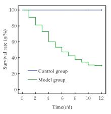
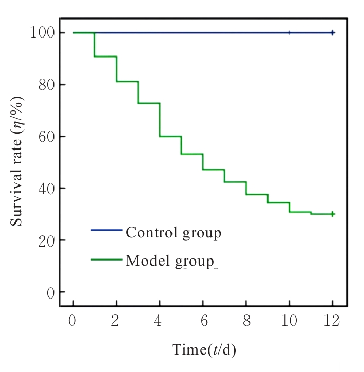
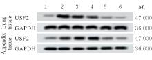
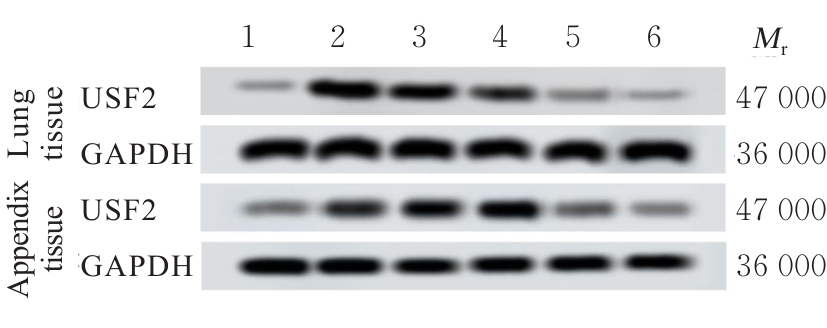
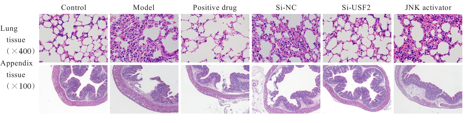
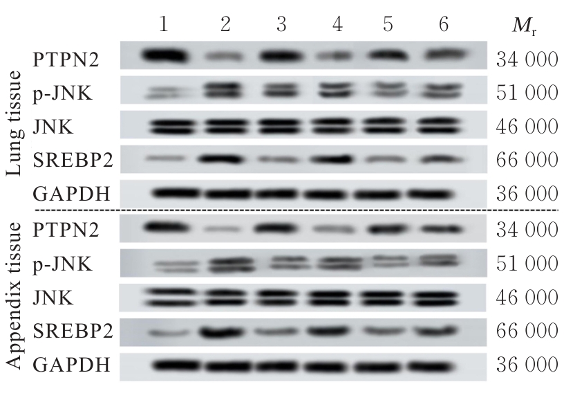
目的 探讨上游转录因子2(USF2)对脓毒症大鼠凝血功能障碍的影响,并基于蛋白质酪氨酸磷酸酶非受体型2(PTPN2)/c-Jun氨基末端激酶(JNK)/甾醇调节元件结合蛋白2(SREBP2)信号通路分析其潜在的作用机制。 方法 从265只健康SD大鼠中随机选取15只作为对照组(不结扎不穿刺),剩余250只大鼠采用盲肠结扎穿刺(CLP)法构建脓毒症模型。将造模成功的75只大鼠随机分为模型组(CLP)、阳性药物组(CLP+20 mg·kg-1辛伐他汀)、小干扰RNA(siRNA)阴性对照(si-NC)组(CLP+转染si-NC)、si-USF2组(CLP+转染USF2-siRNA)和JNK激活剂组(CLP+转染USF2-siRNA+2 mg·kg-1 JNK激活剂Anisomycin),每组15只。采用自动血细胞计数器分析仪评估各组大鼠血小板(PLT)计数;自动凝血分析仪测定各组大鼠凝血酶原时间(PT)、活化部分凝血活酶时间(APTT)和凝血酶时间(TT)以及D-二聚体(DD)和纤维蛋白原(FIB)水平;酶联免疫吸附试验(ELISA)法检测各组大鼠血清中白细胞介素1β(IL-1β)、白细胞介素6(IL-6)、肿瘤坏死因子α(TNF-α)、C反应蛋白(CRP)和降钙素原(PCT)等炎性因子水平;采用试剂盒检测各组大鼠血清中超氧化物歧化酶(SOD)活性以及丙二醛(MDA)和谷胱甘肽(GSH)水平;HE染色观察各组大鼠肺组织和盲肠组织病理形态表现;实时荧光定量PCR(RT-qPCR)和Western blotting法检测各组大鼠肺组织和盲肠组织中USF2 mRNA和蛋白表达水平以及PTPN2、磷酸化JNK(p-JNK)、JNK和SREBP2蛋白表达水平。 结果 造模12 d后,对照组大鼠生存率明显高于模型组。与对照组比较,模型组、阳性药物组、si-NC组、si-USF2组和JNK激活剂组大鼠肺组织及盲肠组织中USF2 mRNA和蛋白表达水平明显升高(P<0.05);与模型组和si-NC组比较,si-USF2组和JNK激活剂组大鼠肺组织及盲肠组织中USF2 mRNA和蛋白表达水平明显降低(P<0.05)。与对照组比较,模型组、阳性药物组、si-NC组、si-USF2组和JNK激活剂组大鼠PLT计数明显降低(P<0.05);与模型组比较,阳性药物组、si-USF2组和JNK激活剂组大鼠PLT计数明显升高(P<0.05);与si-NC组比较,si-USF2组和JNK激活剂组大鼠PLT计数明显升高(P<0.05);与si-USF2组比较,JNK激活剂组大鼠PLT计数明显降低(P<0.05)。与对照组比较,模型组、阳性药物组、si-NC组、si-USF2组和JNK激活剂组大鼠APTT、PT和TT及DD水平明显升高(P<0.05),FIB水平明显降低(P<0.05);与模型组比较,阳性药物组、si-USF2组和JNK激活剂组大鼠APTT、PT和TT及DD水平明显降低(P<0.05),FIB水平明显升高(P<0.05);与si-NC组比较,si-USF2组和JNK激活剂组大鼠APTT、PT和TT及DD水平明显降低(P<0.05),FIB水平明显升高(P<0.05);与si-USF2组比较,JNK激活剂组大鼠APTT、PT和TT及DD水平明显升高(P<0.05),FIB水平明显降低(P<0.05)。与对照组比较,模型组、阳性药物组、si-NC组、si-USF2组和JNK激活剂组大鼠血清中IL-1β、IL-6、TNF-α、CRP、PCT和MDA水平均明显升高(P<0.05),SOD活性和GSH水平明显降低(P<0.05);与模型组比较,阳性药物组、si-USF2组和JNK激活剂组大鼠血清中IL-1β、IL-6、TNF-α、CRP、PCT和MDA水平明显降低(P<0.05),SOD活性和GSH水平明显升高(P<0.05);与si-NC组比较,si-USF2组和JNK激活剂组大鼠血清中IL-1β、IL-6、TNF-α、CRP、PCT及MDA水平明显降低(P<0.05),SOD活性和GSH水平明显升高(P<0.05);与si-USF2组比较,JNK激活剂组大鼠血清中IL-1β、IL-6、TNF-α、CRP、PCT和MDA水平明显升高(P<0.05),SOD活性和GSH水平明显降低(P<0.05)。与对照组比较,模型组大鼠肺组织肺泡结构被破坏,盲肠组织绒毛消失,大量炎性细胞浸润;与模型组比较,阳性药物组、si-USF2组和JNK激活剂组大鼠肺组织肺泡破坏程度及盲肠组织绒毛损坏程度减轻,炎性细胞浸润减少;与si-NC组比较,si-USF2组和JNK激活剂组大鼠肺组织和盲肠组织上述病理变化程度明显减轻;与si-USF2组比较,JNK激活剂组大鼠肺组织和盲肠组织上述病理变化加重。与对照组比较,模型组、阳性药物组、si-NC组、si-USF2组和JNK激活剂组大鼠肺组织和盲肠组织中SREBP2蛋白表达水平及p-JNK/JNK比值明显升高(P<0.05),PTPN2蛋白表达水平明显降低(P<0.05);与模型组比较,阳性药物组、si-USF2组和JNK激活剂组大鼠肺组织和盲肠组织中SREBP2蛋白表达水平及p-JNK/JNK比值明显降低(P<0.05),PTPN2蛋白表达水平明显升高(P<0.05);与si-NC组比较,si-USF2组和JNK激活剂组大鼠肺组织及盲肠组织中SREBP2蛋白表达水平和p-JNK/JNK比值明显降低(P<0.05),PTPN2蛋白表达水平明显升高(P<0.05);与si-USF2组比较,JNK激活剂组大鼠肺组织和盲肠组织中SREBP2蛋白表达水平及p-JNK/JNK比值明显升高(P<0.05),PTPN2蛋白表达水平明显降低(P<0.05)。 结论 敲低USF2基因能够明显改善脓毒症大鼠的肺部和盲肠组织病理形态,缓解凝血功能障碍,并降低机体炎性因子和氧化应激水平,其作用机制可能与其调控PTPN2/JNK/SREBP2信号通路有关。

关键词: 上游转录因子2, 脓毒症, 凝血功能障碍, 蛋白质酪氨酸磷酸酶非受体型2, c-Jun氨基末端激酶, 甾醇调节元件结合蛋白-2

Abstract:

Objective To discuss the effect of upstream transcription factor 2 (USF2) on coagulation dysfunction in the septic rats, and to clarify its potential mechanism based on the protein tyrosine phosphatase non-receptor type 2 (PTPN2)/c-Jun N-terminal kinase (JNK)/sterol regulatory element-binding protein 2 (SREBP2) signaling pathway. Methods Fifteen healthy SD rats were randomly selected from 265 rats as control group (no ligation or puncture); the remaining 250 rats were used to establish the sepsis models by cecal ligation and puncture (CLP). Seventy-five successfully modeled rats were randomly divided into model group (CLP), positive drug group (CLP+20 mg·kg?1 simvastatin), small interfering RNA (siRNA) negative control (si-NC) group (CLP+transfection with si-NC), si-USF2 group (CLP+transfection with USF2-siRNA), and JNK activator group (CLP+transfection with USF2-siRNA+2 mg·kg?1 JNK activator Anisomycin), with 15 rats in each group. An automatic hematology analyzer was used to detect the platelet (PLT) count of the rats in various groups; an automatic coagulation analyzer was used to detect the prothrombin time (PT), activated partial thromboplastin time (APTT), thrombin time (TT), and the levels of D-dimer (DD) and fibrinogen (FIB) of the rats in various groups; enzyme-linked immunosorbent assay (ELISA) was used to detect the levels of inflammatory factors including interleukin-1β (IL-1β), interleukin-6 (IL-6), tumor necrosis factor-α (TNF-α), C-reactive protein (CRP), and procalcitonin (PCT) in serum of the rats in various groups; kits were used to detect the superoxide dismutase (SOD) activity and the levels of malondialdehyde (MDA) and glutathione (GSH) in serum of the rats in various groups; HE staining was used to observe the pathomorphology of lung tissue and cecal tissue of the rats in various groups; real-time fluorescence quantitative PCR (RT-qPCR) and Western blotting methods were used to detect the expression levels of USF2 mRNA and protein and the expression levels of PTPN2, phosphorylated JNK (p-JNK), JNK, and SREBP2 proteins in lung tissue and cecal tissue of the rats in various groups. Results After 12 d of modeling, the survival rate of the rats in control group was significantly higher than that in model group. Compared with control group, the expression levels of USF2 mRNA and protein in lung tissue and cecal tissue of the rats in model group, positive drug group, si-NC group, si-USF2 group, and JNK activator group were significantly increased (P<0.05); compared with model group and si-NC group, the expression levels of USF2 mRNA and protein in lung tissue and cecal tissue of the rats in si-USF2 group and JNK activator group were significantly decreased (P<0.05). Compared with control group, the PLT counts of the rats in model group, positive drug group, si-NC group, si-USF2 group, and JNK activator group were significantly decreased (P<0.05); compared with model group, the PLT counts of the rats in positive drug group, si-USF2 group, and JNK activator group were significantly increased (P<0.05); compared with si-NC group, the PLT counts of the rats in si-USF2 group and JNK activator group were significantly increased (P<0.05); compared with si-USF2 group, the PLT count of the rats in JNK activator group was significantly decreased (P<0.05). Compared with control group, the APTT, PT, TT and DD levels of the rats in model group, positive drug group, si-NC group, si-USF2 group, and JNK activator group were significantly increased (P<0.05), and the FIB level was significantly decreased (P<0.05); compared with model group, the APTT, PT, TT and DD levels of the rats in positive drug group, si-USF2 group, and JNK activator group were significantly decreased (P<0.05), and the FIB level was significantly increased (P<0.05); compared with si-NC group, the APTT, PT, TT and DD levels of the rats in si-USF2 group and JNK activator group were significantly decreased (P<0.05), and the FIB level was significantly increased (P<0.05); compared with si-USF2 group, the APTT, PT, TT and DD levels of the rats in JNK activator group were significantly increased (P<0.05), and the FIB level was significantly decreased (P<0.05). Compared with control group, the levels of IL-1β, IL-6, TNF-α, CRP, PCT and MDA in serum of the rats in model group, positive drug group, si-NC group, si-USF2 group, and JNK activator group were significantly increased (P<0.05), and the SOD activity and GSH level were significantly decreased (P<0.05); compared with model group, the levels of IL-1β, IL-6, TNF-α, CRP, PCT and MDA in serum of the rats in positive drug group, si-USF2 group, and JNK activator group were significantly decreased (P<0.05), and the SOD activity and GSH level were significantly increased (P<0.05); compared with si-NC group, the levels of IL-1β, IL-6, TNF-α, CRP, PCT and MDA in serum of the rats in si-USF2 group and JNK activator group were significantly decreased (P<0.05), and the SOD activity and GSH level were significantly increased (P<0.05); compared with si-USF2 group, the levels of IL-1β, IL-6, TNF-α, CRP, PCT and MDA in serum of the rats in JNK activator group were significantly increased (P<0.05), and the SOD activity and GSH level were significantly decreased (P<0.05). Compared with control group, the alveolar structure of the lung tissue of the rats in model group rats was damaged, the villi of the cecal tissue disappeared, and a large number of inflammatory cells infiltrated; compared with model group, the lung tissue alveolar damage and cecal villi damage of the rats in positive drug group, si-USF2 group, and JNK activator group were alleviated, and inflammatory cell infiltration was reduced; compared with si-NC group, the above pathological changes in the lung and cecal tissues of the rats in si-USF2 group and JNK activator group were significantly alleviated; compared with si-USF2 group, the pathological changes in the lung and cecal tissues of rats in JNK activator group were aggravated.Compared with control group, the expression level of SREBP2 protein and the p-JNK/JNK ratio in lung tissue and cecal tissue of the rats in model group, positive drug group, si-NC group, si-USF2 group, and JNK activator group were significantly increased (P<0.05), and the expression level of PTPN2 protein was significantly decreased (P<0.05); compared with model group and si-NC group, the expression level of SREBP2 protein and the p-JNK/JNK ratio in lung tissue and cecal tissue of the rats in positive drug group, si-USF2 group, and JNK activator group were significantly decreased (P<0.05), and the expression level of PTPN2 protein was significantly increased (P<0.05); compared with si-NC group, the expression level of SREBP2 protein and the p-JNK/JNK ratio in lung tissue and cecal tissue of the rats in si-USF2 group and JNK activator group were significantly decreased (P<0.05), and the expression level of PTPN2 protein was significantly increased (P<0.05); compared with si-USF2 group, the expression level of SREBP2 protein and the p-JNK/JNK ratio in lung tissue and cecal tissue of the rats in JNK activator group were significantly increased (P<0.05), and the expression level of PTPN2 protein was significantly decreased (P<0.05). Conclusion Knockdown of USF2 gene can significantly improve the pathomorphology of lung tissue and cecal tissue, alleviate coagulation dysfunction, and reduce the levels of inflammatory factors and oxidative stress in septic rats; its mechanism may be related to the regulation of PTPN2/JNK/SREBP2 signaling pathway.

Key words: Upstream transcription factor 2, Sepsis, Coagulation dysfunction, Protein tyrosine phosphatase non-receptor type 2, c-Jun N-terminal kinase, Sterol regulatory element-binding protein 2

中图分类号: 

  • R631.2