吉林大学学报(医学版) ›› 2025, Vol. 51 ›› Issue (4): 855-865.doi: 10.13481/j.1671-587X.20250401

• 基础研究 •    下一篇

右美托咪定对肠源性脓毒症大鼠小肠黏膜损伤的改善作用及其机制

杨堃1,付茜瑶1,孙永强1,杨坤1,蒙俊2()   

  1. 1.昆明医科大学第一附属医院麻醉科,云南 昆明 650000
    2.云南省文山州人民医院心血管外科,云南 文山 663000
  • 收稿日期:2024-09-27 接受日期:2024-11-07 出版日期:2025-07-28 发布日期:2025-08-25
  • 通讯作者: 蒙俊 E-mail:mengjun888@163.com
  • 作者简介:杨 堃(1978-),女,云南省大理市人,主任医师,医学博士,主要从事肠源性脓毒症方面的研究。
  • 基金资助:
    国家自然科学基金项目(82260371);云南省科技厅基础研究计划项目(202201AY070001-075)

Protective effect of dexmedetomidine on intestinal mucosal injury in rats with enterogenous sepsis and its mechanism

Kun YANG1,Qianyao FU1,Yongqiang SUN1,Kun YANG1,Jun MENG2()   

  1. 1.Department of Anesthesiology,First Affiliated Hospital,Kunming Medical University,Kunming 650000,China
    2.Department of Cardiovascular Surgery,People’s Hospital,Wenshan Prefecture,Yunan Province,Wenshan 663000,China
  • Received:2024-09-27 Accepted:2024-11-07 Online:2025-07-28 Published:2025-08-25
  • Contact: Jun MENG E-mail:mengjun888@163.com

摘要:

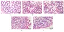
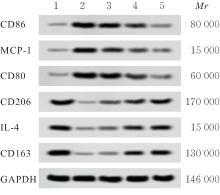
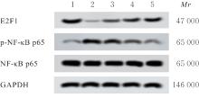
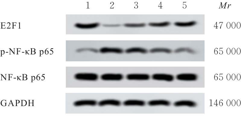
目的 探讨右美托咪定(DEX)对保护肠源性脓毒症大鼠肠道功能的影响,并基于E2F转录因子1(E2F1)/核因子κB(NF-κB)信号通路初步探讨其潜在作用机制。 方法 60只SD大鼠,其中50只大鼠以盲肠结扎穿孔法建立肠源性脓毒症大鼠模型,其余10只大鼠作为假手术组,假手术组大鼠仅分离盲肠远端,不结扎和穿孔。将40只造模成功的大鼠随机分为模型组、低剂量DEX组、中剂量DEX组和高剂量DEX组,每组10只。低、中和高剂量DEX组大鼠即刻腹腔注射20、40及60 μg·kg-1 DEX,假手术组和模型组大鼠腹腔注射等剂量生理盐水。给药24 h后检测各组大鼠肠道肌电活动情况,检测各组大鼠盲肠中大肠埃希菌、乳酸杆菌和双歧杆菌菌落数,HE染色检测各组大鼠小肠组织的病理形态表现,试剂盒检测各组大鼠小肠组织匀浆上清中分泌型免疫球蛋白A(sIgA)水平和血清中二胺氧化酶(DAO)及D-乳酸水平,实时荧光定量PCR(RT-qPCR)法检测各组大鼠小肠组织中巨噬细胞极化标志物mRNA表达水平,Western blotting法检测各组大鼠小肠组织中巨噬细胞极化标志物和E2F1、磷酸化NF-κB p65(p-NF-κB p65)及NF-κB p65蛋白表达水平。 结果 与假手术组比较,模型组大鼠肠道平滑肌慢波频率和振幅降低(P<0.05);与模型组比较,低剂量DEX组大鼠肠道平滑肌慢波振幅升高(P<0.05),中和高剂量DEX组大鼠肠道平滑肌慢波频率及振幅升高(P<0.05);与低剂量DEX组比较,中和高剂量DEX组大鼠肠道平滑肌慢波频率及振幅升高(P<0.05);与中剂量DEX组比较,高剂量DEX组大鼠肠道平滑肌慢波频率和振幅升高(P<0.05)。与假手术组比较,模型组大鼠肠道大肠埃希菌菌落数增加(P<0.05),双歧杆菌和乳酸杆菌菌落数减少(P<0.05);与模型组比较,低剂量DEX组大鼠肠道双歧杆菌菌落数增加(P<0.05),中和高剂量DEX组大鼠肠道大肠埃希菌菌落数减少(P<0.05),双歧杆菌和乳酸杆菌菌落数增加(P<0.05);与低剂量DEX组比较,中和高剂量DEX组大鼠肠道大肠埃希菌菌落数减少(P<0.05),双歧杆菌和乳酸杆菌菌落数增加(P<0.05);与中剂量DEX组比较,高剂量DEX组大鼠肠道大肠埃希菌菌落数减少(P<0.05),双歧杆菌和乳酸杆菌菌落数增加(P<0.05)。HE染色,假手术组大鼠小肠黏膜组织结构正常且完好;模型组大鼠小肠黏膜上皮细胞坏死,绒毛受损、塌陷、排列紊乱;与模型组比较,低、中和高剂量DEX组大鼠小肠组织的病理学明显改善。与假手术组比较,模型组大鼠小肠组织匀浆上清中sIgA水平降低(P<0.05),血清中DAO和D-乳酸蛋白水平升高(P<0.05);与模型组比较,低剂量DEX组大鼠血清中DAO水平降低(P<0.05),中和高剂量DEX组大鼠小肠组织匀浆上清中sIgA水平升高(P<0.05),血清中DAO和D-乳酸蛋白水平降低(P<0.05);与低剂量DEX组比较,中和高剂量DEX组大鼠小肠组织匀浆上清中sIgA水平升高(P<0.05),血清中DAO和D-乳酸蛋白水平降低(P<0.05);与中剂量DEX组比较,高剂量DEX组大鼠小肠组织匀浆上清中sIgA水平升高(P<0.05),血清中DAO和D-乳酸蛋白水平降低(P<0.05)。与假手术组比较,模型组大鼠小肠组织中CD86、单核细胞趋化蛋白1(MCP-1)和CD80 mRNA及蛋白表达水平升高(P<0.05),CD206、白细胞介素(IL-4)和CD163 mRNA及蛋白表达水平降低(P<0.05);与模型组比较,低剂量DEX组大鼠小肠组织中CD80 mRNA、CD86蛋白和MCP-1蛋白表达水平降低(P<0.05),IL-4 mRNA、CD163 mRNA、CD206蛋白和CD163蛋白表达水平降低(P<0.05),中和高剂量DEX组大鼠小肠组织中CD86、MCP-1和CD80 mRNA及蛋白表达水平降低(P<0.05),CD206、IL-4和CD163 mRNA及蛋白表达水平升高(P<0.05);与低剂量DEX组比较,中和高剂量DEX组大鼠小肠组织中CD86、MCP-1和CD80 mRNA及蛋白表达水平降低(P<0.05),CD206、IL-4和CD163 mRNA及蛋白表达水平升高(P<0.05);与中剂量DEX组比较,高剂量DEX组大鼠小肠组织中CD86、MCP-1和CD80 mRNA及蛋白表达水平降低(P<0.05),CD206、IL-4和CD163 mRNA及蛋白表达水平升高(P<0.05)。与假手术组比较,模型组大鼠小肠组织中E2F1蛋白表达水平降低(P<0.05),p-NF-κB p65/NF-κB p65比值升高(P<0.05);与模型组比较,低、中和高剂量DEX组大鼠小肠组织中E2F1蛋白表达水平和p-NF-κB p65/NF-κB p65比值降低(P<0.05);与低剂量DEX组比较,中和高剂量DEX组大鼠小肠组织中E2F1蛋白表达水平升高(P<0.05),p-NF-κB p65/NF-κB p65比值降低(P<0.05);与中剂量DEX组比较,高剂量DEX组大鼠小肠组织中E2F1蛋白表达水平升高(P<0.05),p-NF-κB p65/NF-κB p65比值降低(P<0.05)。 结论 DEX对肠源性脓毒症大鼠小肠黏膜损伤具有改善作用,并促进小肠组织中巨噬细胞向M2型极化转变,其机制可能与DEX调控E2F1/NF-κB信号通路有关。

关键词: 右美托咪定, E2F转录因子1, 核因子κB, 巨噬细胞极化, 肠源性脓毒症

Abstract:

Objective To discuss the protective effect of dexmedetomidine (DEX) on intestinal function in rats with enterogenous sepsis, and to clarify its potential mechanism based on E2F transcription factor 1 (E2F1)/nuclear factor kappa B (NF-κB) signaling pathway. Methods Sixty SD rats were selected, among which 50 rats were used to establish enterogenous sepsis models by cecal ligation and puncture (CLP), and the remaining 10 rats were used as sham operation group (only cecal separation without ligation or puncture). The 40 successfully modeled rats were randomly divided into model group, low dose of DEX group, medium, doses of DEX group, and high dose of DEX group, with 10 rats in each group. The rats in low, medium, and high dose of DEX groups were intraperitoneally injected with 20, 40 and 60 μg·kg-1 DEX immediately after modeling, while the rats in sham operation group and model group were intraperitoneally injected with the same volume of saline. After 24 h of administration, the intestinal myoelectric activities of the rats in various groups were detected; the colony counts of Escherichia coliLactobacillus and Bifidobacterium in cecal contents of the rats in various groups were detected; the pathomorphology of small intestinal tissue of the rats was observed by HE staining; the levels of secretory immunoglobulin A (sIgA) in supernatant of small intestinal tissue homogenate and the levels of diamine oxidase (DAO) and D-lactic acid in serum of the rats in various groups were detected by kit; real-time fluorescence quantitative PCR (RT-qPCR) method was used to detect the mRNA expression levels of macrophage polarization markers in small intestinal tissues of the rats in various groups; Western blotting method was used to detect the protein expression levels of macrophage polarization markers, E2F1, phosphorylated NF-κB p65 (p-NF-κB p65), and NF-κB p65 in small intestinal tissue of the rats in various groups. Results Compared with sham operation group, the slow wave frequency and amplitude of intestinal smooth muscle of the rats in model group were decreased (P<0.05); compared with model group, the slow wave amplitude of intestinal smooth muscle of the rats in low dose of DEX groups was increased (P<0.05), the slow wave frequency and amplitude of intestinal smooth muscle of the rats in medium and high doses of DEX groups were increased (P<0.05); compared with low dose of DEX group, the slow wave frequency and amplitude of the rats in medium and high doses of DEX groups were increased (P<0.05); compared with medium dose of DEX group, the slow wave frequency and amplitude of intestinal smooth muscle of the rats in high dose of DEX group were increased (P<0.05). Compared with sham operation group, the colony count of Escherichia coli in intestinal tract of the rats in model group was increased (P<0.05), while the colony counts of Bifidobacterium and Lactobacillus were decreased (P<0.05); compared with model group, the colony count of Bifidobacterium in intestinal tract of the rats in low dose of DEX group was decreased (P<0.05), the colony count of Escherichia coli in intestinal tract of the rats in medium, and high doses of DEX groups was decreased (P<0.05), while the colony counts of Bifidobacterium and Lactobacillus were increased (P<0.05); compared with low dose of DEX group, the colony count of Escherichia coli in intestinal tract of the rats in medium and high dose of DEX groups was decreased (P<0.05), while the colony counts of Bifidobacterium and Lactobacillus were increased (P<0.05); compared with medium dose of DEX group, the colony count of Escherichia coli in intestinal tract of the rats in high dose of DEX group was decreased (P<0.05), while the colony counts of Bifidobacterium and Lactobacillus were increased (P<0.05). The HE staining results showed that the small intestinal mucosal structure in sham operation group was normal and intact; the small intestinal mucosal epithelial cells in model group were necrotic, with damaged, collapsed and disordered villi; Compared with model groups, the pathological changes of small intestinal tissues in low, medium, and high doses of DEX groups were improved. Compared with sham operation group, the level of sIgA in supernatant of small intestinal tissue homogenate of the rats in model group was decreased (P<0.05), while the protein expression levels of DAO and D-lactic acid in serum were increased (P<0.05); compared with model group, the level of DAO in serum of the rats in low dose of DEX groups was decreased (P<0.05), the level of sIgA in supernatant of small intestinal tissue homogenate of the rats in medium and high doses of DEX groups was increased (P<0.05), while the protein expression levels of DAO and D-lactic acid in serum were decreased (P<0.05); compared with low dose of DEX group, the level of sIgA in supernatant of small intestinal tissue homogenate of the rats in medium and high doses of DEX groups was increased (P<0.05), while the protein expression levels of DAO and D-lactic acid in serum were decreased (P<0.05); compared with medium dose of DEX group, the level of sIgA in supernatant of small intestinal tissue homogenate of the rats in high dose of DEX group was significantly increased (P<0.05), while the protein expression levels of DAO and D-lactic acid in serum were decreased (P<0.05). The RT-qPCR results and Western blotting results showed that compared with sham operation group, the mRNA and protein expression levels of CD86, monocyte chemoattractant protein-1 (MCP-1), and CD80 in small intestinal tissue of the rats in model group were increased (P<0.05), while the mRNA and protein expression levels of CD206, interleukin-4 (IL-4) and, CD163 were decreased (P<0.05); compared with model group, the expression levels of CD80 mRNA, CD86 protein and MCP-1 protein in small intestinal tissue of the rats in low dose of DEX group were decreased (P<0.05), and the expression levels of IL-4 mRNA, CD163 mRNA, CD206 protein, and CD163 protein were decreased (P<0.05), the mRNA and protein expression levels of CD86, MCP-1, and CD80 in small intestinal tissue of the rats in medium and high doses of DEX groups were decreased (P<0.05), while the mRNA and protein expression levels of CD206, IL-4 and CD163 were increased (P<0.05); compared with low dose of DEX group, the mRNA and protein expression levels of CD86, MCP-1, and CD80 in small intestinal tissue of the rats in medium and high doses of DEX groups were decreased (P<0.05), while the mRNA and protein expression levels of CD206, IL-4, and CD163 were increased (P<0.05); compared with medium dose of DEX group, the mRNA and protein expression levels of CD86, MCP-1, and CD80 in small intestinal tissue of the rats in high dose of DEX group were decreased (P<0.05), while the mRNA and protein expression levels of CD206, IL-4, and CD163 were increased (P<0.05). The Western blotting results showed that compared with sham operation group, the protein expression level of E2F1 in small intestinal tissue of the rats in model group was decreased (P<0.05), while the ratio of p-NF-κB p65/NF-κB p65 was increased (P<0.05); compared with model group, the protein expression levels of E2F1 and ratio of p-NF-κB p65/NF-κB p65 in small intestinal tissue of the rats in low, medium and high doses of DEX groups were decreased (P<0.05); compared with low dose of DEX group, the protein expression level of E2F1 in small intestinal tissue of the rats in medium and high doses of DEX groups was increased (P<0.05), while the ratio of p-NF-κB p65/NF-κB p65 was decreased (P<0.05); compared with medium dose of DEX group, the protein expression level of E2F1 in small intestinal tissue of the rats in high dose of DEX group was increased (P<0.05), while the ratio of p-NF-κB p65/NF-κB p65 was decreased (P<0.05). Conclusion DEX can improve the small intestinal mucosal injury in the rats with enterogenous sepsis and promote the polarization of macrophages to M2 type in small intestinal tissues, and its mechanism may be related to the regulation of E2F1/NF-κB signaling pathway by DEX.

Key words: Dexmedetomidine, E2F transcription factor 1, Nuclear factor kappa B, Macrophage polarization, Enterogenous sepsis

中图分类号: 

  • R614