吉林大学学报(医学版) ›› 2026, Vol. 52 ›› Issue (2): 530-535.doi: 10.13481/j.1671-587X.20260226

• 临床医学 • 上一篇    下一篇

呈黄褐混浊态的双侧胸腔积液快速交替进展患者1例报告及文献复习

丁艺,贾文慧,杨晨露,董春玲()   

  1. 吉林大学第二医院呼吸与危重症医学科,吉林 长春 130041
  • 收稿日期:2025-06-02 接受日期:2025-07-14 出版日期:2026-03-28 发布日期:2026-04-15
  • 通讯作者: 董春玲 E-mail:cldong@jlu.edu.cn
  • 作者简介:丁 艺(1998-),男,山东省滕州市人,在读硕士研究生,主要从事间质性肺疾病和肺部感染性疾病等方面的研究。
  • 基金资助:
    吉林省财政厅卫生专项项目(2020SCZT0230);北京市医学奖励基金会项目(YXJL-2024-0129-0028)

Bilateral pleural effusion with rapid alternating progression in patient with yellow-brown turbid appearance:A case report and literature review

Yi DING,Wenhui JIA,Chenlu YANG,Chunling DONG()   

  1. Department of Respiratory and Critical Care Medicine,Second Hospital,Jilin University,Changchun 130041,China
  • Received:2025-06-02 Accepted:2025-07-14 Online:2026-03-28 Published:2026-04-15
  • Contact: Chunling DONG E-mail:cldong@jlu.edu.cn

摘要:

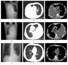
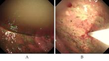
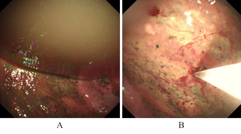
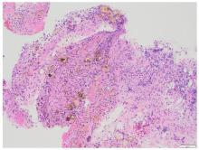
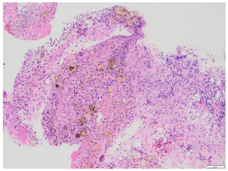
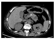
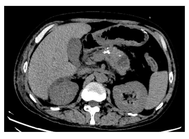
胰源性胸腔积液常由胰腺假性囊肿、胰胸膜瘘(PPF)和胰腺炎等引发。该疾病的临床表现以胸部症状为主,腹部症状较少见,缺乏特异性,故极易出现诊治延误。呈黄褐混浊态的双侧胸腔积液快速交替进展者较为罕见,国内外未见相关报道。本文作者报道1例胰源性胸腔积液患者,对其临床表现、胸膜病理特点及诊治方案进行总结,并结合相关文献进行复习。患者,男性,40岁,因“咳嗽、咳痰、胸痛伴阵发性呼吸困难”就诊,其胸腔积液呈快速进展的黄褐混浊态,排查常见病因后未能明确诊断,治疗期间突发腹痛。结合患者既往有长期饮酒史并曾行胰腺假性囊肿胃吻合术,遂完善胸腔积液淀粉酶检查,明确诊断继发于胰腺假性囊肿的胰源性胸腔积液,并行内镜逆行胰胆管造影(ERCP)及相关介入治疗。定期随访后,胸腔积液无复发迹象。对于有胰腺相关病史的胸腔积液患者,无论是否并发腹部症状,均应考虑胰源性胸腔积液的可能,并警惕血胸、脓胸、纵隔炎和呼吸衰竭等相关严重并发症。对于内科治疗效果不佳者,应细致评估胰液引流路径及解剖异常,以指导精准个体化治疗,改善患者预后。

关键词: 胸腔积液, 胰腺假性囊肿, 胸腔镜检查, 胰胸膜瘘, 病例报告

Abstract:

Pancreatic-derived pleural effusion is commonly caused by pancreatic pseudocysts, pancreaticopleural fistula (PPF), and pancreatitis. Its clinical presentation is primarily characterized by thoracic symptoms, with abdominal symptoms being less common and lacking specificity, leading to frequent diagnostic and therapeutic delays. Bilateral pleural effusions presenting as yellowish-brown turbid fluid with rapid alternating progression are rare, and no related cases were reported domestically or internationally. This article reported a case of patient with pancreatic-derived pleural effusion, summarized its clinical presentation, pleural pathological features, and management strategy, and the relevant literatures were reviewed. The patient, a 40-year-old male, presented with “cough, sputum production, chest pain accompanied by paroxysmal dyspnea”. His pleural effusion exhibited rapid progression with a yellowish-brown turbid appearance. After ruling out common causes, a definitive diagnosis remained elusive. During treatment, the patient developed sudden abdominal pain. Given his history of chronic alcohol consumption and prior gastrostomy for pancreatic pseudocyst, amylase testing of the pleural effusion was performed. This confirmed a diagnosis of pancreatic-derived pleural effusion secondary to pancreatic pseudocyst. The patient subsequently underwent endoscopic retrograde cholangiopancreatography (ERCP) and related interventional procedures. Regular follow-up revealed no recurrence of pleural effusion. For the patients with pleural effusion and a history of pancreatic disease, pancreatic origin should be considered regardless of abdominal symptoms, while vigilance is warranted for severe complications including hemothorax, empyema, mediastinitis, and respiratory failure. When medical management proves inadequate, meticulous evaluation of pancreatic drainage pathways and anatomical anomalies is essential to guide precise and individualized treatment and improve the patient’s prognosis.

Key words: Pleural effusion, Pancreatic pseudocyst, Thoracoscopy, Pancreatopleural fistula, Case report

中图分类号: 

  • R561.3