Journal of Jilin University(Medicine Edition) ›› 2021, Vol. 47 ›› Issue (2): 284-291.doi: 10.13481/j.1671-587X.20210205
• Research in basic medicine • Previous Articles Next Articles
Effect of mitochondrial fission/fussion protein expression imbalance in pathological changes of kidney tissue in IgA nephropathy mice
Xu ZHANG1,2,Naimeng LIU3,Jiaoyan MA2,Nan LIN2,Mindan SUN4( )
)
- 1.Department of Gynecology and Pediatrics,Clinical Medicine Center,Changchun Medical College,Changchun 130031,China
2.Department of Pathophysiology,School of Basic Medical Sciences,Jilin University,Changchun 130021,China
3.Department of Breast Surgery,First Hospital,Jilin University,Changchun 130021,China
4.Department of Nephropathy,First Hospital,Jilin University,Changchun 130021,China
-
Received:2020-09-12Online:2021-03-28Published:2021-03-25 -
Contact:Mindan SUN E-mail:mindansun@sina.com
CLC Number:
- R692
Cite this article
Xu ZHANG,Naimeng LIU,Jiaoyan MA,Nan LIN,Mindan SUN. Effect of mitochondrial fission/fussion protein expression imbalance in pathological changes of kidney tissue in IgA nephropathy mice[J].Journal of Jilin University(Medicine Edition), 2021, 47(2): 284-291.
share this article
Tab. 1
Primer sequences of RT-qPCR"
| Primer | Sequence (5'-3') |
|---|---|
| DRP1 | Forward: TCTACATGGCGTATAAGGACCTG |
| Reverse: CTGCATGGACTAAGATCACAGC | |
| MFN1 | Forward: CCTACTGCTCCTTCTAACCCA |
| Reverse: AGGGACGCCAATCCTGTGA | |
| MFN2 | Forward: GTGGGCTGGAGACTCATCG |
| Reverse: CTCACTGGCGTATTCCGAA | |
| GAPDH | Forward: GTCGTGGAGTCTACTGGTGTC |
| Reverse: GAGCCCTTCCACAATGCCAAA |

Fig. 1
IgA deposition in glumerulus of mice in various groups (Immunofluorescence,×200)"

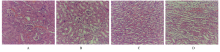
Fig. 2
Pathomorphology of kidney tissue of mice in various groups (HE,×200)"


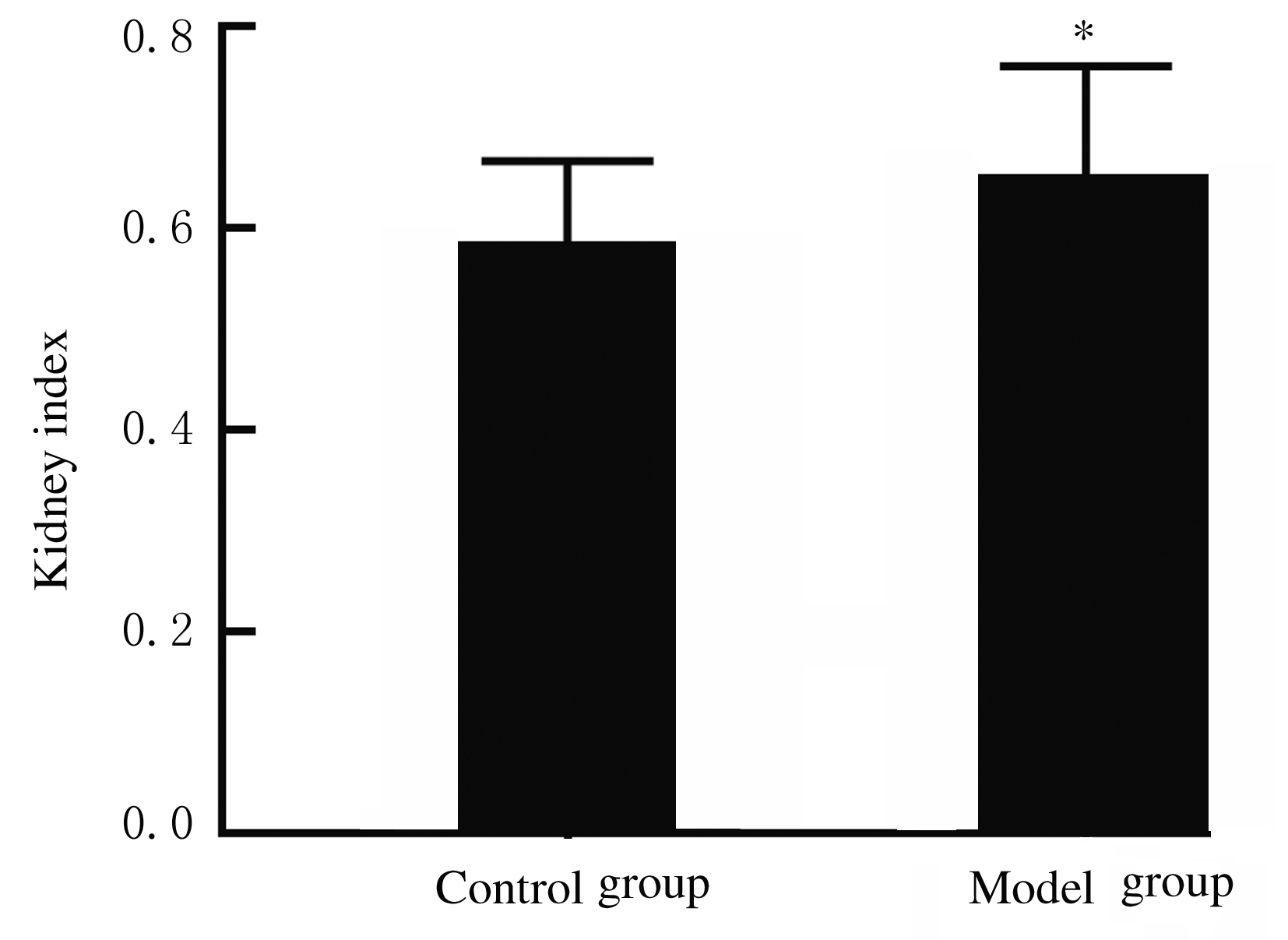
Fig. 3
Kidney indexes of mice in various groups"
Tab. 2
Levels of serum creatinine and urea nitrogen of mice in various groups"
| Group | Creatinine[cB/(μmol·L-1)] | Urea nitrogen[cB/(mmol·L-1)] |
|---|---|---|
| Control | 40.7±2.6 | 7.4±1.4 |
| Model | 75.3±5.8* | 21.6±2.1* |
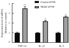
Fig. 4
Levels of TNF-α,IL-1β, and IL-6 in serum of mice in various groups"


Fig. 5
Expression levels of Drp1, Mfn1, and Mfn2 mRNA in kidney tissue of mice in various groups"
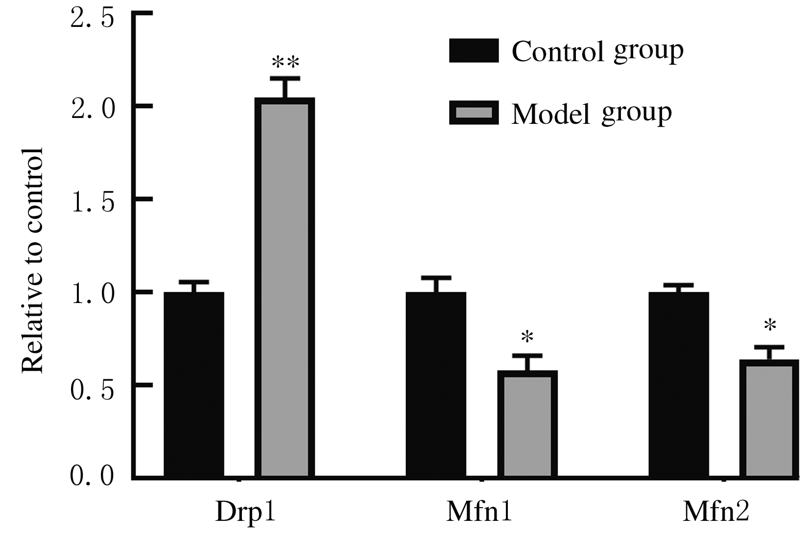

Fig. 6
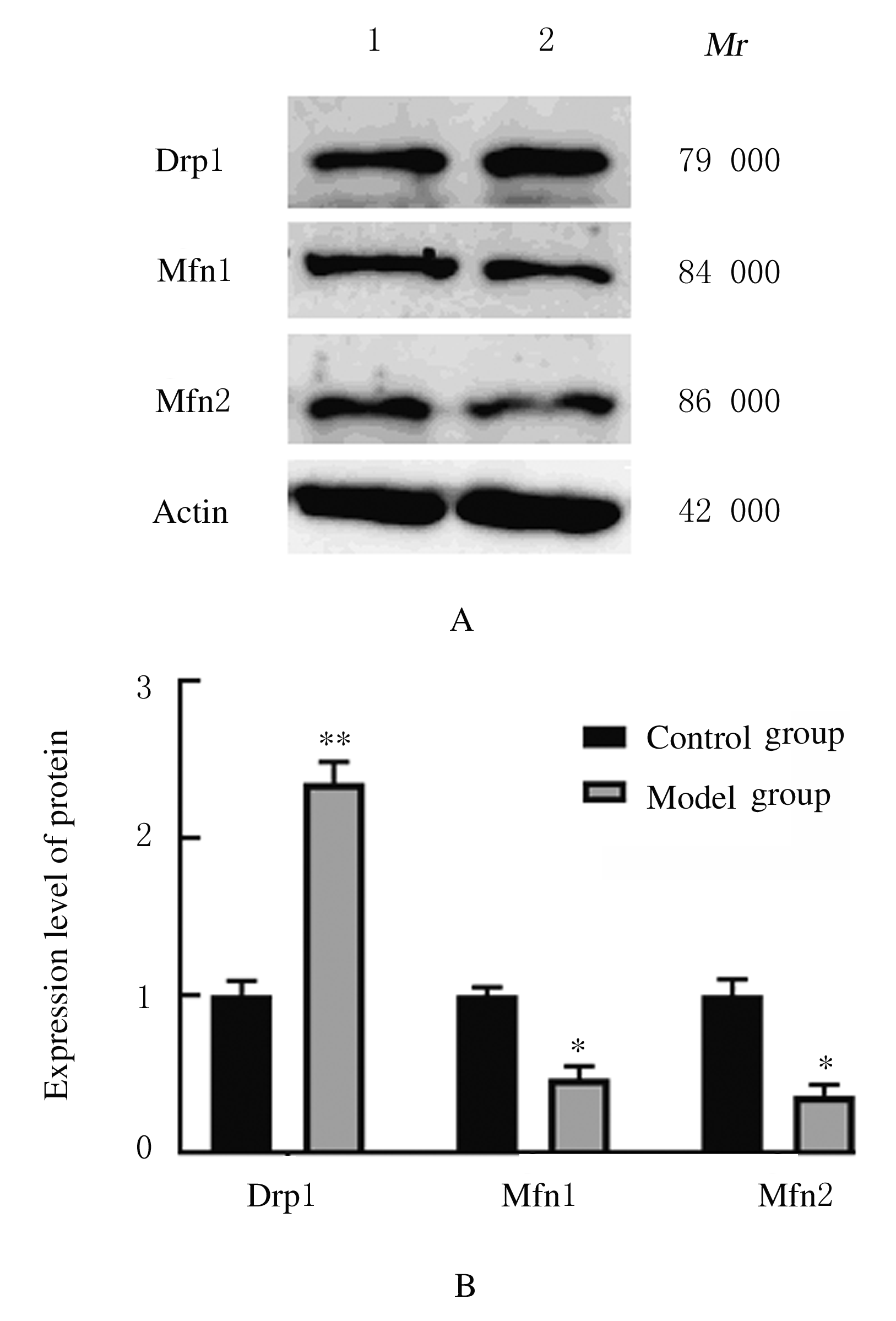
Electrophoregram (A) and histogram (B) of expressions of Drp1, Mfn1, and Mfn2 proteins in kidney tissue of mice in various groups"
| 1 | WYATT R J, JULIAN B A. IgA nephropathy [J]. N Engl J Med, 2013, 368(25): 2402-2414. |
| 2 | HUSSAIN T A L, HUSSEIN M H, MANA H A L, et al. Pathophysiology of IgA nephropathy [J]. Adv Anat Pathol, 2017, 24(1):56-62. |
| 3 | SCHENA F P. A retrospective analysis of the natural history of primary IgA nephropathy worldwide [J]. Am J Med, 1990, 89(2) :209-215. |
| 4 | RAUEN T, FLOEGE J. Inflammation in IgA nephropathy [J]. Pediatr Nephrol Berlin Ger, 2017, 32(12): 2215-2224. |
| 5 |
PERSE M, VECERIC-HALER Z. The role of IgA in the pathogenesis of IgA nephropathy [J]. Int J Mol Sci, 2019, 20(24).DOI:10.3390/JJMS 20246199.
doi: 10.3390/JJMS 20246199 |
| 6 | CLARK A J, PARIKH S M. Mitochondrial metabolism in acute kidney injury [J]. Semin Nephrol, 2020, 40(2):101-113. |
| 7 | BHARGAVA P, SCHNELLMANN R G. Mitochondrial energetics in the kidney [J]. Nat Rev Nephrol, 2017, 13(10):629-646. |
| 8 | BLIEK A MVAN D E R, SEDENSKY M M, MORGAN P G. Cell biology of the mitochondrion [J]. Genetics, 2017, 207(3): 843-871. |
| 9 | LIN M T, BEAL M F. Mitochondrial dysfunction and oxidative stress in neurodegenerative diseases [J]. Nature, 2006, 443(7113): 787-795. |
| 10 | 关毅鸣, 王丽妍, 刘文虎. 线粒体功能及其与急性肾损伤和糖尿病肾病的关系[J].医学研究杂志,2020, 49(7):5-8. |
| 11 | 汤 颖, 娄探奇, 成彩联, 等.实验性IgA肾病模型的改进 [J]. 中山大学学报(医学科学版),2006,27(2):184-187. |
| 12 | BERTHOUX F, SUZUKI H, THIBAUDIN L, et al. Autoantibodies targeting galactose-deficient IgA1 associate with progression of IgA nephropathy [J]. J Am Soc Nephrol, 2012, 23(9):1579-1587. |
| 13 | HATTON G E, DU R E, PEDROZA C, et al. Choice of reference creatinine for post-traumatic acute kidney injury diagnosis [J]. J Am Coll Surg, 2019, 229(6): 580-588.e4. |
| 14 | OGUNDIPE D J, AKOMOLAFE R O, SANUSI A A, et al. Ocimum gratissimum ameliorates gentamicin-induced kidney injury but decreases creatinine clearance following sub-chronic administration in rats [J]. J Evid Based Complementary Altern Med, 2017,22(4):592-602. |
| 15 | ZHAO Y F, ZHU L, LIU L J, et al. TREM-1 contributes to inflammation in IgA nephropathy [J]. Kidney Dis (Basel), 2018,4(1):29-36. |
| 16 | 赵成波, 曾 欢, 王润秀,等. IgA肾病相关的免疫细胞膜分子及系膜细胞激活的研究进展[J]. 赣南医学院学报,2020,40(8):848-852. |
| 17 | 欧阳彦,谢静远.IgA肾病遗传学机制进展[J].中国实用内科杂志,2020,40(7):529-532. |
| 18 | 杨琼琼.IgA肾病诊治相关的生物标志物[J].中国实用内科杂志,2020,40(7):533-538. |
| 19 | DI LERNIA V. IgA nephropathy during treatment with TNF-alpha blockers: Could it be predicted? [J]. Med Hypotheses, 2017, 107:12-13. |
| 20 | HE L, PENG X, LIU G, et al. Anti-inflammatory effects of triptolide on IgA nephropathy in rats[J]. Immunopharmacol Immunotoxicol,2015,37(5):421-427. |
| 21 | MAKITA Y, SUZUKI H, KANO T, et al. TLR9 activation induces aberrant IgA glycosylation via APRIL- and IL-6-mediated pathways in IgA nephropathy [J]. Kidney Int, 2020, 97(2):340-349. |
| 22 | TOPF U, USZCZYNAKA-RATAJCZAK B, CHACINSKA A. Mitochondrial stress-dependent regulation of cellular protein synthesis [J]. J Cell Sci, 2019, 32(8):jcs226258. |
| 23 | YOULE R J, BLIEK A MVAN D E R. Mitochondrial fission,fusion,and stress[J]. Science,2012,337(6098):1062-1065. |
| 24 | YU B C, CHO N J, PARK S, et al. IgA nephropathy is associated with elevated urinary mitochondrial DNA copy numbers [J]. Sci Rep, 2019, 9(1):16068. |
| 25 | GONG X, DUAN Y, ZHENG J, et al. Tetramethylpyrazine prevents contrast-induced nephropathy via modulating tubular cell mitophagy and suppressing mitochondrial fragmentation, CCL2/CCR2-mediated inflammation, and intestinal injury [J]. Oxid Med Cell Longev, 2019, 2019:7096912. |
| [1] | Fei LIU,Shuo LIANG,Yuexuan WANG,Lijing QIN,Wei GUO,Zhicheng WANG. Effect of low dose ionizing radiation on hippocampal neurons in STZ-induced diabetic rats [J]. Journal of Jilin University(Medicine Edition), 2021, 47(1): 1-7. |
| [2] | Haiying ZHANG,Wenxin ZHANG,Wenjing ZHAO,Wei LI. Induction effect of gallic acid on apoptosis of human hepatocellular carcinoma HepG-2 cells through regulating mitochondrial oxidative phosphorylation [J]. Journal of Jilin University(Medicine Edition), 2021, 47(1): 125-132. |
| [3] | LIN Jiuhan, TIAN Lin, ZHANG Nana, DONG Ying, LYU Hang, WANG Dan, QU Meng, ZHANG Yong, SUN Liankun, YU Chunyan, LIU Xi. Inhibitory effect of mitochondrial dynamics changes on growth of transplanted melanoma in APP/PS1 mice [J]. Journal of Jilin University(Medicine Edition), 2020, 46(03): 476-481. |
| [4] | LOU Tingting, HUANG Qingxia, LI Xiangyan, ZHAO Daqing. Protective effect of ginseng extract on cardiomyocyte injury induced by palmitic acidand its mechanism [J]. Journal of Jilin University(Medicine Edition), 2019, 45(06): 1248-1255. |
| [5] | GUO Qingbang, FENG Wenhua, ZHANG Zhao. Inhibitory effect of coenzyme Q10 on apoptosis of human coronary endothelial cells induced by high glucose and its mechanism [J]. Journal of Jilin University(Medicine Edition), 2019, 45(05): 1031-1035. |
| [6] | WANG Aifu, ZHANG Qi, ZHANG Daoming, XIU Yuting, DING Yaming, LIU Linlin. Effect of silencing mitochondrial ribosomal protein L35 gene on growth of human esophageal cancer TE-1 cells [J]. Journal of Jilin University(Medicine Edition), 2019, 45(01): 28-32. |
| [7] | CHI Ming, GAO Ling, WU Weiwei, ZHANG Boru, WANG Lei. Improvement effect of berberine on acute lung injury and inflammation induced by lipopolysaccharide and its mechanism [J]. Journal of Jilin University(Medicine Edition), 2018, 44(06): 1194-1199. |
| [8] | YU Yang, XU Lu, LIU Shibing, LI Songyan, XU Ye. Induction effect of myricetin on apoptosis of human ovarian cancer SKOV3 cells by promoting DRP1-dependent mitochondrial fission [J]. Journal of Jilin University(Medicine Edition), 2018, 44(05): 903-907. |
| [9] | ZHENG Guilang, GUO Yuxiong, LYU Juanjuan, HUANG Jinda, LIU Cui, ZENG Qiyi. Protective effect of overexpression of uncoupling protein 2 on mitochondria in cardiomyocytes of rats with sepsis [J]. Journal of Jilin University Medicine Edition, 2018, 44(04): 698-703. |
| [10] | LI Xin, MA Yunfei, TANG Geng, WEI Qi, JI Hongchi, TIAN Jiaan, SHEN Yannan, WANG Zhicheng. Enhancement of ROS induced by mitochondria-targeted KillerRed in proliferation inhibition of radiation on HeLa cells [J]. Journal of Jilin University Medicine Edition, 2018, 44(04): 718-723. |
| [11] | YANG Yi, BAO Kangda, ZHOU Yanbing, YUAN Chao, LI Yong, LIU Xiaoming, XU Jinrui. Regulation of Wnt5a in apoptosis of human lung adenocarcinoma A549 cells and its mechanism [J]. Journal of Jilin University Medicine Edition, 2018, 44(02): 229-234. |
| [12] | XU Xuemei, HUANG Xiaoxia, JIN Ou, ZHANG Hailin, SHI Hongxue. Protective effect of epithelial growth factor on oxidative damage of skeletal muscle cells in model rats of oxygen-glucose deprivation [J]. Journal of Jilin University Medicine Edition, 2018, 44(02): 310-314. |
| [13] | QI Yali, LIU Yang, LIANG Shuo, GONG Shouliang, WANG Zhicheng, LIU Weiwu. Effects of Smac overexpression mediated by triple-targeting on apoptosis and cell cycle progression of breast cancer MDA-MB-231 cells [J]. Journal of Jilin University Medicine Edition, 2018, 44(01): 90-94. |
| [14] | YU Yang, LIU Shibing, LI Songyan, XU Lu, XU Ye. Induction effect of myricetin on autophagy in SKOV3 cells and promoting effect on mitochondrial fission [J]. Journal of Jilin University Medicine Edition, 2017, 43(04): 685-689. |
| [15] | WEI Yanhong, LIU Xiaodong, LIU Yan, LU Lijuan. Effect of PI3K-Akt signaling pathway on openness degree of mitochondrial permeability transition pore in aged model rats of myocardial ischemic preconditioning [J]. Journal of Jilin University Medicine Edition, 2016, 42(04): 648-652. |
|