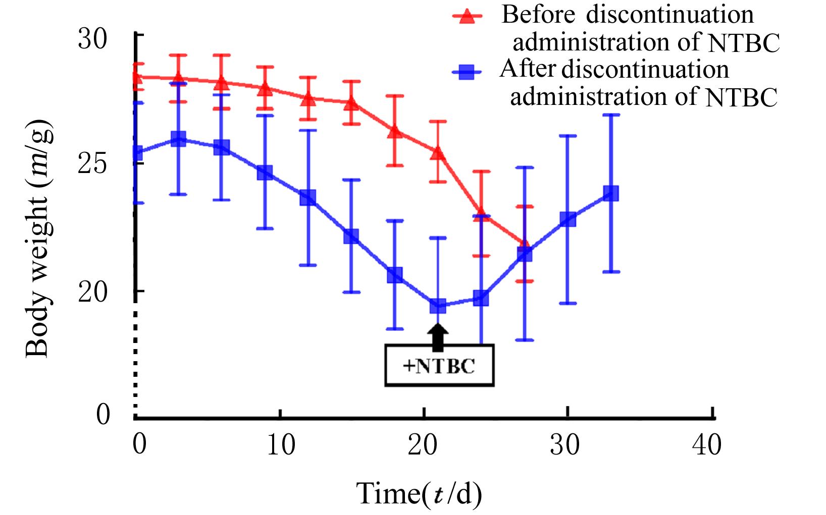
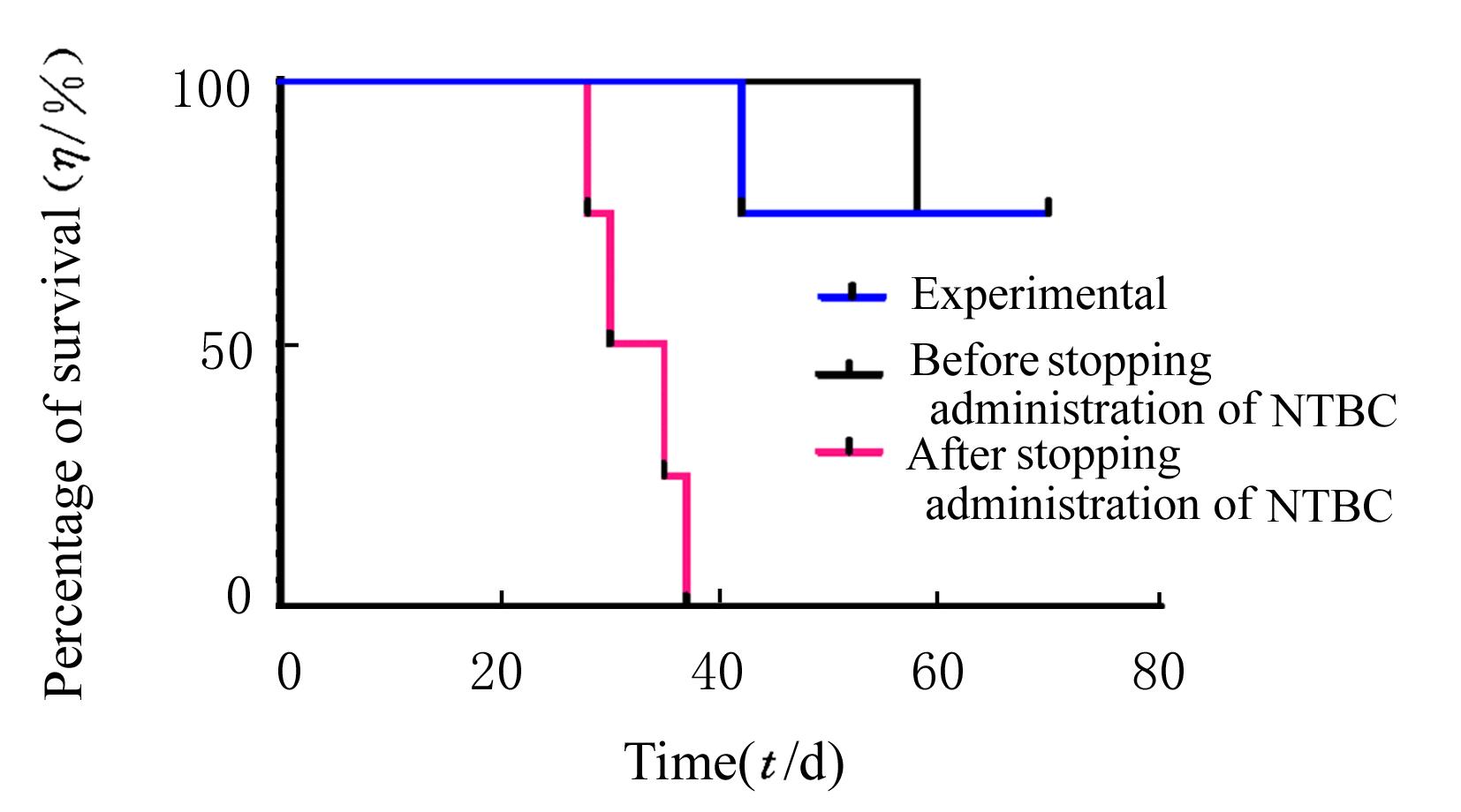
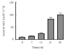
| 1 |
OLIVO R, GUARRERA J V, PYRSOPOULOS N T. Liver transplantation for acute liver failure[J]. Clin Liver Dis, 2018, 22(2): 409-417.
|
| 2 |
KOBAYASHI N, FUJIWARA T, WESTERMAN K A,et al. Prevention of acute liver failure in rats with reversibly immortalized human hepatocytes[J]. Science, 2000, 287(5456): 1258-1262.
|
| 3 |
IWASE H, LIU H, SCHMELZER E, et al. Transplantation of hepatocytes from genetically engineered pigs into baboons[J]. Xenotransplantation, 2017, 24(2): 10.1111/xen.12289.
|
| 4 |
IBRAHIM Z, BUSCH J, AWWAD M, et al. Selected physiologic compatibilities and incompatibilities between human and porcine organ systems[J]. Xenotransplantation, 2006, 13(6): 488-499.
|
| 5 |
REID L M, JEFFERSON D M. Culturing hepatocytes and other differentiated cells[J].Hepatology,1984,4(3): 548-559.
|
| 6 |
SUEMIZU H, HASEGAWA M, KAWAI K J, et al. Establishment of a humanized model of liver using NOD/Shi-scid IL2Rgnull mice[J]. Biochem Biophys Res Commun, 2008, 377(1): 248-252.
|
| 7 |
UEHARA S, HIGUCHI Y, YONEDA N, et al. An improved TK-NOG mouse as a novel platform for humanized liver that overcomes limitations in both male and female animals[J]. Drug Metab Pharmacokinet, 2022, 42: 100410.
|
| 8 |
HASEGAWA M, KAWAI K J, MITSUI T, et al. The reconstituted ‘humanized liver’ in TK-NOG mice is mature and functional[J]. Biochem Biophys Res Commun, 2011, 405(3): 405-410.
|
| 9 |
ZHANG L D, GE J Y, ZHENG Y W, et al. Survival-assured liver injury preconditioning (SALIC) enables robust expansion of human hepatocytes in fah-/- Rag2-/- IL2rg-/- rats[J].Adv Sci,2021,8(19):e2101188.
|
| 10 |
LI Y, WU Q, WANG Y J, et al. Porcine hepatocytes: isolation and liver tissue engineering for xenotransplantation[J]. Methods Mol Biol, 2020, 2110: 267-287.
|
| 11 |
CHARNI-NATAN M, GOLDSTEIN I. Protocol for primary mouse hepatocyte isolation[J]. STAR Protoc, 2020, 1(2): 100086.
|
| 12 |
FOQUET L, WILSON E M, VERHOYE L, et al. Successful engraftment of human hepatocytes in uPA-SCID and FRG® KO mice[J]. Methods Mol Biol, 2017, 1506: 117-130.
|
| 13 |
AZUMA H, PAULK N, RANADE A, et al. Robust expansion of human hepatocytes in Fah-/-/Rag2-/-/ Il2rg-/- mice[J]. Nat Biotechnol, 2007,25(8): 903-910.
|
| 14 |
ZHANG K, ZHANG L D, LIU W M, et al. In vitro expansion of primary human hepatocytes with efficient liver repopulation capacity[J]. Cell Stem Cell, 2018, 23(6): 806-819.e4.
|
| 15 |
PAN X, DU W, YU X, et al. Establishment and characterization of immortalized porcine hepatocytes for the study of hepatocyte xenotransplantation[J]. Transplant Proc, 2010, 42(5): 1899-1906.
|
| 16 |
URRUNAGA N H, MAGDER L S, WEIR M R,et al. Prevalence, severity, and impact of renal dysfunction in acute liver failure on the US liver transplant waiting list[J]. Dig Dis Sci, 2016, 61(1): 309-316.
|
| 17 |
LI X X, WANG Y, YANG H Y, et al. Liver and hepatocyte transplantation: what can pigs contribute?[J]. Front Immunol, 2021, 12: 802692.
|
| 18 |
GROMPE M, STROM S. Mice with human livers[J]. Gastroenterology, 2013, 145(6): 1209-1214.
|
| 19 |
GROMPE M, AL-DHALIMY M, FINEGOLD M,et al.Loss of fumarylacetoacetate hydrolase is responsible for the neonatal hepatic dysfunction phenotype of lethal albino mice[J]. Genes Dev, 1993, 7(12a): 2298-2307.
|
| 20 |
QI Z P, WANG X, WEI H M, et al. Infiltrating neutrophils aggravate metabolic liver failure in fah-deficient mice[J]. Liver Int, 2015, 35(3): 774-785.
|
| 21 |
GROMPE M, LACONI E, SHAFRITZ D. Principles of therapeutic liver repopulation[J]. Semin Liver Dis, 1999, 19(1): 7-14.
|
| 22 |
LI F, COWLEY D O, BANNER D, et al. Efficient genetic manipulation of the NOD-Rag1-/-IL2RgammaC-null mouse by combining in vitro fertilization and CRISPR/Cas9 technology[J]. Sci Rep, 2014, 4: 5290.
|
| 23 |
TRAGGIAI E, CHICHA L, MAZZUCCHELLI L,et al.Development of a human adaptive immune system in cord blood cell-transplanted mice[J].Science, 2004, 304(5667): 104-107.
|
| 24 |
PAN T C, TAO J W, CHEN Y, et al. Robust expansion and functional maturation of human hepatoblasts by chemical strategy[J]. Stem Cell Res Ther, 2021, 12(1): 151.
|
| 25 |
CARBONARO M, LEE J, PEFANIS E, et al. Efficient engraftment and viral transduction of human hepatocytes in an FRG rat liver humanization model[J]. Sci Rep, 2022, 12(1): 14079.
|
| 26 |
BISSIG K D, WIELAND S F, TRAN P, et al. Human liver chimeric mice provide a model for hepatitis B and C virus infection and treatment[J]. J Clin Invest, 2010, 120(3): 924-930.
|
| 27 |
JOSEPH B, MALHI H, BHARGAVA K K, et al. Kupffer cells participate in early clearance of syngeneic hepatocytes transplanted in the rat liver[J]. Gastroenterology, 2002, 123(5): 1677-1685.
|
| 28 |
WILSON E M, BIAL J, TARLOW B, et al. Extensive double humanization of both liver and hematopoiesis in FRGN mice[J]. Stem Cell Res, 2014, 13(3): 404-412.
|
 ),Zheng HU(