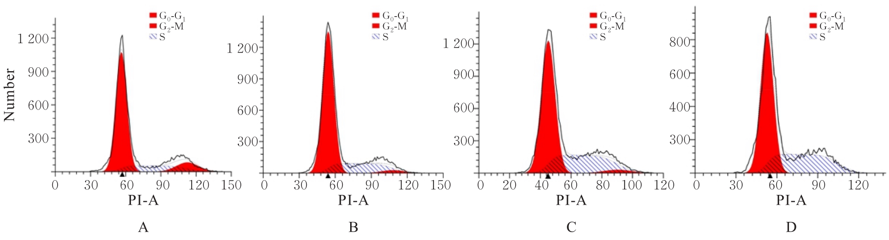
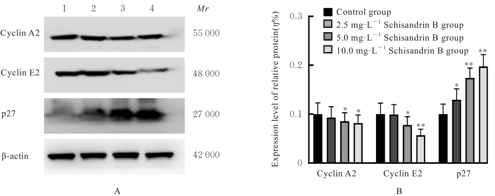
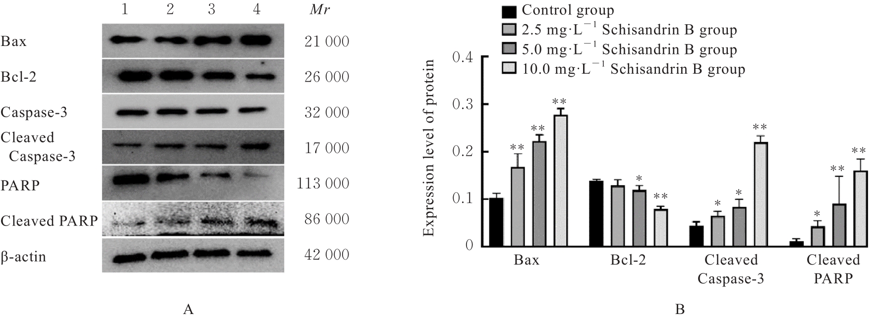
| 1 |
KLEIN A P. Pancreatic cancer epidemiology: understanding the role of lifestyle and inherited risk factors[J]. Nat Rev Gastroenterol Hepatol, 2021, 18(7): 493-502.
|
| 2 |
TORPHY R J, FUJIWARA Y, SCHULICK R D. Pancreatic cancer treatment: better, but a long way to go[J]. Surg Today, 2020, 50(10): 1117-1125.
|
| 3 |
MCGUIGAN A, KELLY P, TURKINGTON R C, et al. Pancreatic cancer: a review of clinical diagnosis, epidemiology, treatment and outcomes[J]. World J Gastroenterol, 2018, 24(43): 4846-4861.
|
| 4 |
JAAKS P, COKER E A, VIS D J, et al. Effective drug combinations in breast, colon and pancreatic cancer cells[J]. Nature, 2022, 603(7899): 166-173.
|
| 5 |
HO W J, JAFFEE E M, ZHENG L. The tumour microenvironment in pancreatic cancer—clinical challenges and opportunities[J]. Nat Rev Clin Oncol, 2020, 17: 527-540.
|
| 6 |
HU Y W, LI H T, LI R L, et al. Protective effects of Schisandrin B against D-GalN-induced cell apoptosis in human hepatocyte (L02) cells via modulating Bcl-2 and Bax[J]. Bioengineered, 2021, 12(1): 7205-7214.
|
| 7 |
LUO W, LIN K, HUA J Y, et al. Schisandrin B attenuates diabetic cardiomyopathy by targeting MyD88 and inhibiting MyD88-dependent inflammation[J]. Adv Sci, 2022, 9(31): e2202590.
|
| 8 |
YAN L S, ZHANG S F, LUO G, et al. Schisandrin B mitigates hepatic steatosis and promotes fatty acid oxidation by inducing autophagy through AMPK/mTOR signaling pathway[J]. Metabolism, 2022, 131: 155200.
|
| 9 |
LI Z M, ZHAO L J, XIA Y S, et al. Schisandrin B attenuates hepatic stellate cell activation and promotes apoptosis to protect against liver fibrosis[J]. Molecules, 2021, 26(22): 6882.
|
| 10 |
TAN S R, ZHENG Z, LIU T Q, et al. Schisandrin B induced ROS-mediated autophagy and Th1/Th2 imbalance via selenoproteins in Hepa1-6 cells[J]. Front Immunol, 2022, 13: 857069.
|
| 11 |
LI S P, WANG H, MA R D, et al. Schisandrin B inhibits epithelial-mesenchymal transition and stemness of large-cell lung cancer cells and tumorigenesis in xenografts via inhibiting the NF-κB and p38 MAPK signaling pathways[J]. Oncol Rep, 2021, 45(6): 115.
|
| 12 |
JIANG Y, ZHANG Q L, BAO J S, et al. Schisandrin B inhibits the proliferation and invasion of glioma cells by regulating the HOTAIR-micoRNA-125a-mTOR pathway[J]. Neuroreport, 2017, 28(2): 93-100.
|
| 13 |
NASSER M I, HAN T Y, ADLAT S, et al. Inhibitory effects of Schisandrin B on human prostate cancer cells[J]. Oncol Rep, 2019, 41(1): 677-685.
|
| 14 |
PFAB C, ABGARYAN A, DANZER B, et al. Ceftazidime and cefepime antagonize 5-fluorouracil’s effect in colon cancer cells[J]. BMC Cancer, 2022, 22(1): 125.
|
| 15 |
ICARD P, FOURNEL L, WU Z R, et al. Interconnection between metabolism and cell cycle in cancer[J]. Trends Biochem Sci, 2019, 44(6): 490-501.
|
| 16 |
JR R R. Cyclin-dependent protein serine/threonine kinase inhibitors as anticancer drugs[J]. Pharmacol Res, 2019, 139: 471-488.
|
| 17 |
LIU Y X, DONG Y Y, ZHAO L P, et al. TRIM59 overexpression correlates with poor prognosis and contributes to breast cancer progression through AKT signaling pathway[J]. Mol Carcinog, 2018, 57(12): 1792-1802.
|
| 18 |
KUMARI S, KUMAR P, KUMAR M, et al. Expression of p27 and p16 and their clinical significance in gastric cancer[J]. Clin Transl Oncol, 2021, 23(4): 856-865.
|
| 19 |
SINGH R, LETAI A, SAROSIEK K. Regulation of apoptosis in health and disease: the balancing act of BCL-2 family proteins[J]. Nat Rev Mol Cell Biol, 2019, 20(3): 175-193.
|
| 20 |
WARREN C F A, WONG-BROWN M W, BOWDEN N A. BCL-2 family isoforms in apoptosis and cancer[J]. Cell Death Dis, 2019, 10(3): 177.
|
| 21 |
SONG L J, CHEN X, MI L, et al. Icariin-induced inhibition of SIRT6/NF-κB triggers redox mediated apoptosis and enhances anti-tumor immunity in triple-negative breast cancer[J]. Cancer Sci, 2020, 111(11): 4242-4256.
|
| 22 |
ABARIKWU S O, FAROMBI E O. Atrazine induces apoptosis of SH-SY5Y human neuroblastoma cells via the regulation of Bax/Bcl-2 ratio and caspase-3-dependent pathway[J]. Pestic Biochem Physiol, 2015, 118: 90-98.
|
| 23 |
WAGNER A K, AMIN K B, NIYONKURU C, et al. CSF Bcl-2 and cytochrome C temporal profiles in outcome prediction for adults with severe TBI[J]. J Cereb Blood Flow Metab, 2011, 31(9): 1886-1896.
|
| 24 |
PEÑA-BLANCO A, GARCÍA-SÁEZ A J. Bax, Bak and beyond - mitochondrial performance in apoptosis[J]. FEBS J, 2018, 285(3): 416-431.
|
| 25 |
SIDDIQUI W A, AHAD A, AHSAN H. The mystery of BCL2 family: Bcl-2 proteins and apoptosis: an update[J]. Arch Toxicol, 2015, 89(3): 289-317.
|
 ),Ling QI2(