Journal of Jilin University(Medicine Edition) ›› 2024, Vol. 50 ›› Issue (4): 939-946.doi: 10.13481/j.1671-587X.20240407

• Research in basic medicine • Previous Articles     Next Articles

Biocompatibility of PTMC/PVP temperature-controlled shrinkage nanofiber membrane with mouse fibroblasts and its repairment effect on full-thickness skin defects in rats

Liping LIU1,Chiyu LI2,Tao YANG1,Shaoru WANG1,Yun LIU1,Guomin LIU2,Zhiqiang CHENG3,Yungang LUO4(),Zhihui LIU1()   

  1. 1.Department of Prosthodontics, Stomatology Hospital, Jilin University, Changchun 130021, China
    2.Department of Orthopedics, Second Hospital, Jilin University, Changchun 130041, China
    3.School of Resources and Environment, Jilin Agricultural University, Changchun 130118, China
    4.Stomatology Center, Jingyue Campus, First Hospital, Jilin University, Changchun 130118, China
  • Received:2023-07-22 Online:2024-07-28 Published:2024-08-01
  • Contact: Yungang LUO,Zhihui LIU E-mail:luoygjlu@sina.com;liu_zh@jlu.edu.cn

Abstract:

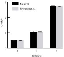
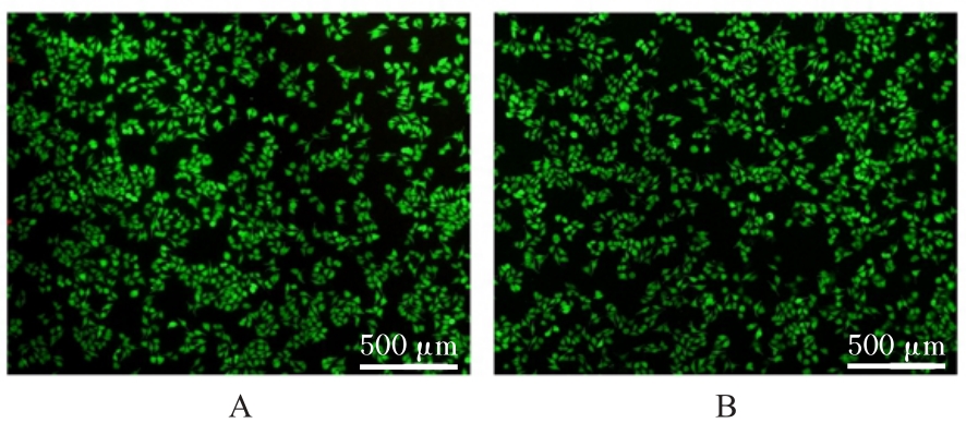
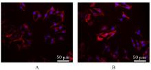
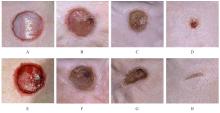
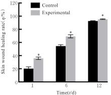
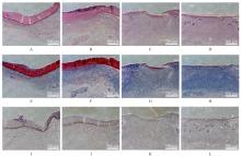
Objective To discuss the effect of temperature-controlled shrinkage polytrimethylene carbonate (PTMC)/polyvinylpyrrolidone (PVP) nanofiber membrane on the biological behavior of mouse fibroblasts and the repairment effect on full-thickness skin defects in the rats, and to clarify the potential mechanism. Methods The murine L929 fibroblast cells were used in the in vitro experiments and were divided into control group and experimental group (treated with PTMC/PVP nanofiber membranes). The proliferation activities of the cells in two groups were detected by CCK-8 assay; the numbers of live/dead cells in two groups were observed by live/dead cell staining; the morphology of the cells was observed by cytoskeletal staining. A total of 12 six-week-old male SD rats were selected in the in vivo experiment, and were randomly divided into control group and experimental group, and there were six rats in each group. The full-thickness skin defect model was established, and the rats in experimental group were treated with PTMC/PVP nanofiber membranes. The photographs were taken after operation, and the wound healing rates of the cells in two groups were calculated on the 0, 3rd, 6th, and 12th days. On the 6th and 12th days after operation, the skin samples around the wound of the rats in two groups were taken, and the histopathology of the would skin and adjacent tissue was detected by HE staining; the collagen deposition in wound skin tissue of the rats in two groups was observed by Masson trichrome staining; the numbers of angiogenesis in wound skin tissue of the rats were detected by CD31 immunohistochemical staining. Results The CCK-8 assay results showed that the proliferation activity of the cells in experimental group showed an increasing trend on the 1st, 3st, and 5st days, and there was no significant difference in the proliferation activities of the cells bewteen experimental group and control group (P>0.05). The live/dead cell staining experiment results showed that compared with control group, the cell density and number of the cells in experimental group had no significant changes, and were predominantly live cells. The cytoskeletal staining results showed that the cells in experimental and control groups appeared spindle-shaped and well-spread. In the in vivo experiments, on the 3rd, 6th, and 12th days, compared with control group, the wound healing rates of the cells in experimental group were increased (P<0.01), and the wound healing rate of the cells was 95.45% on the 12th day, indicating nearly complete healing of the wound. The HE staining showed that on the 12th day, the wound skin structure of the cells in experimental group was more similar to the normal skin, and there was abundant granulation tissue, regular epidermal structure, and new blood vessel formation. The Masson trichrome staining results showed that compared with control group, the collagen deposition in wound tissue of the rats in experimental group was increased. The immunohistochemical staining results showed that the expression of CD31 in wound tissue of the rats in experimental group was increased, indicating the increasing of the number of angiogenesis. Conclusion The PTMC/PVP thermoresponsive nanofiber membranes exhibit good biocompatibility and can promote the repairment of full-thickness skin defects in the rats; its mechanism may be related to the enhancement of proliferation activity of the basal cells.

Key words: Wound dressing, Mechanical mechanics, Temperature-controlled shrinkage, Polytrimethylene carbonate /polyvinylpyrrolidone, Nanofiber, Electrospinning

CLC Number: 

  • R318.08