Journal of Jilin University(Medicine Edition) ›› 2024, Vol. 50 ›› Issue (6): 1691-1702.doi: 10.13481/j.1671-587X.20240623

• Research in clinical medicine • Previous Articles    

Eosinophil infiltration in cervical lesion and cervical cancer tissues and their clinical significances

Yanyan LU1,2,Xiangbo XU1,3,Yamei WU2,Yuqi LIU1,Han WANG1,Lijuan YANG1,Zhenjiang WANG1,Zishen XIAO1,Yanbo LIU1()   

  1. 1.Department of Pathophysiology,School of Basic Medical Sciences,Beihua University,Jilin 132013,China
    2.Department of Pathology,Gynaecology and Obstetrics Hospital,Changchun City,Jilin Province,Changchun 130028,China
    3.Department of Gynaecology and Obstetrics,People’s Hospital,Jilin City,Jilin Province,Jilin 132011,China
  • Received:2024-01-08 Online:2024-11-28 Published:2024-12-10
  • Contact: Yanbo LIU E-mail:liuyanbobeihua@163.com

Abstract:

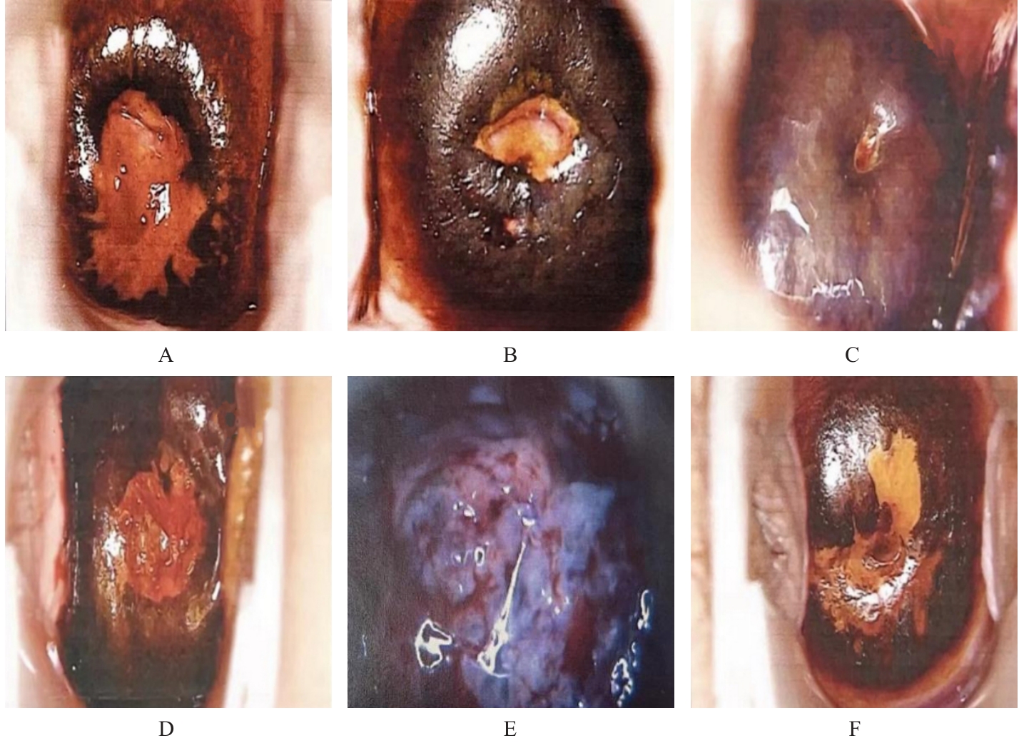
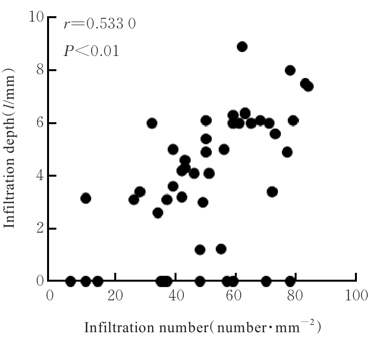
Objective To discuss the differences in eosinophil (EOS) infiltration in cervical tissue and its relationship with cervical-related diseases, and to clarify the effect of EOS on the occurrence and development of cervical intraepithelial neoplasia (CIN) and cervical cancer. Methods The clinical data of 256 patients with cervical diseases were collected and divided into cervical cancer group (n=46, including 26 cases of squamous cell carcinoma, 15 cases of adenocarcinoma, and 5 cases of adenosquamous carcinoma), chronic cervicitis group (n=50), CIN stageⅠ group (n=50), CIN stageⅡ group (n=50), CIN stageⅢ group (n=30), and normal group (adjacent normal cervical tissue, n=30) based on their conditions. Colposcopy was used to observe the morphology of cervical tissue of the patients in various groups; thin-layer liquid-based cytology test (TCT) was used to observe the morphology of the cervical exfoliated cells in various groups; hybrid capture-chemiluminescence method was used to detect the human papillomavirus (HPV) infection in cervical tissue of the patients in various groups; HE staining was used to observe the pathomorphology of cervical tissue of the patients in various groups; Congo red staining was used to detect the numbers of EOS infiltration in cervical tissue of the patients in various groups; Pearson correlation analysis was used to analyze the correlation between the number of EOS infiltration and the malignancy degree of cervical cancer. Results The cervical surface of the patients in normal group was smooth and pink, with uniformly distributed capillaries; the cervical surface of the patients in chronic cervicitis group showed red inflammatory changes, with some accompanied by Nabothian cysts and varying degrees of erosion and ulcers; the patients in CIN stageⅠ, CIN stageⅡ, and CIN stageⅢ groups showed epithelial ulcers, thickening, and irregular morphology, with mosaic and punctate vessels; the cervical surface of the patients in cervical cancer group showed raised areas with neoplasms and necrotic ulcers, and they were fragile and prone to bleeding. After acetic acid staining, no obvious changes of the patients in normal group were observed. The cervix of the patients in chronic cervicitis group showed slight white changes that lasted for a short time; in CIN stageⅠ, CIN stageⅡ, and CIN stageⅢ groups, irregular thin acetowhite epithelium with map-like borders was observed, with increasingly acetowhite reactions and larger areas as the stages advanced. The cervix of the patients in cervical cancer group showed thick acetowhite epithelium that lasted longer, with rigid and clear contours. After iodine staining, the cervix of the patients in normal group was brown, with uniform coloration; the cervix of the patients in chronic cervicitis group showed poor coloration in inflammatory lesion areas; the cervix of the patients in CIN stageⅠ group showed iodine coloration in metaplastic areas, while the cervix of the patients in CIN stageⅢ group showed poor coloration in larger lesion areas; the cervix of the patients in cervical cancer group showed irregular surfaces with cauliflower-like growth and no coloration after iodine staining, appearing orange-yellow or mustard yellow. The TCT observation results showed there were no heteromorphic cells and few inflammatory cells in cervical exfoliated cells of the patients in infiltration in normal group; there were numerous neutrophils and EOS in exfoliated cervical cells without heteromorphic cells in chronic cervicitis group. The heteromorphic binucleated cells with high nuclear-cytoplasmic ratios and deeply stained nuclei were observed in cervical exfoliated cells of the patients in CIN stageⅠ and CIN stageⅡ groups. More heteromorphic cells with high nuclear-cytoplasmic ratios and irregular nuclear membranes were showed in cervical exfoliated cells of the patients in CIN stageⅢ group. The cervical exfoliated cells of the patients in cervical cancer group showed large and prominent nucleoli, clustering into syncytial changes. Compared with normal group, the atypial of cervical exfoliated cells in CIN stageⅠ, CIN stageⅡ, CIN stageⅢ, and cervical cancer groups was increased. The hybrid capture-chemiluminescence results showed that compared with normal and chronic cervicitis groups, the numbers of HPV infection and TCT heteromorphic cells of the patients in CIN stageⅠ, CIN stageⅡ, and CIN stageⅢ groups were increased (P<0.05); compared with CIN stageⅠ, CIN stageⅡ, and CIN stageⅢ groups, the numbers of HPV infection and TCT heteromorphic cells of the patients in cervical cancer group were increased (P<0.05). The HE staining results showed normal cell morphology and structure in normal group, with infiltration of inflammation cells such as neutrophils, monocytes, macrophages, EOS, and lymphocytes; in chronic cervicitis group, the infiltration of inflammatory cells was increased; in CIN group, the cervical cells showed slightly larger nucleoli and heteromorphic cells, with inflammatory cells mainly distributing around the hetermomorphic cells; in cervical cancer group, the cervical cells showed large and deeply stained nucleoli with significant atypia, and the infiltration of inflammatory cells around the cancer cells was increased. Compared with normal group, the numbers of inflammatory cells and EOS infiltration in cervical tissue of the patients in chronic cervicitis group were increased (P<0.05), and the numbers of inflammatory cells and EOS infiltration of the patients in CIN group were increased (P<0.05); compared with chronic cervicitis group, the number of inflammatory cells and EOS infiltration of the patients in CIN group were decreased (P<0.05); compared with chronic cervicitis group and CIN group, the numbers of inflammatory cells and EOS infiltration of the patients in cervical cancer group were increased (P<0.05). The EOS in cervical cancer tissue was mainly distributed around the cancer nests; compared with CIN stageⅠ group, the numbers of EOS infiltration in CIN stageⅡ and CIN stageⅢ groups were increased (P<0.05); compared with CIN stageⅡ group, the number of EOS infiltration in CIN stageⅢ group was increased (P<0.05). The higher the malignancy degree of the tumor, the more EOS infiltration was observed, and the number of EOS infiltration was positively correlated with the invasion depth of cervical cancer (r=0.533 0, P<0.01). Conclusion HPV infection and EOS infiltration play a role in promoting the and occurrence development of cervical precancerous lesions and cervical cancer.

Key words: Eosinophils, Cervical tumors, Cervical intraepithelial neoplasia, Chronic cervicitis, Cell infiltration

CLC Number: 

  • R737.33