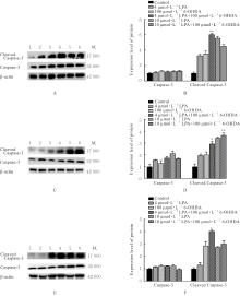
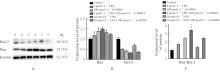
| [1] |
TANSEY M G, WALLINGS R L, HOUSER M C, et al. Inflammation and immune dysfunction in Parkinson disease[J]. Nat Rev Immunol, 2022, 22(11): 657-673.
|
| [2] |
ZHU F D, WANG B D, QIN D L, et al. Carpesii fructus extract exhibits neuroprotective effects in cellular and Caenorhabditis elegans models of Parkinson’s disease[J]. CNS Neurosci Ther, 2024, 30(4): e14515.
|
| [3] |
GRAVANDI M M, ABDIAN S, TAHVILIAN M, et al. Therapeutic targeting of Ras/Raf/MAPK pathway by natural products: a systematic and mechanistic approach for neurodegeneration[J]. Phytomedicine, 2023, 115: 154821.
|
| [4] |
DILNASHIN H, BIRLA H, KESWANI C, et al. Neuroprotective effects of tinospora cordifolia via reducing the oxidative stress and mitochondrial dysfunction against rotenone-induced PD mice[J]. ACS Chem Neurosci, 2023, 14(17): 3077-3087.
|
| [5] |
LIU Z Y, YAO X Q, JIANG W S, et al. Advanced oxidation protein products induce microglia-mediated neuroinflammation via MAPKs-NF-κB signaling pathway and pyroptosis after secondary spinal cord injury[J]. J Neuroinflammation, 2020, 17(1): 90-110.
|
| [6] |
RATIH K, LEE Y R, CHUNG K H, et al. L-Theanine alleviates MPTP-induced Parkinson’s disease by targeting Wnt/β-catenin signaling mediated by the MAPK signaling pathway[J]. Int J Biol Macromol, 2023, 226: 90-101.
|
| [7] |
LIU X D, ZHANG R, FAN J J, et al. The role of ROS/p38 MAPK/NLRP3 inflammasome cascade in arsenic-induced depression-/anxiety-like behaviors of mice[J]. Ecotoxicol Environ Saf, 2023, 261: 115111.
|
| [8] |
MUNOZ L, AMMIT A J. Targeting p38 MAPK pathway for the treatment of Alzheimer’s disease[J]. Neuropharmacology, 2010, 58(3): 561-568.
|
| [9] |
CHAKRABORTY J, CHAKRABORTY S, CHAKRABORTY S, et al. Entanglement of MAPK pathways with gene expression and its omnipresence in the etiology for cancer and neurodegenerative disorders[J]. Biochim Biophys Acta Gene Regul Mech, 2023, 1866(4): 194988.
|
| [10] |
DONG L G, AN M Q, GU H Y, et al. PACAP/PAC1-R activation contributes to hyperalgesia in 6-OHDA-induced Parkinson’s disease model rats via promoting excitatory synaptic transmission of spinal dorsal horn neurons[J]. Acta Pharmacol Sin, 2023, 44(12): 2418-2431.
|
| [11] |
SLÉZIA A, HEGEDÜS P, RUSINA E, et al. Behavioral, neural and ultrastructural alterations in a graded-dose 6-OHDA mouse model of early-stage Parkinson’s disease[J]. Sci Rep, 2023, 13(1): 19478.
|
| [12] |
YU J J, MENG J H, QIN Z W, et al. Dysbiosis of gut microbiota inhibits NMNAT2 to promote neurobehavioral deficits and oxidative stress response in the 6-OHDA-lesioned rat model of Parkinson’s disease[J]. J Neuroinflammation, 2023, 20(1): 117.
|
| [13] |
TIAN Y, LU J, HAO X Q, et al. FTH1 Inhibits Ferroptosis Through Ferritinophagy in the 6-OHDA Model of Parkinson’s Disease[J]. Neurotherapeutics, 2020, 17(4): 1796-1812.
|
| [14] |
PEI J Q, CAI L, WANG F, et al. LPA(2) contributes to vascular endothelium homeostasis and cardiac remodeling after myocardial infarction[J]. Circ Res, 2022, 131(5): 388-403.
|
| [15] |
YANAGIDA K, SHIMIZU T. Lysophosphatidic acid, a simple phospholipid with myriad functions[J]. Pharmacol Ther, 2023, 246: 108421.
|
| [16] |
YUNG Y C, STODDARD N C, MIRENDIL H, et al. Lysophosphatidic Acid signaling in the nervous system[J]. Neuron, 2015, 85(4): 669-682.
|
| [17] |
SALGADO-POLO F, BORZA R, MATSOUKAS M T, et al. Autotaxin facilitates selective LPA receptor signaling[J]. Cell Chem Biol, 2023, 30(1): 69-84.e14.
|
| [18] |
MAGKRIOTI C, ANTONOPOULOU G, FANIDIS D, et al. Lysophosphatidic acid is a proinflammatory stimulus of renal tubular epithelial cells[J]. Int J Mol Sci, 2022, 23(13): 7452.
|
| [19] |
XU L X, SU J, GUO L T, et al. Modulation of LPA1 receptor-mediated neuronal apoptosis by Saikosaponin-d: a target involved in depression[J]. Neuropharmacology, 2019, 155: 150-161.
|
| [20] |
CHANG C L, LIN M E, HSU H Y, et al. Lysophosphatidic acid-induced interleukin-1 bate expressionis mediated through Gi/Rho and the generation of reactive oxygen species in macrophages[J]. J Biomed Sci, 2008, 15(3): 357-363.
|
| [21] |
TAND X Y, WANG X Y, ZHAO Y Y, et al. Doxycycline attenuates breast cancer related inflammation by decreasing plasma lysophosphatidate concentrations and inhibiting NF-κB activation[J]. Mol Cancer, 2017, 16(1): 36.
|
| [22] |
LEE C H, SAPKOTA A, GAIRE B P, et al. NLRP3 inflammasome activation is involved in LPA(1)-mediated brain injury after transient focal cerebral ischemia[J]. Int J Mol Sci, 2020, 21(22): 8595.
|
| [23] |
YANG X Y, ZHAO E Y, ZHUANG W X, et al. LPA signaling is required for dopaminergic neuron development and is reduced through low expression of the LPA1 receptor in a 6-OHDA lesion model of Parkinson’s disease[J]. Neurol Sci, 2015, 36(11): 2027-2033.
|
| [24] |
BIRGBAUER E. Lysophospholipid receptors in neurodegeneration and neuroprotection[J]. Explor Neuroprotective Ther, 2024, 4(4): 349-365.
|
| [25] |
牟笑笑, 管清燕, 孔 荐, 等. 溶血磷脂酸下调盐酸阿霉素诱导的卵巢癌SKOV3细胞的凋亡[J]. 中国生物化学与分子生物学报, 2021, 37(10): 1401-1407.
|
| [26] |
CHOI J H, OH J, LEE M J, et al. Inhibition of lysophosphatidic acid receptor 1-3 deteriorates experimental autoimmune encephalomyelitis by inducing oxidative stress[J]. J Neuroinflammation, 2021, 18(1): 240.
|
| [27] |
PLASTIRA I, BERNHART E, GOERITZER M, et al. Lysophosphatidic acid via LPA-receptor 5/protein kinase D-dependent pathways induces a motile and pro-inflammatory microglial phenotype[J]. J Neuroinflammation, 2017, 14(1): 253.
|
| [28] |
XIA Q, HU Q, WANG H, et al. Induction of COX-2-PGE2 synthesis by activation of the MAPK/ERK pathway contributes to neuronal death triggered by TDP-43- depleted microglia[J]. Cell Death Dis, 2015, 6(3): e1702.
|
| [29] |
IBA M, KIM C, KWON S, et al. Inhibition of p38α MAPK restores neuronal p38γ MAPK and ameliorates synaptic degeneration in a mouse model of DLB/PD[J]. Sci Transl Med, 2023, 15(695): eabq6089.
|
| [30] |
RANSOHOFF R M, PERRY V H. Microglial physiology: unique stimuli, specialized responses[J]. Annu Rev Immunol, 2009, 27: 119-145.
|
| [31] |
NORRIS G T, KIPNIS J. Immune cells and CNS physiology: microglia and beyond[J]. J Exp Med, 2019, 216(1): 60-70.
|
| [32] |
NAGATSU T, MOGI M, ICHINOSE H, et al. Cytokines in Parkinson’s disease[J]. J Neural Transm Suppl, 2000(58): 143-151.
|
| [33] |
HARMS A S, FERREIRA S A, ROMERO-RAMOS M. Periphery and brain, innate and adaptive immunity in Parkinson’s disease[J]. Acta Neuropathol, 2021, 141(4): 527-545.
|
| [34] |
PLASTIRA I, BERNHART E, JOSHI L, et al. MAPK signaling determines lysophosphatidic acid (LPA)-induced inflammation in microglia[J]. J Neuroinflammation, 2020,17(1): 127.
|
 ),Xiaoyun YANG1,2(