| [1] |
MCGLYNN K A, PETRICK J L, EL-SERAG H B. Epidemiology of hepatocellular carcinoma [J]. Hepatology, 2021,73(): 4-13.
|
| [2] |
黄 勇, 黄声稀, 刘秀峰. 肝细胞癌系统治疗进展与展望[J]. 临床肝胆病杂志, 2025, 41(8): 1491-1496.
|
| [3] |
YETISkin E, GÜNDOĞDU Ö, METE D, et al. Synthesis, cytotoxicity, and antibacterial studies of 2,4,5,6-substituted hexahydro-1H-isoindole-1,3(2H)- dione[J]. Chem Biol Drug Des, 2023, 102(6): 1448-1457.
|
| [4] |
WANG Y N, CHEN H, SHENG R L, et al. Synthesis and bioactivities of marine pyran-isoindolone derivatives as potential antithrombotic agents [J]. Mar Drugs, 2021, 19(4): 218.
|
| [5] |
GAO C L, TANG S M, ZHANG H X, et al. A novel marine pyran-isoindolone compound enhances fibrin lysis mediated by single-chain urokinase-type plasminogen activator [J]. Mar Drugs, 2022, 20(8): 495.
|
| [6] |
KRZYŻAK E, MARCINIAK A, SZKATUŁA D, et al. A series of novel 1-H-isoindole-1,3(2H)-dione derivatives as acetylcholinesterase and butyrylcholinesterase inhibitors: in silico, synthesis and in vitro studies[J]. Molecules, 2024, 29(15): 3528.
|
| [7] |
SPANÒ V, BARRECA M, ROCCA R, et al. Insight on [1,3] thiazolo [4,5-e] isoindoles as tubulin polymerization inhibitors[J]. Eur J Med Chem, 2021, 212: 113122.
|
| [8] |
TAGHIZAD S, BEHBAHANINIA K, JAHROMY M H,et al. Pyrrolopyridine and isoindole as potential anticonvulsant agents: design, synthesis, and pharmacological evaluation[J]. Curr Comput Aided Drug Des, 2022, 18(3): 159-167.
|
| [9] |
HAWKINSON J E, SZOKE B G, GAROFALO A W, et al. Pharmacological, pharmacokinetic, and primate analgesic efficacy profile of the novel bradykinin B1 Receptor antagonist ELN441958[J]. J Pharmacol Exp Ther, 2007, 322(2): 619-630.
|
| [10] |
TSOU P S, LU C Y, GURREA-RUBIO M, et al. Soluble CD13 induces inflammatory arthritis by activating the bradykinin receptor B1[J]. J Clin Invest, 2022, 132(11): e151827.
|
| [11] |
ALIFORENKO A E, MOTOV V S, BYKOV V V, et al. Analgesic and psychotropic effects of a new bradykinin antagonist[J]. Bull Exp Biol Med, 2024, 176(5): 581-584.
|
| [12] |
DE CARVALHO FRANÇA P R, DE PAIVA J P B, DE CARVALHO R R, et al. R-954, a bradykinin B1 receptor antagonist, as a potential therapy in a preclinical endometriosis model[J]. Peptides, 2024, 181: 171294.
|
| [13] |
SHEN C K, HUANG BR, CHAROENSAENSUK V, et al. Bradykinin B1 receptor affects tumor-associated macrophage activity and glioblastoma progression[J]. Antioxidants (Basel), 2023, 12(8):1533.
|
| [14] |
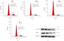
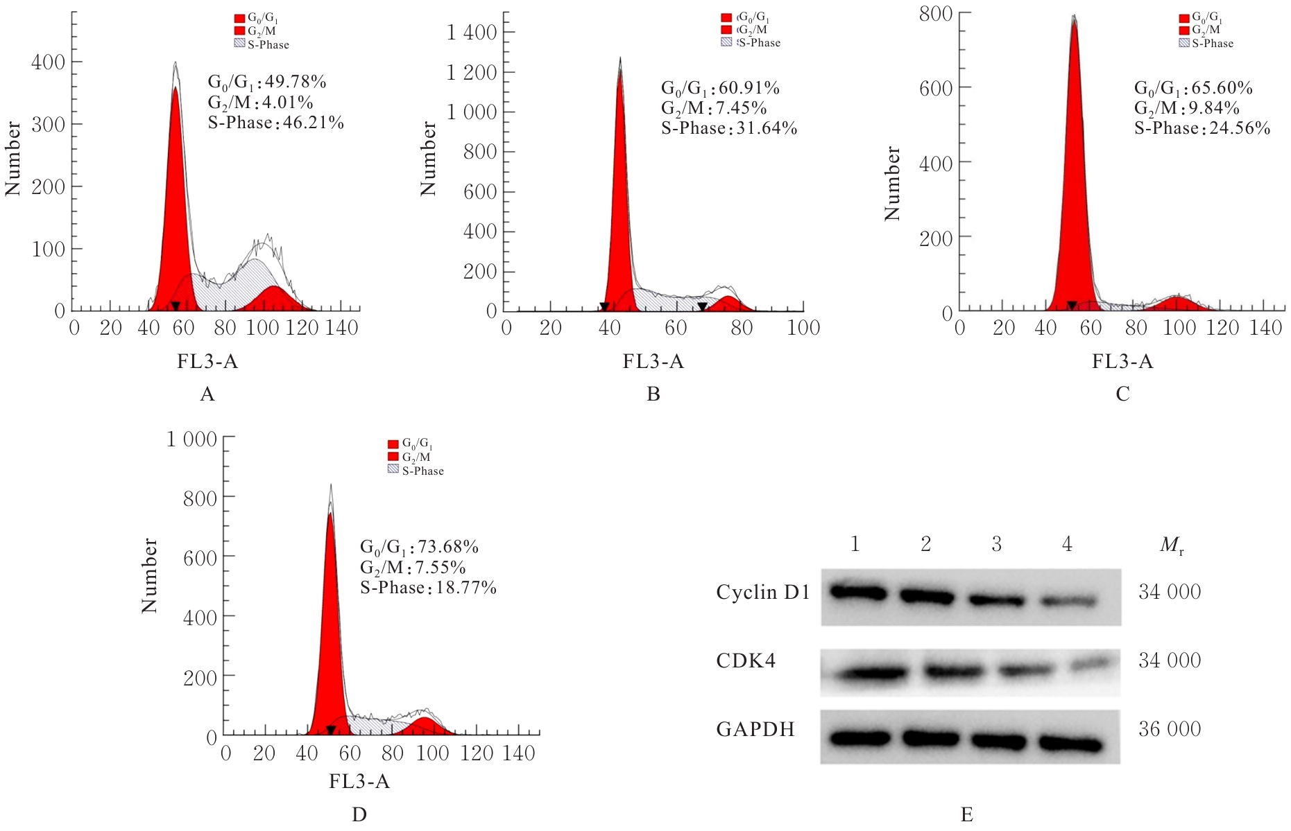
WANG Y O, ZHANG B X, HUANG Y B, et al. Novel bradykinin receptor inhibitors inhibit proliferation and promote the apoptosis of hepatocellular carcinoma cells by inhibiting the ERK pathway [J]. Molecules, 2021, 26(13): 3915.
|
| [15] |
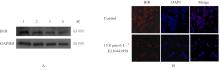
NICOLETTI N F, ERIG T C, ZANIN R F, et al. Mechanisms involved in kinin-induced glioma cells proliferation: the role of ERK1/2 and PI3K/Akt pathways [J]. J Neurooncol, 2014, 120(2): 235-244.
|
| [16] |
LURJE I, CZIGANY Z, BEDNARSCH J, et al. Treatment strategies for hepatocellular carcinoma -a multidisciplinary approach [J]. Int J Mol Sci, 2019, 20(6):1465.
|
| [17] |
张书力, 冯 丹. 槲皮素通过下调缓激肽受体B1表达减轻糖尿病大鼠神经病理性疼痛 [J]. 中国免疫学杂志, 2024, 40(2): 337-342.
|
| [18] |
XIE H Y, LU F, LIU W L, et al. Remimazolam alleviates neuropathic pain via regulating bradykinin receptor B1 and autophagy [J]. J Pharm Pharmacol, 2021, 73(12): 1643-1651.
|
| [19] |
WANG W, ZHOU Y, WEI R, et al. Bradykinin promotes proliferation, migration, and invasion of cervical cancer cells through STAT3 signaling pathways [J]. Oncol Rep, 2019, 42(6): 2521-2527.
|
| [20] |
DEEPAK K, ROY P K, KOLA P, et al. An overview of kinin mediated events in cancer progression and therapeutic applications [J]. Biochim Biophys Acta Rev Cancer, 2022, 1877 (6): 188807.
|
| [21] |
NICOLETTI N F, SÉNÉCAL J, SILVA V D D A, et al. Primary role for kinin B(1) and B(2) receptors in glioma proliferation[J]. Mol Neurobiol, 2017, 54(10):7869-7882.
|
| [22] |
KUMAR S, AGNIHOTRI N. Piperlongumine, a piper alkaloid targets Ras/PI3K/Akt/mTOR signaling axis to inhibit tumor cell growth and proliferation in DMH/DSS induced experimental colon cancer[J]. Biomed Pharmacother, 2019, 109:1462-1477.
|
| [23] |
MUÑOZ J P, CARRILLO-BELTRÁN D, AAEDO-AGUILERA V, et al. Tobacco exposure enhances human papillomavirus 16 oncogene expression via EGFR/PI3K/Akt/c-Jun signaling pathway in cervical cancer cells[J]. Front Microbiol, 2018, 9: 3022.
|
| [24] |
ZHANG H, XIA P, LIU J, et al. ATIC inhibits autophagy in hepatocellular cancer through the Akt/FOXO3 pathway and serves as a prognostic signature for modeling patient survival[J]. Int J Biol Sci, 2021, 17(15): 4442-4458.
|
| [25] |
LU M, HARTMANN D, BRAREN R, et al. Oncogenic Akt-FOXO3 loop favors tumor-promoting modes and enhances oxidative damage-associated hepatocellular carcinogenesis[J]. BMC Cancer, 2019, 19(1): 887.
|
 )
)