Journal of Jilin University(Medicine Edition) ›› 2021, Vol. 47 ›› Issue (4): 896-903.doi: 10.13481/j.1671-587X.20210411
• Research in basic medicine • Previous Articles Next Articles
Tianjiao MAO1,Duo SUN1,Xing GAO1,Wei WEI1,Xiheng LI1,Kexin JIANG1,Qiu JIANG2( ),Jiang LI1,3()
- 1.Department of Prosthodontics,Stomatology Hospital,Jilin University,Changchun 130021,China
2.Department of Pediatric Dentistry,Stomatology Hospital,Jilin University,Changchun 130021,China
3.Department of Prosthodontics,Affiliated Stomatology Hospital,Guangzhou Medical University,Guangzhou 510150,China
-
Received:2020-12-02Online:2021-07-28Published:2021-07-22 -
Contact:Qiu JIANG,Jiang LI E-mail:jiangqiu@163.com;ljiang@gzhmu.edu.cn
CLC Number:
- R285.5
Cite this article
Tianjiao MAO,Duo SUN,Xing GAO,Wei WEI,Xiheng LI,Kexin JIANG,Qiu JIANG,Jiang LI. [J].Journal of Jilin University(Medicine Edition), 2021, 47(4): 896-903.
share this article
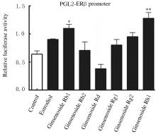
Fig.1
Activities of luciferase in HEK293T cells in various groups"

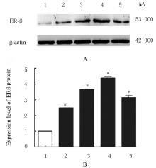
Fig.2
Electrophoregram(A) and histogram(B) of expressions of ERβ protein in MC3T3-E1 cells in various groups"

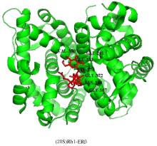
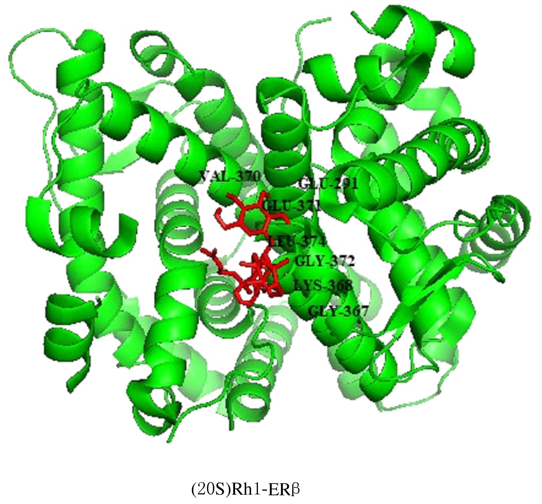
Fig. 3
Docking sites of ginsenoside Rh1 and ERβ"
Tab.1
Proliferation rates of MC3T3-E1 cells in various groups"
| Group | Proliferation rate | ||
|---|---|---|---|
| (t/h) 24 | 48 | 72 | |
| Control | 100.000±0.116 | 100.000±0.011 | 100.000±0.013 |
| Ginsenoside Rh1(mmol·L-1) | |||
| 5×10⁻⁶ | 103.579±0.072 | 103.954±0.013 | 104.952±0.021 |
| 1×10⁻⁵ | 108.301±0.071* | 108.302±0.012* | 109.731±0.023* |
| 5×10⁻⁵ | 110.159±0.078* | 112.582±0.009* | 113.025±0.023* |
| 1×10⁻⁴ | 117.323±0.067* | 120.016±0.011* | 122.282±0.021* |
| 1×10⁻³ | 107.158±0.068* | 109.711±0.013* | 107.632±0.019* |
| 5×10⁻³ | 103.107±0.103 | 103.292±0.015 | 102.107±0.014 |
| Estrodiol | 109.663±0.071* | 110.770±0.009* | 111.819±0.009* |

Fig. 4
Results of ALP staining of MC3T3-E1 cells during osteogenic differentiation in various groups (Bar=500 μm)"
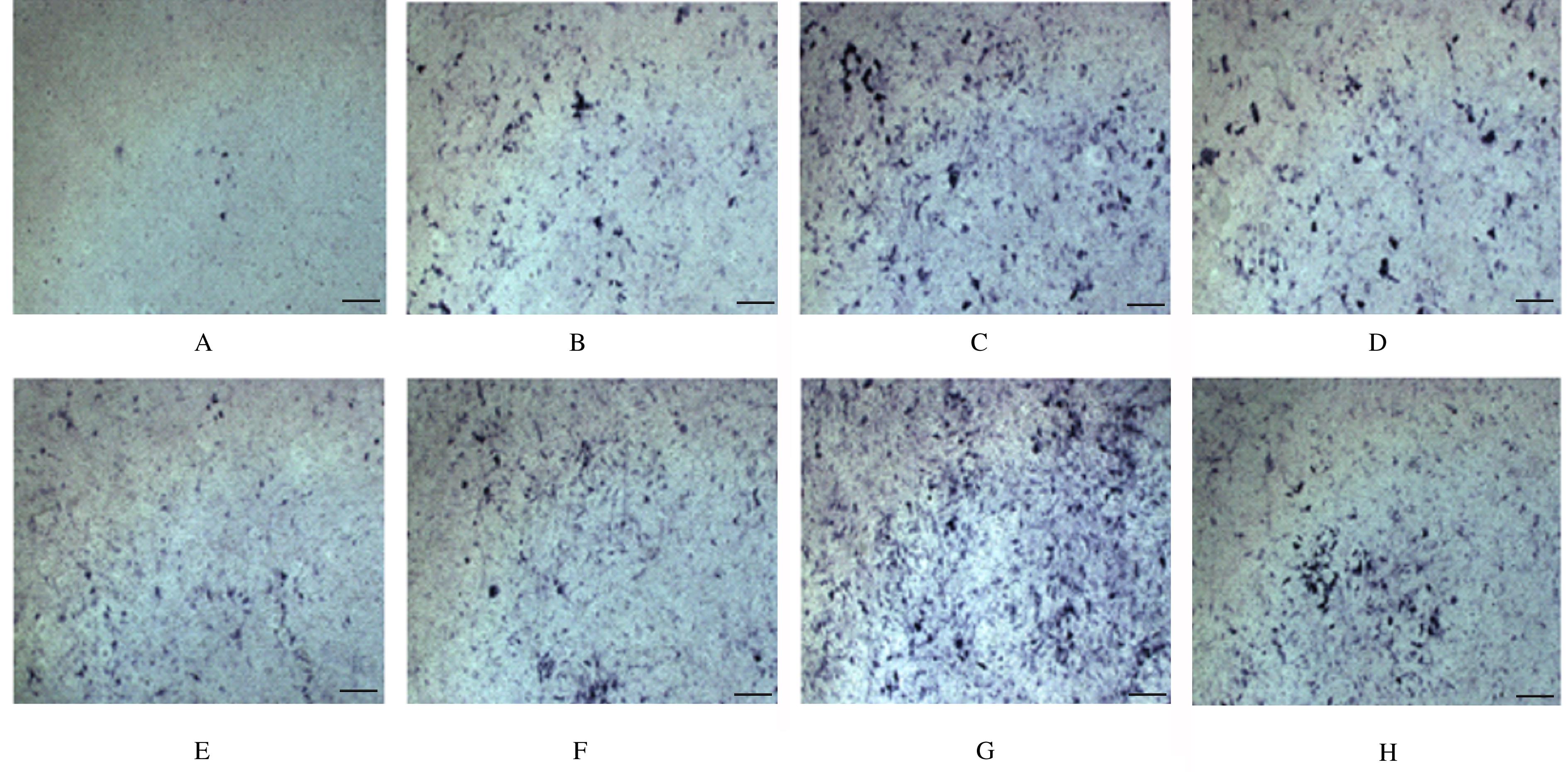
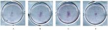
Fig. 5
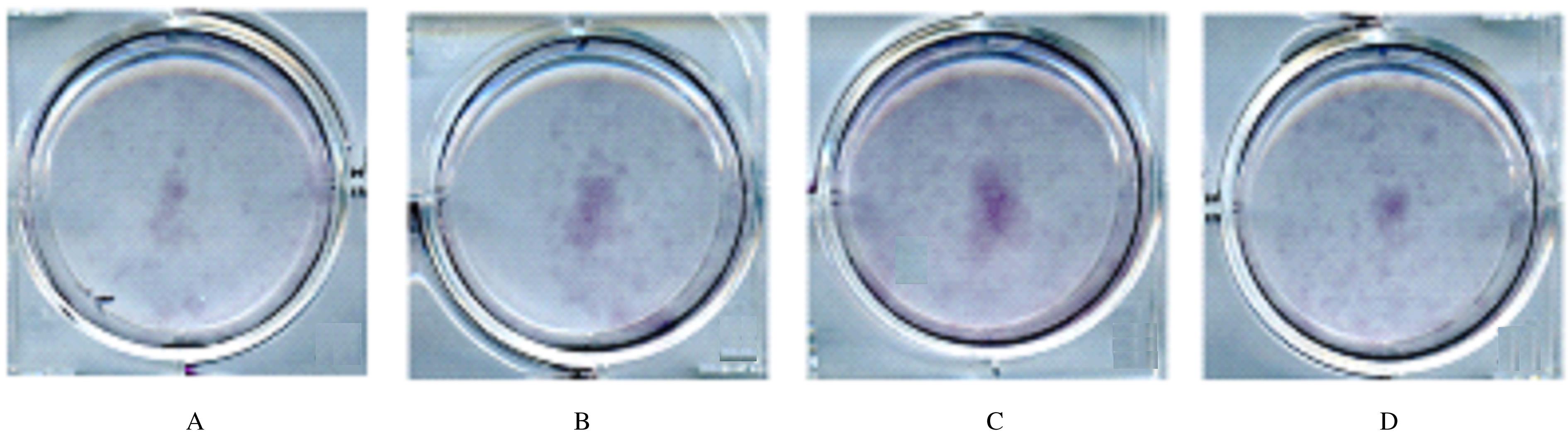
Calcification of bone matrix during osteogenic differentiation of MC3T3-E1 cells in various groups (Alizarin red)"
| 1 | YANG D, LIU T, JIANG G, et al. Senkyunolide H attenuates osteoclastogenesis and postmenopausal osteoporosis by regulating the NF-κB, JNK and ERK signaling pathways[J]. Biochem Biophys Res Commun, 2020,533(3):510-518. |
| 2 | CHEN C H, WANG L, SERDAR TULU U, et al. An osteopenic/osteoporotic phenotype delays alveolar bone repair[J]. Bone, 2018,112:212-219. |
| 3 | IGUACEL I, MIGUEL-BERGES M L, GÓMEZ-BRUTON A, et al. Veganism, vegetarianism, bone mineral density, and fracture risk: a systematic review and meta-analysis[J]. Nutr Rev, 2019, 77(1): 1-18. |
| 4 | KANIS J A, COOPER C, RIZZOLI R, et al. Scientific Advisory Board of the European Society for Clinical and Economic Aspects of Osteoporosis (ESCEO) and the Committees of Scientific Advisors and National Societies of the International Osteoporosis Foundation (IOF). European guidance for the diagnosis and management of osteoporosis in postmenopausal women[J]. Osteoporos Int, 2019,30(1):3-44. |
| 5 | ZHU H, HE Y S, MA J, et al. The dual roles of ginsenosides in improving the anti-tumor efficiency of cyclophosphamide in mammary carcinoma mice[J]. J Ethnopharmacol, 2021,265:113271. |
| 6 | ZHANG X N, HUANG F L, CHEN X Y, et al. Ginsenoside Rg3 attenuates ovariectomy-induced osteoporosis via AMPK/mTOR signaling pathway[J]. Drug Dev Res, 2020, 81(7): 875-884. |
| 7 |
YANG B, ZHAO S C, YAN L. The role of ginsenoside Rb1 in bone homeostasis[J]. Curr Stem Cell Res Ther, 2020. DOI:10.2174/1574888x15666200628141743 .
doi: 10.2174/1574888x15666200628141743 |
| 8 | LIU Y, XU Z J, WANG Q Q, et al. Selective regulation of RANKL/RANK/OPG pathway by heparan sulfate through the binding with estrogen receptor β in MC3T3-E1 cells[J]. Int J Biol Macromol, 2020, 161: 1526-1534. |
| 9 | JIANG L T, ZHANG W J, WEI L, et al. Early effects of parathyroid hormone on vascularized bone regeneration and implant osseointegration in aged rats[J]. Biomaterials, 2018, 179: 15-28. |
| 10 | KHAJURIA D K, PATIL O N, KARASIK D, et al. Development and evaluation of novel biodegradable chitosan based metformin intrapocket dental film for the management of periodontitis and alveolar bone loss in a rat model[J]. Arch Oral Biol, 2018,85:120-129. |
| 11 | PATERNI I, GRANCHI C, KATZENELLENBOGEN J A,et al.Estrogen receptors alpha (ERα) and beta (ERβ):subtype-selective ligands and clinical potential[J]. Steroids, 2014, 90: 13-29. |
| 12 | COMPSTON J E, MCCLUNG M R, LESLIIE W D. Osteoporosis[J]. Lancet, 2019,393(10169):364-376. |
| 13 | KIM J H, YI Y S, KIM M Y, et al. Role of ginsenosides, the main active components of Panax ginseng, in inflammatory responses and diseases[J]. J Ginseng Res, 2017,41(4):435-443. |
| 14 | ZHANG T, ZHONG S, LI T, et al. Saponins as modulators of nuclear receptors[J]. Crit Rev Food Sci Nutr, 2020,60(1):94-107. |
| 15 | THOMAS T, MARTIN A. Formation osseuse, facteurs de régulation[J]. J Soc Biol, 2008, 202(4): 257-264. |
| 16 | SCHMOLDT A, BENTHE H F, HABERLAND G. Digitoxin metabolism by rat liver microsomes[J]. Biochem Pharmacol, 1975,24(17):1639-1641. |
| 17 | WIREN K M, CHAPMAN EVANS A, ZHANG X W. Osteoblast differentiation influences androgen and estrogen receptor-alpha and-beta expression[J]. J Endocrinol, 2002,175(3):683-694. |
| 18 | LIN Y T, PENG S W, IMTIYAZ Z, et al. In vivo and in vitro evaluation of the osteogenic potential of Davallia mariesii T. Moore ex Baker[J]. J Ethnopharmacol, 2021,264:113126. |
| 19 | 顾艺婧,傅稼耀,武文婧,等.抗骨相关细胞衰老作用治疗雌激素缺乏骨质疏松症的初步研究[J].同济大学学报(医学版),2019,40(3):274-280. |
| 20 | KUO T R, CHEN C H. Bone biomarker for the clinical assessment of osteoporosis: recent developments and future perspectives[J]. Biomark Res, 2017, 5: 18. |
| [1] | Yunfeng MA,Xiaofei HAN. Effects of salvianolate on osteoclast differentiation and bone resorption in osteoporotic rats by regulating SMAD2/FKBP1A/NF-κB axis [J]. Journal of Jilin University(Medicine Edition), 2022, 48(1): 111-121. |
| [2] | Leihua CUI,Yubo HOU,Chang SU,Minghe LI,Xin NIE. Effect of N-acetylcysteine on apoptosis of MC3T3-E1 cells induced by nicotine and its mechanism [J]. Journal of Jilin University(Medicine Edition), 2022, 48(1): 26-32. |
| [3] | Xuanchen LIU,Xiaoying TIE,Yulin LIU, WangNing. Effect of polygonum multiflorum extract on osteoporosis and differentiation of bone marrow mesenchymal stem cells in agravic mice [J]. Journal of Jilin University(Medicine Edition), 2021, 47(6): 1386-1396. |
| [4] | Xining LI,Wei WENG,Zheyuan SHEN,Xiaojie DOU,Jikang MIN. Therapeutic effect of estradiol combined with 1,25-dihydroxyvitamin D3 on postmenopausal osteoporosis in rats [J]. Journal of Jilin University(Medicine Edition), 2021, 47(4): 857-864. |
| [5] | Qian MAO, Cuicui CHEN, Huankun LIANG, Penge LIU, Shuhai ZHONG, Laiqing LI. Establishment and evaluation of a double?labeled time? resolved immunofluorescence analysis method for detecting levels of β?CTX and N?MID [J]. Journal of Jilin University(Medicine Edition), 2021, 47(3): 770-776. |
| [6] | SUN Jingchun, JIN Hui, YANG Wenqi, XU Hui. Effect of total flavonids of Rhizomadrynariae on sclerostin expression in bone tissue of osteoporosis rats and its mechanism [J]. Journal of Jilin University(Medicine Edition), 2020, 46(05): 911-916. |
| [7] | XU Dongliang, PENG Zhaohui, XIONG Meicai. Promotion effect of curcumin on implant osseointegration in osteoporosis rats [J]. Journal of Jilin University(Medicine Edition), 2019, 45(04): 877-881. |
| [8] | LI Xining, CHEN Xiaochun, ZHU Yunfei, CAO Jingnan, ZOU Yiting, DU Xiao, WANG Qichun. Effect of PERK signaling pathway on osteoblast differentiation of female rats with experimental postmenopausal osteoporosis [J]. Journal of Jilin University Medicine Edition, 2018, 44(02): 260-264. |
| [9] | ZHU Tongtong, HUANG Liandi, LI Junwei, ZHAO Benzheng, JIANG Mengyang, LI Na, ZHAO Fan. Protective effect of icariin on osteoporosis in ovariectomized rats and its mechanism [J]. Journal of Jilin University Medicine Edition, 2016, 42(05): 915-919. |
| [10] | FANG Tengjiaozi, LIU Jie, GU Zhongyi, GONG Haihuan, BU Wenhuan, DONG Yue, SUN Hongchen. Osteogenesis differentiation of MC3T3-E1 cells induced by miRNA-2861 mimic transfection mediated by polyethylenimine [J]. Journal of Jilin University Medicine Edition, 2016, 42(05): 848-854. |
| [11] | Temuribagen,Tubuxin,QIN Xiong,Hanbagenna,BAI Hai-hua. Preventive effect of Mongolian medicine Tonglaga -5 Pill on osteoporosis induced by retinoic acid in rats and its mechanism [J]. Journal of Jilin University Medicine Edition, 2014, 40(04): 763-767. |
| [12] | ZHANG Lei,ZHANG Shi-tao,ZENG Xian-yang,ZHANG Xiao-ping,HAO Fan-fan,WANG Feng,LI Wan-nan,FU Xue-qi. Cloning and expression of megakaryocyte protein tyrosine phosphatase 2 and its relationship with osteoporosis [J]. Journal of Jilin University Medicine Edition, 2013, 39(2): 227-231. |
| [13] | FU Jian-song, ZHUANG Xin-ming, FU Chang-feng, QU Zhi-ga,JIANG Yi-kun, LIU Yi. Curative effect and security evaluation of ballon kyphoplasty in treatment for osteoporosis vertebral compression fractures [J]. J4, 2012, 38(5): 1003-1007. |
| [14] | ZHANG Xiao-bo, DENG Shi-lin . Association between habitual physical activity and onset of postmenopausal osteoporosis [J]. J4, 2012, 38(4): 775-778. |
| [15] | CHEN Qing-Fu, YANG Shu-Yu, YAN Bing, LIU Chang-Qin, SHI Xiu-Lin, ZHANG Hui-Jie, YU Ya-xin, WANG Li-Ying, LI Xue-Jun. Comparative analysis of bone mineral density and incidence of osteoporosis in |vegetarians |and omnivores [J]. J4, 2010, 36(4): 794-796. |