Journal of Jilin University(Medicine Edition) ›› 2021, Vol. 47 ›› Issue (2): 249-256.doi: 10.13481/j.1671-587X.20210201
• Research in basic medicine • Next Articles
Influence of PGE2 on PD-1 expression in infiltrating T lymphocytes in non-small cell lung cancer tissue and its mechanism
Ye YUAN1,Jinbao ZHUANG1,Xu SHI1,Changyuan LI2( )
)
- 1.Department of Laboratory,First Hospital,Jilin University,Changchun 130021,China
2.Department of Thoracic Surgery,First Hospital,Jilin University,Changchun 130031,China
-
Received:2020-09-09Online:2021-03-28Published:2021-03-25 -
Contact:Changyuan LI E-mail:lichangyuanjlu@163.com
CLC Number:
- R34
Cite this article
Ye YUAN,Jinbao ZHUANG,Xu SHI,Changyuan LI. Influence of PGE2 on PD-1 expression in infiltrating T lymphocytes in non-small cell lung cancer tissue and its mechanism[J].Journal of Jilin University(Medicine Edition), 2021, 47(2): 249-256.
share this article
Tab.1
Clinical parameters of NSCLC patients"
| Parameter | Stage Ⅰ (n=20) | Stage Ⅱ (n=25) | Stage Ⅲ (n=17) | Stage Ⅳ (n=13) |
|---|---|---|---|---|
TNM stage (number) | T1aN0M0 (11) T1bN0M0 (9) | T1bN1M0 (7) T2aN1M0 (10) T2bN0M0 (8) | T1N2M0 (6) T2N2M0 (6) T3N1M0 (5) | T2N2M1a (8) T3N2M1a (5) |
| Age (year) | 56 (42-63) | 58 (46-67) | 57 (44-65) | 56 (45-68) |
| Gender(Female/Male) | 14/6 | 17/8 | 12/5 | 9/4 |
Tab. 2
Primer sequences of target genes in RT-qPCR"
| Primer | Sequence (5'-3') |
|---|---|
PD-1 Forward Reverse | AAGCTTATGTGGGTCCGGC GGATCCTCAAAGAGGCC |
EP2 Forward Reverse | CCACGATGCTCCTGCTGCTT TCCACAAAGGTCAGTCTGTTT |
EP4 Forward Reverse | GGTCATCTTACTCATCGCCACCTCTC TCCCACTAACCTCATCCACCAACAG |
GAPDH Forward Reverse | GGTGGTCTCCTCTGACTTCAACA GTGGTCGTTGAGGGCAATG |

Fig. 1
Percentages of infiltrating CD4+ and CD8+ T lymphocytes in NSCLC patients at different stages detected by flow cytometry"


Fig. 2
Expression levels of PD-1 mRNA in infiltrating CD4+(A) and CD8+(B) T lymphocytes in NSCLC patients at different stages"
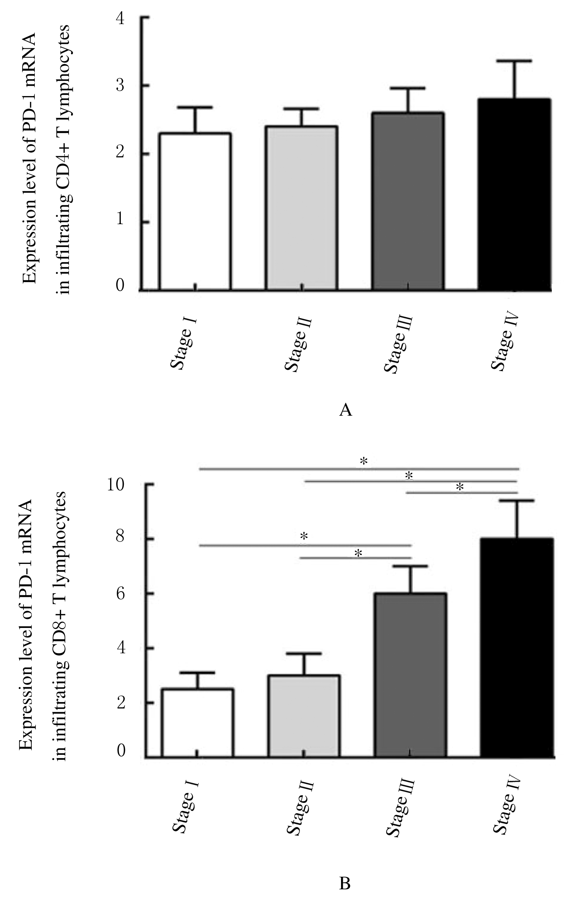

Fig. 3
Levels of PGE2 in serum of NSCLC patients at different stages"
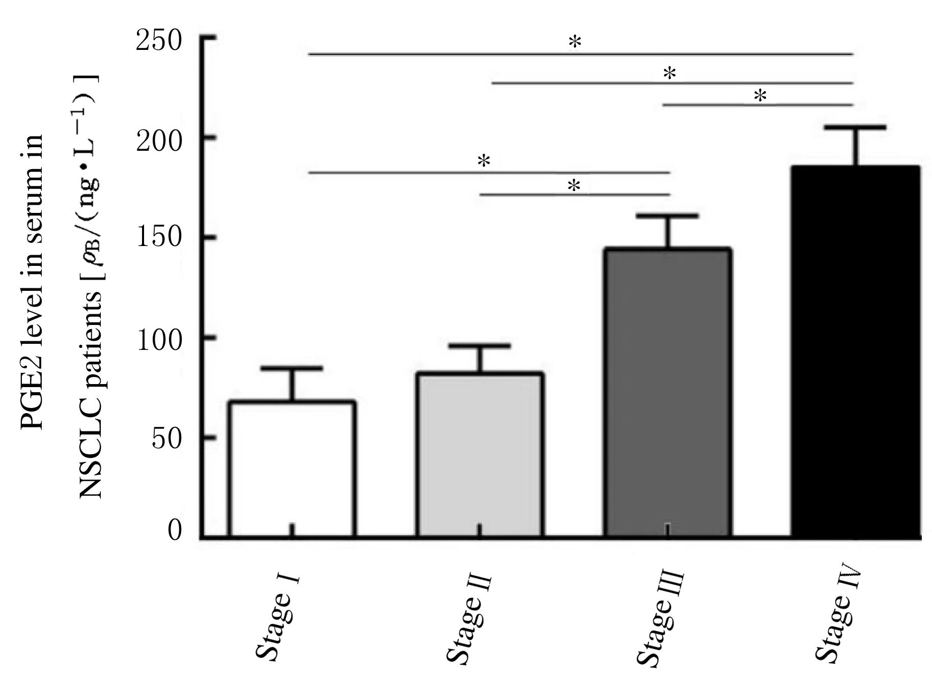

Fig. 4
Correlation between PD-1 mRNA expression level in CD8+T lymphocytes and serum PGE2 level"
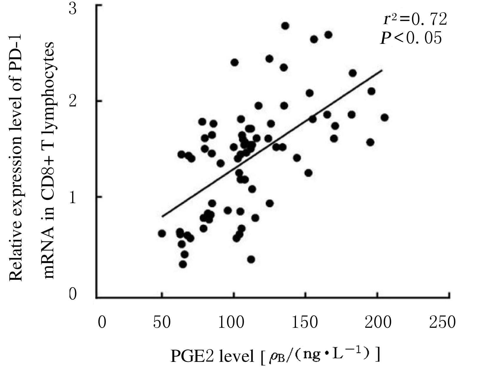

Fig. 5
Expression levels of PD-1 mRNA in CD8+ T lymphocytes after treated with PGE2"
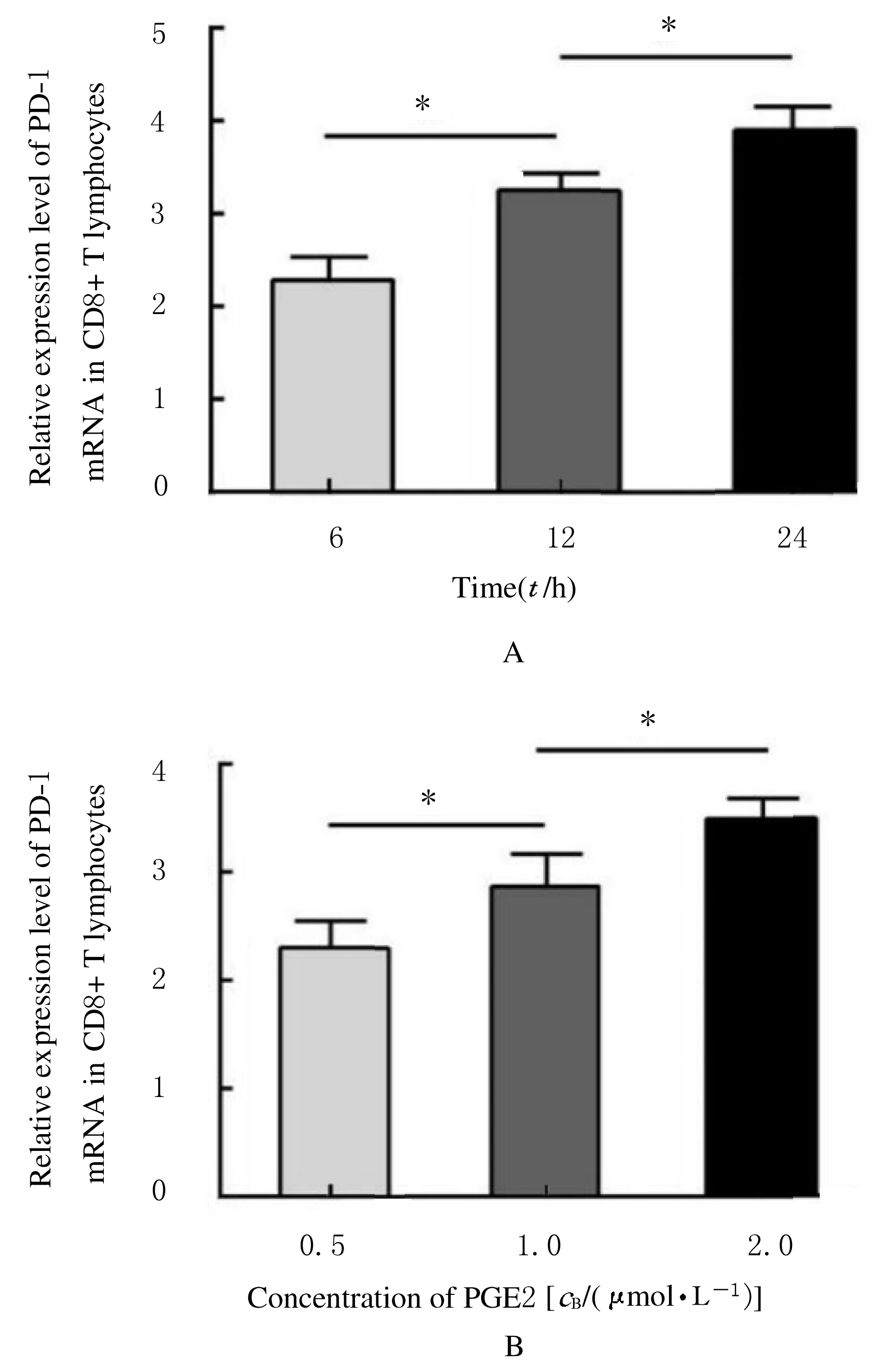
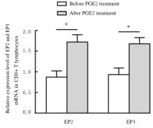
Fig. 6
Expression levels of EP2 and EP4 mRNA in CD8+ T lymphocytes after treated with PGE2"

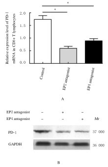
Fig. 7
Expression levels of PD-1 mRNA (A) and expression amounts of PD-1 protein (B) in CD8+T lymphocytes after treated with EP2 and EP4 antagonists"

| 1 | 国家统计局. 中国统计年鉴2019 [M]. 北京: 中国统计出版社, 2019. |
| 2 | HERBST R S, MORGENSZTERN D, BOSHOFF C. The biology and management of non-small cell lung cancer [J]. Nature, 2018, 553(7689):446-454. |
| 3 | COMEN E A, BOWMAN R L, KLEPPE M. Underlying causes and therapeutic targeting of the inflammatory tumor microenvironment [J]. Front Cell Dev Biol, 2018, 6:56. |
| 4 | LIN E W, KARAKASHEVA T A, HICKS P D, et al. The tumor microenvironment in esophageal cancer [J]. Oncogene, 2016, 35(41):5337-5349. |
| 5 | TSUGE K, INAZUMI T, SHIMAMOTO A, et al. Molecular mechanisms underlying prostaglandin E2-exacerbated inflammation and immune diseases [J]. Int Immunol, 2019, 31(9):597-606. |
| 6 | MARTINEZ-COLÓN G J, MOORE B B. Prostaglandin E(2) as a regulator of immunity to pathogens [J]. Pharmacol Ther, 2018, 185:135-146. |
| 7 | ALSAAB H O, SAU S, ALZHRANI R, et al. PD-1 and PD-L1 checkpoint signaling inhibition for cancer immunotherapy: mechanism, combinations, and clinical outcome [J]. Front Pharmacol, 2017, 8:561. |
| 8 | DERMANI F K, SAMADI P, RAHMANI G, et al. PD-1/PD-L1 immune checkpoint: potential target for cancer therapy [J]. J Cell Physiol, 2019, 234(2):1313-1325. |
| 9 | LUEN S J, SAVAS P, FOX S B, et al. Tumour-infiltrating lymphocytes and the emerging role of immunotherapy in breast cancer [J]. Pathology, 2017, 49(2):141-155. |
| 10 | REISER J, BANERJEE A. Effector, memory, and dysfunctional CD8(+) T cell fates in the antitumor immune response [J]. J Immunol Res, 2016, 2016:8941260. |
| 11 | MCGRANAHAN N, SWANTON C. Clonal heterogeneity and tumor evolution: past, present, and the future [J]. Cell, 2017, 168(4):613-628. |
| 12 | BUCHAN S L, FALLATAH M, THIRDBOROUGH S M, et al. PD-1 blockade and CD27 stimulation activate distinct transcriptional programs that synergize for CD8(+) T-cell-driven antitumor immunity [J]. Clin Cancer Res, 2018, 24(10):2383-2394. |
| 13 | ARRIETA O, MONTES-SERVIN E, HERNANDEZ-MARTINEZ J M, et al. Expression of PD-1/PD-L1 and PD-L2 in peripheral T-cells from non-small cell lung cancer patients [J]. Oncotarget, 2017, 24;8(60):101994-102005. |
| 14 | MILDNER F, SOPPER S, AMANN A, et al. Systematic review: Soluble immunological biomarkers in advanced non-small-cell lung cancer (NSCLC) [J]. Crit Rev Oncol Hematol, 2020, 153:102948. |
| 15 | RAPHAEL I, NALAWADE S, EAGAR T N, et al. T cell subsets and their signature cytokines in autoimmune and inflammatory diseases [J]. Cytokine, 2015, 74(1):5-17. |
| 16 | PICCIRILLO C A. Transcriptional and translational control of Foxp3+ regulatory T cell functional adaptation to inflammation [J]. Curr Opin Immunol, 2020, 67:27-35. |
| 17 | CHEN J H, PERRY C J, TSUI Y C, et al. Prostaglandin E2 and programmed cell death 1 signaling coordinately impair CTL function and survival during chronic viral infection [J]. Nat Med, 2015, 21(4):327-334. |
| 18 | GORCHS L, FERNANDEZ MOREO C, BANKHEAD P, et al. Human pancreatic carcinoma-associated fibroblasts promote expression of co-inhibitory markers on CD4+ and CD8+T-Cells [J]. Front Immunol, 2019, 10:847. |
| 19 | PENG L, YE Y, MULLIKIN H, et al. Expression of trophoblast derived prostaglandin E2 receptor 2 (EP2) is reduced in patients with recurrent miscarriage and EP2 regulates cell proliferation and expression of inflammatory cytokines [J]. J Reproductive Immunol, 2020, 142:103210. |
| 20 | BRADBURY P, RUMZHUM N N, AMMIT A J. EP(2) and EP(4) receptor antagonists: Impact on cytokine production and β(2) -adrenergic receptor desensitization in human airway smooth muscle [J]. J Cell Physiol, 2019, 234(7):11070-11077. |
| [1] | WANG Tianyue, ZHOU Qianlan, SHANG Yunxiao. Effect of epigallocatechin-3-gallate on airway inflammation and Treg/Th17 immune balance of mice with obese asthma [J]. Journal of Jilin University(Medicine Edition), 2019, 45(03): 491-497. |
| [2] | WANG Zhijing, SU Rongjian, DU Xiaoyuan. Effects of GRP78 on sensibility of gemcitabine on patients with NSCLC [J]. Journal of Jilin University(Medicine Edition), 2019, 45(03): 595-600. |
| [3] | LI Lijing, ZANG Yaru, YU Wenhui, LIANG Junwei, TONG Yanjun. Therapeutic effect of chemotherapy combined with sodium cantharidinate vitamin B6 injection in patientswith non-small cell lung cancer and its mechanism [J]. Journal of Jilin University(Medicine Edition), 2018, 44(06): 1286-1290. |
| [4] | CHEN Fei, SHI Jinxian, JIAO Peng, JIN Quan, XU Lishuo, ZHANG Li, MA Ning. Detection of levels of serum visfatin and PGE2 in patients with periodontitis and their relationships with activity of periodontitis [J]. Journal of Jilin University Medicine Edition, 2018, 44(03): 563-567. |
| [5] | YANG Wenyan, LIU Qiang, SUN Zhijuan, DU Liqing, XU Chang, WANG Yan, LIU Yang, WANG Qin. Effect of melatonin on radiosensitivity of non-small cell lung cancer H1299 cells [J]. Journal of Jilin University Medicine Edition, 2018, 44(03): 532-536. |
| [6] | XI Yanli, XU Na, LI Shu, WANG Di, WANG Shuran, NIU Fenglan. Inhibitory effect of gallic acid on proliferation of human non-small cell lung cancer A549 cells [J]. Journal of Jilin University Medicine Edition, 2016, 42(06): 1092-1098. |
| [7] | YUN Fen, JIA Yongfeng, HAN Zhao, SUN Qinnuan, LI Xiuxia, YU Huiling. Expression of HIF-2αin non-small cell lung cancer tissue and its relationships with MVD,Ki67,and GST-π [J]. Journal of Jilin University Medicine Edition, 2016, 42(05): 954-957. |
| [8] | LA Zong,WANG Jian-xia,CUI Ni,LI Ming-yue,XING Shu-gang,REN Bo,ZHANG Li-hong,LI Wei,LI Yu-lin. Correlation of CD4 and CD8 positive lymphocytes with tumor angiogenesis in microenvironment of breast invasive ductal carcinoma [J]. Journal of Jilin University Medicine Edition, 2014, 40(05): 1064-1073. |
| [9] | HUANG Xiong,CAI Chuan-shu. Relationship between acute radiation pneumonitis after intensity modulated radiation therapy in patients with non-small cell lung cancer and parameters of dose-volume histogram [J]. Journal of Jilin University Medicine Edition, 2014, 40(04): 892-894. |
| [10] | ZHANG Hui,SHAO Guo-guang,YAN Xu,SUN Hong-wei,WANG Xing-xing,SHAO Ming-xin,MA Ke-wei . Influence of visceral pleural invasion on prognosis of patients with early non-small cell lung cancer after operation [J]. Journal of Jilin University Medicine Edition, 2014, 40(02): 369-373. |
| [11] | LIU Huan-xin,BAI Yi-feng,WANG Wei,GUO Lin-lang. Expressions of LRIG1 and EGFR mRNA in peripheral blood of non-small cell lung cancer patients and their clinical significances [J]. Journal of Jilin University Medicine Edition, 2014, 40(02): 363-368. |
| [12] | TAO Tao,CHEN Hong,GU Xue-shuang,ZHOU Jun-hao,PENG Dan-yi,ZHANG Li-jun. [J]. Journal of Jilin University Medicine Edition, 2013, 39(3): 559-564. |
| [13] | LI En-xi,YIN Wei-min,WANG Xu,MA Ke-wei. Efficacy and influencing factor analysis of GP regimen in postoperative adjuvant chemotherapy for non-small-cell lung cancer [J]. Journal of Jilin University Medicine Edition, 2013, 39(1): 122-127. |
| [14] | WANG Jing,ZHAO Jian-jun,LI Yun-xia,TAN Ping,XU Shan-shan. Clinical application of ATP - tumor chemosensitivity assay of in vitro susceptibility test in treatment of pleural fluid in |patients |with non-small cell lung cancer [J]. J4, 2012, 38(1): 135-139. |
| [15] | XU Zhi-Gang, CHEN Li, LU Xiao-Yan, LI Wei, XIANG Dan, LI Zhi-Jun. Inhibitory effect of celecoxib on proliferation of Lewis lung cancer |cells and its mechanism [J]. J4, 2010, 36(1): 58-62. |