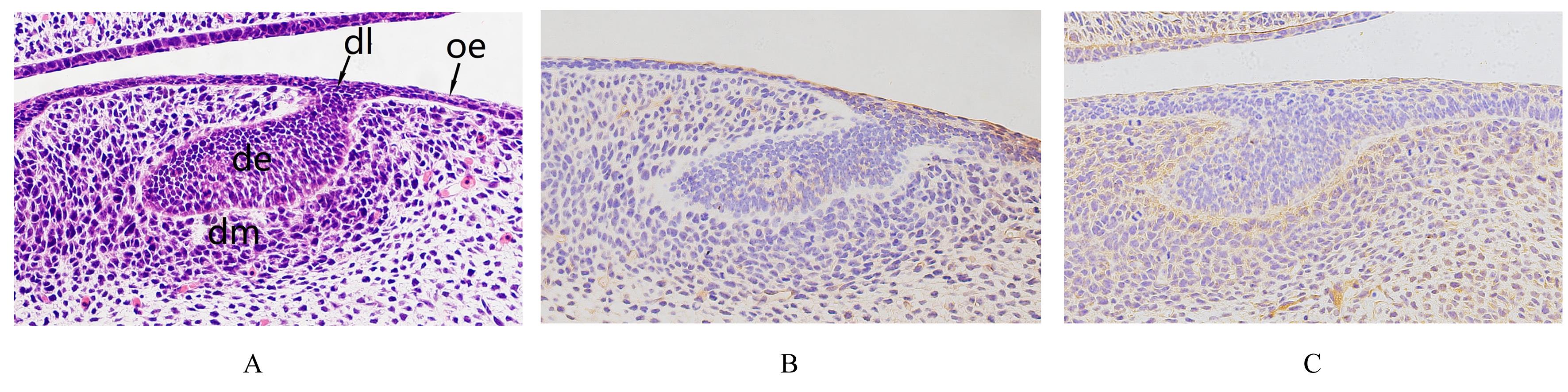
| 1 |
NEVES V C M, SHARPE P T. Regulation of reactionary dentine formation[J]. J Dent Res, 2018, 97(4): 416-422.
|
| 2 |
PEI F, LIU J L, ZHANG L, et al. The functions of mechanosensitive ion channels in tooth and bone tissues[J]. Cell Signal, 2021, 78: 109877.
|
| 3 |
HE P P, ZHANG Y, KIM S O, et al. Ameloblast differentiation in the human developing tooth: effects of extracellular matrices[J]. Matrix Biol, 2010, 29(5): 411-419.
|
| 4 |
YANG J, LU X, LIU S F, et al. The involvement of genes related to bile secretion pathway in rat tooth germ development[J]. J Mol Histol, 2020, 51(1): 99-107.
|
| 5 |
BALIC A, THESLEFF I. Tissue interactions regulating tooth development and renewal[J]. Curr Top Dev Biol, 2015, 115: 157-186.
|
| 6 |
BLOCH D, YALOVSKY S. Cell polarity signaling[J]. Curr Opin Plant Biol, 2013, 16(6): 734-742.
|
| 7 |
CHANG B, SVOBODA K K H, LIU X H. Cell polarization: from epithelial cells to odontoblasts[J]. Eur J Cell Biol, 2019, 98(1): 1-11.
|
| 8 |
TJÄDERHANE L, KOIVUMÄKI S, PÄÄKKÖNEN V,et al. Polarity of mature human odontoblasts[J]. J Dent Res, 2013, 92(11): 1011-1016.
|
| 9 |
KYPRIANOU C, CHRISTODOULOU N, HAMILTON R S, et al. Basement membrane remodelling regulates mouse embryogenesis[J]. Nature, 2020, 582(7811): 253-258.
|
| 10 |
ÜSTÜN Y, REIBETANZ M, BRACHVOGEL B,et al.Dual role of laminin‑511 in regulating melanocyte migration and differentiation[J]. Matrix Biol, 2019, 80: 59-71.
|
| 11 |
KAWAS SAL, WARSHAWSKY H. Ultrastructure and composition of basement membrane separating mature ameloblasts from enamel[J]. Arch Oral Biol, 2008, 53(4): 310-317.
|
| 12 |
ARIMORI T, MIYAZAKI N, MIHARA E, et al. Structural mechanism of laminin recognition by integrin[J]. Nat Commun, 2021, 12(1): 4012.
|
| 13 |
SUN G Y, GUILLON E, HOLLEY S A. Integrin intra-heterodimer affinity inversely correlates with integrin activatability[J]. Cell Rep, 2021, 35(10): 109230.
|
| 14 |
LEE J L, STREULI C H. Integrins and epithelial cell polarity[J]. J Cell Sci, 2014, 127(Pt 15): 3217-3225.
|
| 15 |
LI R, PENDERGAST A M. Arg kinase regulates epithelial cell polarity by targeting β1-integrin and small GTPase pathways[J]. Curr Biol, 2011, 21(18): 1534-1542.
|
| 16 |
PENG J L, LI X L, ZHANG Y, et al. Par3/integrin β1 regulates embryo adhesion via changing endometrial luminal epithelium polarity[J].Biol Reprod,2021,104(6):1228-1238.
|
| 17 |
CHEN B, GOODMAN E, LU Z, et al. Function of beta1 integrin in oral epithelia and tooth bud morphogenesis[J]. J Dent Res, 2009, 88(6): 539-544.
|
| 18 |
SAITO K, FUKUMOTO E, YAMADA A, et al. Interaction between fibronectin and β1 integrin is essential for tooth development[J]. PLoS One, 2015, 10(4): e0121667.
|
| 19 |
CLOUTIER G, SALLENBACH-MORRISSETTE A, BEAULIEU J F. Non-integrin laminin receptors in epithelia[J]. Tissue Cell, 2019, 56: 71-78.
|
| 20 |
HU D W, WANG Y C, LI A X, et al. LAMR1 restricts Zika virus infection by attenuating the envelope protein ubiquitination[J]. Virulence,2021,12(1): 1795-1807.
|
| 21 |
NELSON J, MCFERRAN N V, PIVATO G, et al. The 67 kDa laminin receptor: structure, function and role in disease[J]. Biosci Rep, 2008, 28(1): 33-48.
|
 ),Hong LIU1,2(