Journal of Jilin University(Medicine Edition) ›› 2025, Vol. 51 ›› Issue (2): 307-316.doi: 10.13481/j.1671-587X.20250204
• Research in basic medicine • Previous Articles
Effect of securinine on proliferation and apoptosis of human colon cancer SW620 cells and its mechanism
Jing DENG,Xuan WANG,Changyu SHI,Siqi YANG,Qinling ZOU,Ming JIN( )
)
- Department of Biochemistry and Molecular Biology,School of Medical Sciences,Yanbian University,Yanji 133000,China
-
Received:2024-05-09Accepted:2024-06-26Online:2025-03-28Published:2025-04-22 -
Contact:Ming JIN E-mail:jinming@ybu.edu.cn
CLC Number:
- R33
Cite this article
Jing DENG,Xuan WANG,Changyu SHI,Siqi YANG,Qinling ZOU,Ming JIN. Effect of securinine on proliferation and apoptosis of human colon cancer SW620 cells and its mechanism[J].Journal of Jilin University(Medicine Edition), 2025, 51(2): 307-316.
share this article
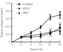
Fig. 1
Subcutaneous transplanted tumor volumes of nude mice in various groups"

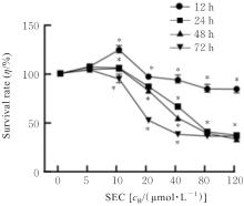
Fig. 2
Survival rates of SW620 cells after treated with different doses of SEC for different time"
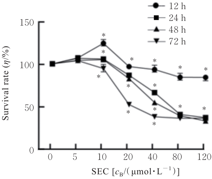
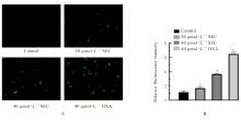
Fig. 3
Apoptosis of SW620 cells in various groups detected by TUNEL assay"
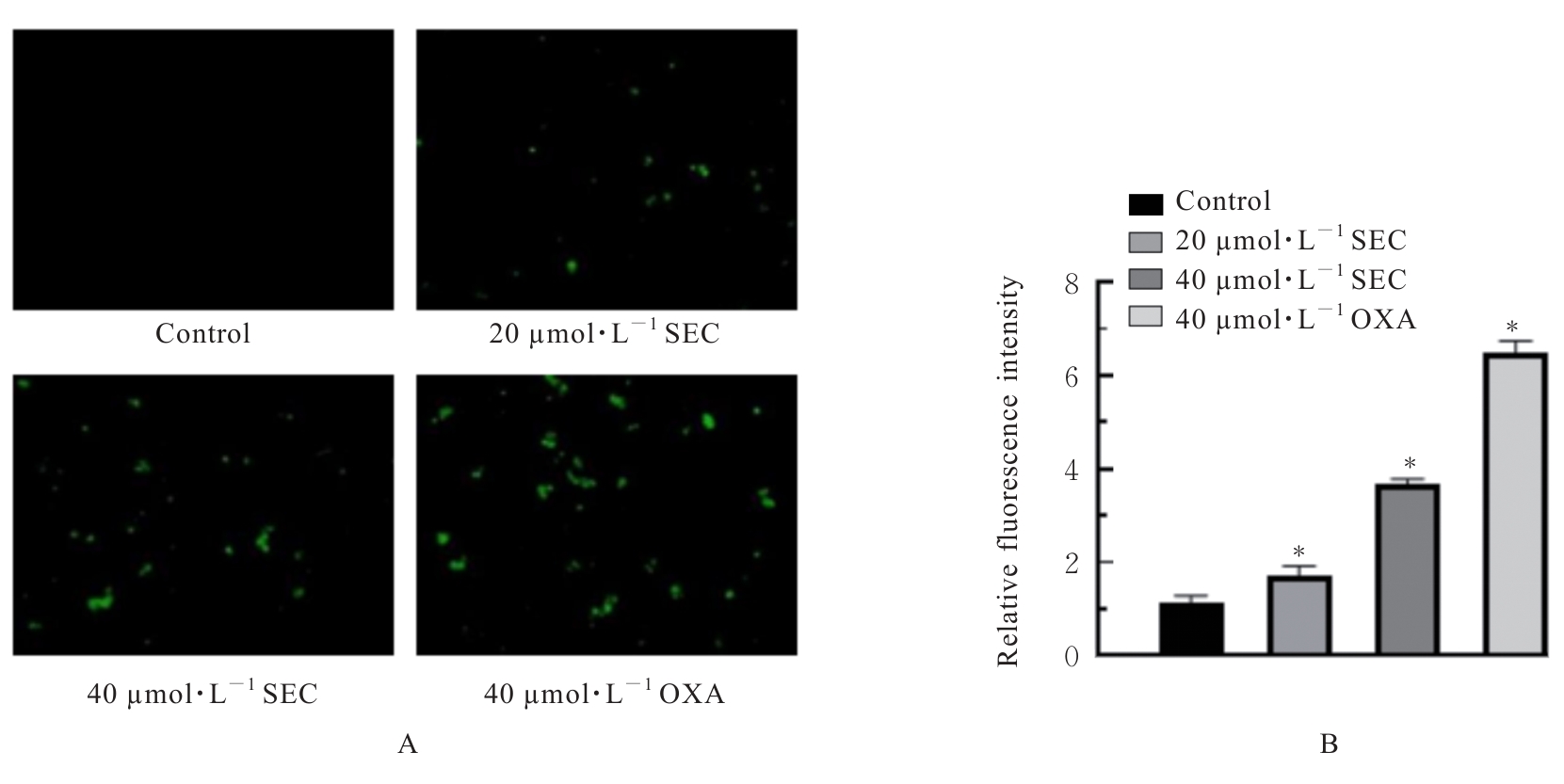
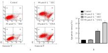
Fig. 4
Apoptosis of SW620 cells in various groups detected by flow cytometry"

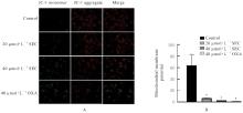
Fig. 5
Mitochondrial membrane potentials of SW620 cells in various groups"
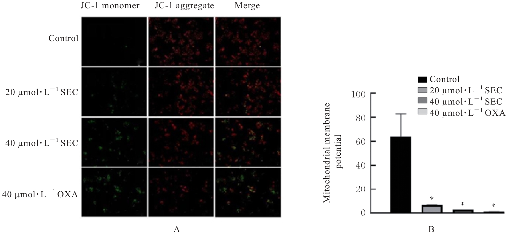
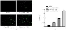
Fig. 6
Levels of ROS in SW620 cells in various groups detected by DCFH-DA fluorescence staining"
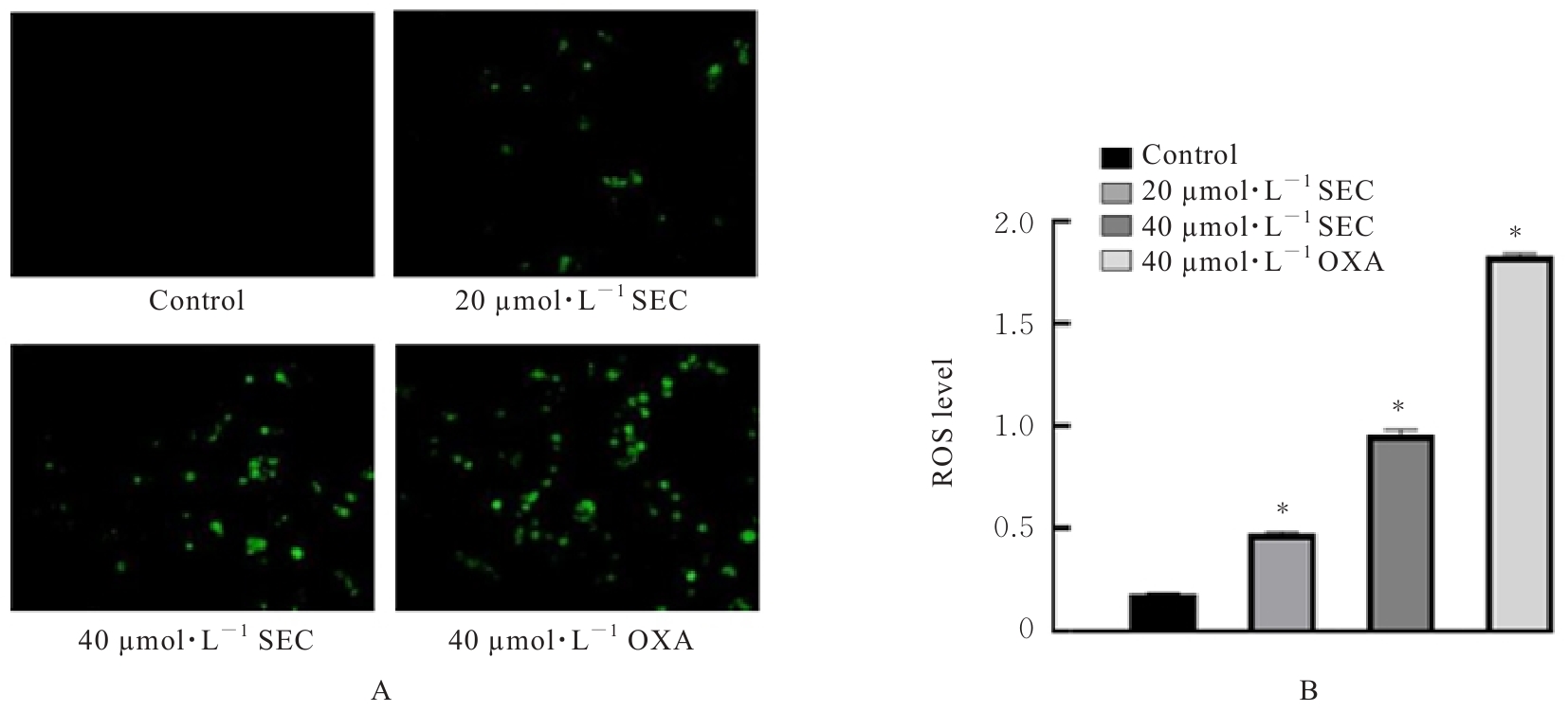
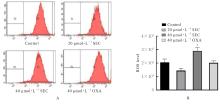
Fig. 7
ROS levels in SW620 cells in various groups detected by flow cytometry"

Fig. 8
Electrophoregram(A) and histogram(B) of expressions of cytochrome C, Bax and Bcl-2 proteins in SW620 cells in various groups"
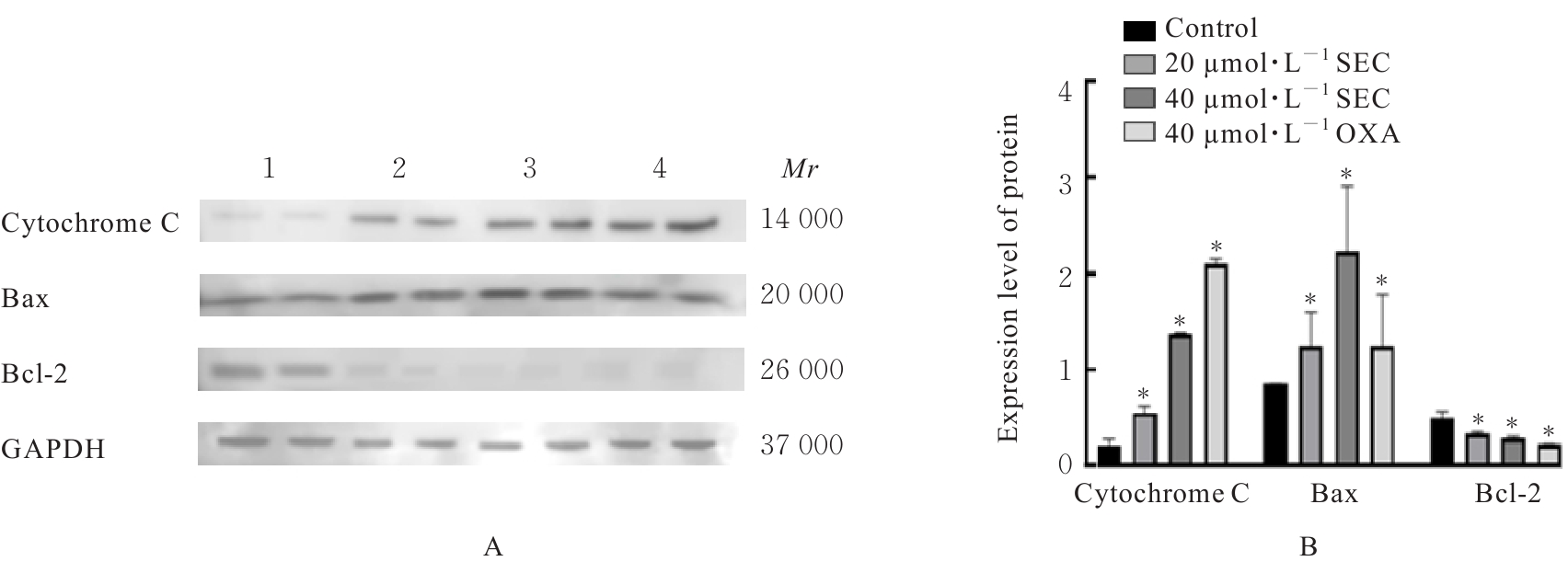

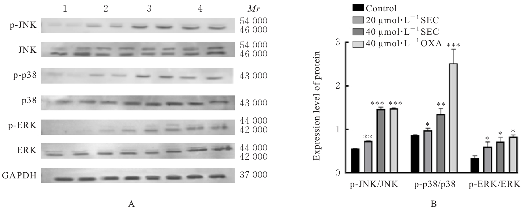
Fig. 9
Electrophoregram(A) and histogram(B) of expressions of MAPK signaling pathway related proteins in SW620 cells in various groups"
| 1 | SOBRERO A F, PUCCINI A, SHI Q, et al. A new prognostic and predictive tool for shared decision making in stage Ⅲ colon cancer[J]. Eur J Cancer, 2020, 138: 182-188. |
| 2 | PAGÈS F, ANDRÉ T, TAIEB J, et al. Prognostic and predictive value of the Immunoscore in stage Ⅲ colon cancer patients treated with oxaliplatin in the prospective IDEA France PRODIGE-GERCOR cohort study[J]. Ann Oncol, 2020, 31(7): 921-929. |
| 3 | SIEGEL R L, MILLER K D, JEMAL A. Cancer statistics, 2018[J]. CA A Cancer J Clin, 2018, 68(1): 7-30. |
| 4 | ONEDA E, ZANIBONI A. Adjuvant treatment of colon cancer with microsatellite instability-the state of the art[J]. Crit Rev Oncol Hematol, 2022, 169: 103537. |
| 5 | KLOCHKOV S, NEGANOVA M. Unique indolizidine alkaloid securinine is a promising scaffold for the development of neuroprotective and antitumor drugs[J]. RSC Adv, 2021, 11(31): 19185-19195. |
| 6 | MUN J G, JEON H D, YOON D H, et al. Supercritical extract of Cannabis sativa inhibits lung metastasis in colorectal cancer cells by increasing AMPK and MAPKs-mediated apoptosis and cell cycle arrest[J]. Nutrients, 2022, 14(21): 4548. |
| 7 | LI M D, HAN S W, ZHANG G, et al. Antiproliferative activity and apoptosis-inducing mechanism of L-securinine on human breast cancer MCF-7 cells[J]. Pharmazie, 2014, 69(3): 217-223. |
| 8 | HAN S W, ZHANG G, LI M D, et al. L-securinine induces apoptosis in the human promyelocytic leukemia cell line HL-60 and influences the expression of genes involved in the PI3K/AKT/mTOR signaling pathway[J]. Oncol Rep, 2014, 31(5): 2245-2251. |
| 9 | ZHANG G, LI M D, HAN S W, et al. Induction of human chronic myeloid leukemia K562 cell apoptosis by virosecurinine and its molecular mechanism[J]. Mol Med Rep, 2014, 10(5): 2365-2371. |
| 10 | STEFANOWICZ-HAJDUK J, SPARZAK-STEFANOWSKA B, KRAUZE-BARANOWSKA M, et al. Securinine from phyllanthus glaucus induces cell cycle arrest and apoptosis in human cervical cancer HeLa cells[J]. PLoS One, 2016, 11(10): e0165372. |
| 11 | YIP K L, TSAI T N, YANG I P, et al. Metformin enhancement of therapeutic effects of 5-fluorouracil and oxaliplatin in colon cancer cells and nude mice[J]. Biomedicines, 2022, 10(5): 955. |
| 12 | XIE L P, LIANG S Q, JIWA H B, et al. Securinine inhibits the tumor growth of human bladder cancer cells by suppressing Wnt/β-catenin signaling pathway and activating p38 and JNK signaling pathways[J]. Biochem Pharmacol, 2024, 223: 116125. |
| 13 | ZHANG W, BHAGWATH A S, RAMZAN Z, et al. Itraconazole exerts its antitumor effect in esophageal cancer by suppressing the HER2/AKT signaling pathway[J]. Mol Cancer Ther, 2021, 20(10): 1904-1915. |
| 14 | MA L, LI X, ZHAO X P, et al. Oxaliplatin promotes siMAD2L2-induced apoptosis in colon cancer cells[J]. Mol Med Rep, 2021, 24(3): 629. |
| 15 | SIEGEL R L, MILLER K D, FUCHS H E,et al. Cancer statistics, 2022[J]. CA A Cancer J Clin, 2022,72(1):7-33. |
| 16 | LICHTENSTEIN P, HOLM N V, VERKASALO P K, et al. Environmental and heritable factors in the causation of cancer: analyses of cohorts of twins from Sweden, Denmark, and Finland[J]. N Engl J Med, 2000, 343(2): 78-85. |
| 17 | SONG M Y, GARRETT W S, CHAN A T. Nutrients, foods, and colorectal cancer prevention[J]. Gastroenterology, 2015, 148(6): 1244-1260.e16. |
| 18 | PATEL S G, KARLITZ J J, YEN T, et al. The rising tide of early-onset colorectal cancer: a comprehensive review of epidemiology, clinical features, biology, risk factors, prevention, and early detection[J]. Lancet Gastroenterol Hepatol, 2022, 7(3): 262-274. |
| 19 | GREENWELL M, RAHMAN P M. Medicinal plants: their use in anticancer treatment[J]. Int J Pharm Sci Res, 2015, 6(10): 4103-4112. |
| 20 | STRÖHLE A, MAIKE W, HAHN A. Diet and tumor diseases of the colon and rectum: what is scientifically proven?[J]. Med Monatsschr Pharm, 2007, 30(1):25-32. |
| 21 | ZHANG D X, LIU H X, YANG B B, et al. L-securinine inhibits cell growth and metastasis of human androgen-independent prostate cancer DU145 cells via regulating mitochondrial and AGTR1/MEK/ERK/STAT3/PAX2 apoptotic pathways[J]. Biosci Rep, 2019, 39(5): BSR20190469. |
| 22 | RANA S, GUPTA K, GOMEZ J, et al. Securinine induces p73-dependent apoptosis preferentially in p53-deficient colon cancer cells[J]. FASEB J, 2010, 24(6): 2126-2134. |
| 23 | BURKE P J. Mitochondria, bioenergetics and apoptosis in cancer[J]. Trends Cancer, 2017, 3(12): 857-870. |
| 24 | RAY P D, HUANG B W, TSUJI Y. Reactive oxygen species (ROS) homeostasis and redox regulation in cellular signaling[J]. Cell Signal, 2012, 24(5): 981-990. |
| 25 | HERRERA B, ALVAREZ A M, SÁNCHEZ A, et al. Reactive oxygen species (ROS) mediates the mitochondrial-dependent apoptosis induced by transforming growth factor (beta) in fetal hepatocytes[J]. FASEB J, 2001, 15(3): 741-751. |
| 26 | MA C G, SONG M M, ZHANG Y, et al. Nickel nanowires induce cell cycle arrest and apoptosis by generation of reactive oxygen species in HeLa cells[J]. Toxicol Rep, 2014, 1: 114-121. |
| 27 | PIETENPOL J A, STEWART Z A. Cell cycle checkpoint signaling: cell cycle arrest versus apoptosis[J]. Toxicology, 2002, 181/182: 475-481. |
| 28 | BRAICU C, BUSE M, BUSUIOC C, et al. A comprehensive review on MAPK: a promising therapeutic target in cancer[J]. Cancers (Basel), 2019, 11(10): 1618. |
| 29 | PLOTNIKOV A, FLORES K, MAIK-RACHLINE G, et al. The nuclear translocation of ERK1/2 as an anticancer target[J]. Nat Commun, 2015, 6: 6685. |
| 30 | SU L J, ZHANG J H, GOMEZ H, et al. Mitochondria ROS and mitophagy in acute kidney injury[J]. Autophagy, 2023, 19(2): 401-414. |
| 31 | AGGELI I S, GAITANAKI C, BEIS I. Involvement of JNKs and p38-MAPK/MSK1 pathways in H2O2-induced upregulation of heme oxygenase-1 mRNA in H9c2 cells[J]. Cell Signal, 2006, 18(10): 1801-1812. |
| 32 | LEE P J, CAMHI S L, CHIN B Y, et al. AP-1 and STAT mediate hyperoxia-induced gene transcription of heme oxygenase-1[J]. Am J Physiol Lung Cell Mol Physiol, 2000, 279(1): L175-L182.[PubMed] |
| 33 | KOUL H K, PAL M, KOUL S. Role of p38 MAP kinase signal transduction in solid tumors[J]. Genes Cancer, 2013, 4(9/10): 342-359. |
| 34 | 雷东旭, 杨 静, 南青山, 等. MAPK信号通路在胃癌中的研究进展[J]. 贵州中医药大学学报, 2023, 45(4): 72-77. |
| 35 | HE H T, QIAO K Y, WANG C, et al. Hydrazinocurcumin induces apoptosis of hepatocellular carcinoma cells through the p38 MAPK pathway[J]. Clin Transl Sci, 2021, 14(5): 2075-2084. |
| 36 | 吴 亭, 张云芳, 邱晓堂, 等. 滋肾固髓汤通过调控p38MAPK/p53信号通路诱导结直肠癌HCT-116细胞凋亡[J]. 现代肿瘤医学, 2023, 31(9): 1608-1613. |
| [1] | Shuyan SUN,Huakun ZHANG,Ziru ZHOU,Feng LI,Xiaobin CUI. Expression of CRNN protein in esophageal squamous cell carcinoma tissue and influence of its overexpression in biological behavior of esophageal squamous cell carcinoma Eca9706 cells [J]. Journal of Jilin University(Medicine Edition), 2025, 51(2): 275-283. |
| [2] | Mengyun LU,Yucheng HAN,Yihong HU,Minhui HE,Yanqun ZHANG,Xianqiong ZOU. Effects of glycolipid transfer protein on proliferation, migration,and invasion of pancreatic cancer PANC-1 cells and their mechanisms [J]. Journal of Jilin University(Medicine Edition), 2025, 51(2): 284-295. |
| [3] | Chongyang ZHANG,Jia LUO,Xue QIN,Panxi SUN,Lili WEI,Xiushi YU. Protective effect of prunetin on cerebral ischemia-reperfusion injury in rats by regulating JNK/p38 pathway [J]. Journal of Jilin University(Medicine Edition), 2025, 51(2): 296-306. |
| [4] | Mengmeng ZHAO,Yalu WANG,Yuxiang XU,Kaige YANG,Yuwen CAO,Wenhu ZHOU,Jing FEI,Wen WANG,Chenghua LUO,Jianming HU. Effects of hydrogen sulfide synthase CBS and CSE on malignant biological behaviour of breast cancer cells [J]. Journal of Jilin University(Medicine Edition), 2025, 51(1): 34-43. |
| [5] | Fang ZHAO,Zhenling LI,Lihua PIAO,Longzhe HAN,Yinji CUI,Chunji QUAN,Xuemei JIN. Effect of Yes-associated proteins on biological behaviors of human cervical cancer SiHa cells [J]. Journal of Jilin University(Medicine Edition), 2025, 51(1): 68-75. |
| [6] | Siqi LI,Guangdao CHEN,Qiyi ZENG. Improvement effect of chrysophanol on hydrogen peroxide-induced apoptosis of EA. hy926 cells and its mechanism [J]. Journal of Jilin University(Medicine Edition), 2024, 50(6): 1512-1518. |
| [7] | Jingshun ZHANG,Yinggang ZOU,Lianwen ZHENG. Effect of over-expression SLC7A5 on apoptosis of ovarian granulosa cells in rats and its mechanism [J]. Journal of Jilin University(Medicine Edition), 2024, 50(6): 1526-1534. |
| [8] | Xuan MA,Kaixiang YANG,Hai DENG,Yucheng HUANG. Effect of parthenolide on apoptosis of chondrocyte under mechanical stretch stress by inhibiting Piezo1 expression and its mechanism [J]. Journal of Jilin University(Medicine Edition), 2024, 50(6): 1621-1631. |
| [9] | Bin ZHAO,Jinye YANG,Zhiyao LI,Chengwei BI,Libo YANG,Zhiyu SHI,Xin LI,Jianpeng ZHANG,Yuanlong SHI,Yong YANG,Guoying ZHANG. Inhibitory effect of miR-30c-5p on proliferation, migration, and invasion of prostate cancer cells and its mechanism [J]. Journal of Jilin University(Medicine Edition), 2024, 50(6): 1632-1643. |
| [10] | Jing LOU,Lei ZHAO,Yanjie ZHU,Shuaiqiang YUAN,Fei WANG,Hangzhou ZHANG,Jiaojiao XU,Xiaoke YU,Liufa HOU. Effect of Fuzheng Ruanjian Anticancer Formula on malignant biological behaviors of hepatocellulars carcinoma HepG2 cells by regulating Akt/MDM2/P53 signaling pathway [J]. Journal of Jilin University(Medicine Edition), 2024, 50(6): 1654-1663. |
| [11] | Qiao WANG,Ziling ZENG,Xing WANG,Ning MA,Zhibin WANG,Guofeng XU,Xiefang YUAN,Xiaoyun WANG,Yuejiao LI,Hongmei TANG,Yun ZHANG. Effect of Aspergillus fumigatus on DNA damage and IL-33 expression in human bronchial epithelial cells and its mechanism [J]. Journal of Jilin University(Medicine Edition), 2024, 50(5): 1205-1216. |
| [12] | Yuxiao SHI,Meilan LIU,Meilin ZHU,Feng WEI. Effects of 5-Aza-CdR on autophagy and apoptosis of papillary thyroid cancer cells in subcutaneous xenograft tumor tissue of nude mice and its mechanism [J]. Journal of Jilin University(Medicine Edition), 2024, 50(5): 1330-1338. |
| [13] | Hua CHEN,Na SHA,Ning LIU,Yang LI,Haijun HU. Effect of human bone marrow mesenchymal stem cells on biological behavior of human liposarcoma SW872 cells through YAP [J]. Journal of Jilin University(Medicine Edition), 2024, 50(4): 1000-1008. |
| [14] | Yongjing YANG,Tianyang KE,Shixin LIU,Xue WANG,Dequan XU,Tingting LIU,Ling ZHAO. Synergistic sensitization of apatinib mesylate and radiotherapy on hepatocarcinoma cells invitro [J]. Journal of Jilin University(Medicine Edition), 2024, 50(4): 1009-1015. |
| [15] | Chaojie GUO,Jiajia ZHANG,Jie ZENG,Huiyu WANG, AIERFATI·Aimaier,Jiang XU. Expressions of PLOD1 in oral squamous cell carcinoma tissue and cells and their significances [J]. Journal of Jilin University(Medicine Edition), 2024, 50(4): 1035-1043. |
|
||