Journal of Jilin University(Medicine Edition) ›› 2026, Vol. 52 ›› Issue (2): 440-450.doi: 10.13481/j.1671-587X.20260216
• Research in basic medicine • Previous Articles Next Articles
Bioinformatic analysis on regulatory mechanism of MAPK-Mcl-1 signaling pathway and macrophage polarization during Bacillus Calmette-Guérin infection and its experimental validation
Ruihan GE1,2,Chen LI1,2,Shengpeng WANG1,3,4,Yang LU1,2,Caixia TAN1,2,Haotian CUI1,3,4,Xinmin WANG3,4( ),Le ZHANG1,2,4()
- 1.Department of Pathophysiology,School of Medicine,Shihezi University,Shihezi 832008,China
2.Xinjiang Provincial and Ethnic High Incidence Key Laboratory,Ministry of Education,Shihezi 832008,China
3.Department of Urology,First Affiliated Hospital,Shihezi University,Shihezi 832008,China
4.Key Laboratory for Prevention and Treatment of High-Incidence Diseases in Central Asia,National Health Commission,Shihezi 832008,China
-
Received:2025-05-05Accepted:2025-06-16Online:2026-03-28Published:2026-04-15 -
Contact:Xinmin WANG,Le ZHANG E-mail:1977602697@qq.com;1257067540@qq.com
CLC Number:
- R363.2
Cite this article
Ruihan GE,Chen LI,Shengpeng WANG,Yang LU,Caixia TAN,Haotian CUI,Xinmin WANG,Le ZHANG. Bioinformatic analysis on regulatory mechanism of MAPK-Mcl-1 signaling pathway and macrophage polarization during Bacillus Calmette-Guérin infection and its experimental validation[J].Journal of Jilin University(Medicine Edition), 2026, 52(2): 440-450.
share this article
Tab.1
Primer sequences of RT-qPCR"
| Gene | Sequence (5'-3') | Length (bp) |
|---|---|---|
| iNOS | F:TGATGTGCTGCCTCTGGTCT | 20 |
| R:GAGCTCCTGGAACCACTCGT | 20 | |
| Fizzl | F:GAGATCCAGAGTGGAGATACTTGC | 24 |
| R:TCTTAGGACAGTTGGCAGCAG | 21 | |
| β-actin | F:GTGACGTTGACATCCGTAAAGA | 22 |
| R:GCCGGACTCATCGTACTCC | 19 |
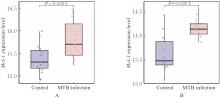
Fig. 1
Expression levels of Mcl-1 gene in macrophages in GEO database"

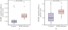
Fig. 2
Activity scores of MAPK signaling pathway in macrophages in GEO database"
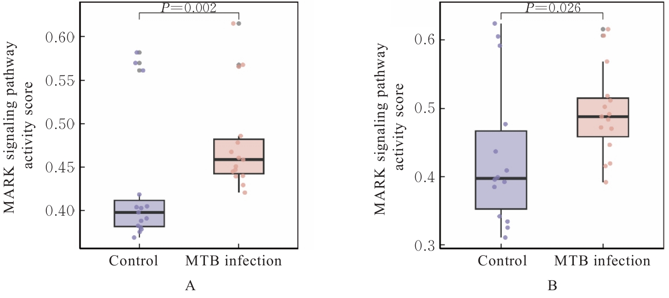
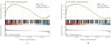
Fig. 3
GSEA enrichment analysis on MAPK signaling pathway in Mcl-1 high expression groups in GEO database"
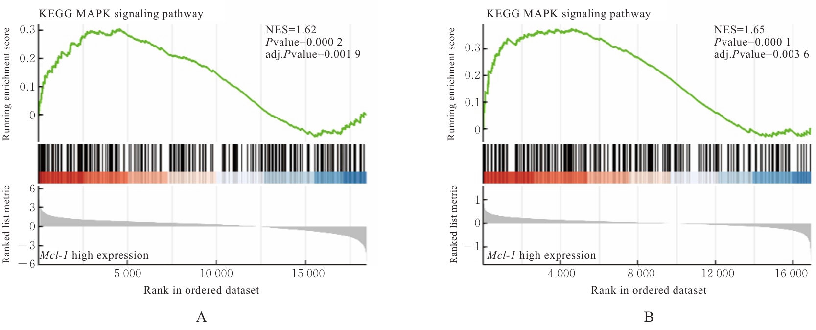
Fig. 4
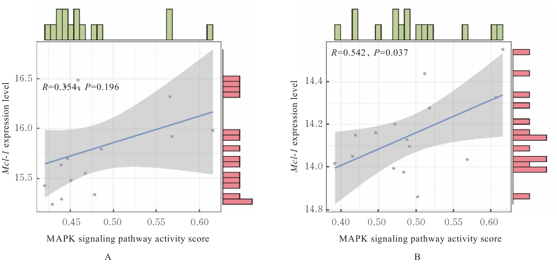
Correlation analysis between Mcl-1 expression level and activity score of MAPK signaling pathway"
Tab.2
Levels of Mcl-1 in supernatant of macrophages in various groups [n=3, x±s, ρB/(μg·L-1)]"
| Group | Mcl-1 level | ||
|---|---|---|---|
| (t/h) 0 | 12 | 24 | |
| Control | 0.48±0.02 | 1.04±0.03 | 1.51±0.14 |
| BCG | 0.46±0.06 | 0.76±0.01* | 1.06±0.04* |
| BCG+PD | - | 0.57±0.04△ | 0.57±0.02△ |
| BCG+SP | - | 0.42±0.05△ | 0.47±0.06△ |
| BCG+SB | - | 0.36±0.12△ | 0.52±0.08△ |
| BCG+PD+SP | - | 0.41±0.06△ | 0.57±0.02△ |
| BCG+PD+SB | - | 0.39±0.03△ | 0.47±0.01△ |
| BCG+SP+SB | - | 0.34±0.07△ | 0.43±0.02△ |
| BCG+PD+SP+SB | - | 0.39±0.08△ | 0.45±0.02△ |
Tab.3
Levels of IL-6 and TNF-α in supernatant of macrophages in various groups [n=3, x±s, ρB/(ng·L-1)]"
| Group | IL-6 | TNF-α | ||||
|---|---|---|---|---|---|---|
| (t/h) 0 | 12 | 24 | 0 | 12 | 24 | |
| Control | 84.14±20.02 | 166.27±7.46 | 51.77±1.22 | 68.08±2.52 | 161.55±13.85 | 158.81±4.73 |
| BCG | 81.64±7.21 | 290.38±9.21* | 59.89±3.17 | 74.82±1.88 | 194.20±5.66* | 177.32±2.87* |
| BCG+PD | - | 190.40±15.20 | 44.75±2.94△ | - | 160.45±41.96△ | 97.91±1.93△ |
| BCG+SP | - | 145.61±13.49△ | 42.28±2.45△ | - | 161.56±20.18△ | 81.86±2.72△ |
| BCG+SB | - | 205.32±10.09 | 47.47±2.44△ | - | 165.15±5.54△ | 86.03±2.07△ |
| BCG+PD+SP | - | 183.54±16.04△ | 43.15±1.22△ | - | 165.91±19.06△ | 64.18±3.37△ |
| BCG+PD+SB | - | 170.24±18.08 | 56.92±1.21 | - | 163.68±18.87△ | 50.81±2.95△ |
| BCG+SP+SB | - | 254.15±16.04 | 53.49±3.64 | - | 195.58±12.93 | 58.43±7.20△ |
| BCG+PD+SP+SB | - | 224.85±17.00 | 82.50±1.20△ | - | 200.75±7.20 | 69.23±2.32△ |
Tab.4
Levels of IL-10 and TGF-β in supernatant of macrophages in various groups [n=3, x±s, ρB/(ng·L-1)]"
| Group | IL-10 | TGF-β | ||||
|---|---|---|---|---|---|---|
| (t/h) 0 | 12 | 24 | 0 | 12 | 24 | |
| Control | 34.69±11.47 | 27.89±3.42 | 53.58±5.07 | 72.74±4.73 | 314.72±9.84 | 424.23±7.36 |
| BCG | 43.57±10.66 | 31.47±5.47 | 71.77±2.68* | 79.52±5.69 | 379.25±17.08* | 464.49±3.65 |
| BCG+PD | - | 38.65±1.85 | 52.26±5.95△ | - | 396.45±18.38 | 289.72±7.69△ |
| BCG+SP | - | 32.45±5.77 | 56.22±2.03△ | - | 424.21±16.88△ | 368.48±9.90△ |
| BCG+SB | - | 35.38±2.26 | 66.16±7.03 | - | 394.31±9.83 | 477.11±18.24 |
| BCG+PD+SP | - | 33.42±1.07 | 63.54±4.16△ | - | 411.40±23.13△ | 320.59±11.41△ |
| BCG+PD+SB | - | 38.66±6.24 | 65.09±2.80△ | - | 426.28±31.43△ | 348.98±7.53△ |
| BCG+SP+SB | - | 37.01±3.60 | 64.42±3.24△ | - | 436.96±16.86△ | 348.98±3.76△ |
| BCG+PD+SP+SB | - | 37.34±1.07 | 56.35±1.65△ | - | 493.91±16.65△ | 469.92±14.51 |
Tab.5
Expression levels of iNOS and Fizz1 mRNA in macrophages in various groups"
| Group | iNOS | Fizz1 | ||||
|---|---|---|---|---|---|---|
| (t/h) 0 | 12 | 24 | 0 | 12 | 24 | |
| Control | 1.00±0.10 | 1.00±0.10 | 1.00±0.10 | 1.00±0.10 | 1.00±0.10 | 1.00±0.10 |
| PD | - | 1.33±0.13 | 0.54±0.05* | - | 1.24±0.66 | 0.31±0.04* |
| SP | - | 1.60±0.13 | 0.84±0.11* | - | 1.49±0.64 | 0.26±0.10* |
| SB | - | 2.31±0.17** | 0.48±0.08* | - | 4.43±2.13* | 0.41±0.11* |
| PD+SP | - | 2.02±0.88** | 1.00±0.14 | - | 8.27±0.64* | 0.77±0.12* |
| PD+SB | - | 2.15±0.04** | 0.74±0.09* | - | 2.69±0.89* | 0.22±0.01* |
| SP+SB | - | 1.35±0.09 | 0.61±0.04* | - | 6.59±0.27* | 0.18±0.08* |
| PD+SP+SB | - | 0.76±0.23 | 0.61±0.06 | - | 2.44±0.90* | 0.83±0.20* |
| BCG | 1.34±0.07* | 0.50±0.03** | 1.86±0.10* | 1.43±0.21* | 2.42±0.48** | 32.32±1.12* |
| BCG+PD | - | 1.57±0.14△△ | 0.64±0.09 | - | 3.48±0.36△ | 24.24±3.66 |
| BCG+SP | - | 1.59±0.15△△ | 0.87±0.03 | - | 1.45±0.63 | 29.51±0.41 |
| BCG+SB | - | 1.94±0.13△△ | 0.52±0.06 | - | 1.49±0.40 | 24.65±2.17 |
| BCG+PD+SP | - | 2.19±0.06△△ | 1.19±0.18 | - | 15.98±0.91△△ | 2.87±0.32△△ |
| BCG+PD+SB | - | 2.56±0.12△△ | 0.91±0.02 | - | 8.16±0.76△△ | 32.88±1.76 |
| BCG+SP+SB | - | 1.35±0.22△△ | 0.37±0.03 | - | 3.52±0.32△ | 14.74±2.40 |
| BCG+PD+SP+SB | - | 1.17±0.15△△ | 0.94±0.08 | - | 7.60±0.79△△ | 22.78±1.76 |
| [1] | LANGE C, AABY P, BEHR M A, et al. 100 years of Mycobacterium bovis Bacille Calmette-Guérin[J]. Lancet Infect Dis, 2022, 22(1): e2-e12. |
| [2] | CHEN J J, GAO L, WU X Y, et al. BCG-induced trained immunity: history, mechanisms and potential applications[J]. J Transl Med, 2023, 21(1): 106. |
| [3] | BICKETT T E, MCLEAN J, CREISSEN E, et al. Characterizing the BCG induced macrophage and neutrophil mechanisms for defense against Mycobacterium tuberculosis [J]. Front Immunol, 2020, 11: 1202. |
| [4] | LIU L M, SHI W J, XIAO X, et al. BCG immunotherapy inhibits cancer progression by promoting the M1 macrophage differentiation of THP-1 cells via the Rb/E2F1 pathway in cervical carcinoma[J]. Oncol Rep, 2021, 46(5): 245. |
| [5] | CHOI Y Y, HAN M S, LEE H J, et al. Mycobacterium bovis osteitis following immunization with Bacille Calmette-Guérin (BCG) in Korea[J]. J Korean Med Sci, 2018, 34(1): e3. |
| [6] | 殷立晗, 李 昕, 徐正中, 等. 结核分枝杆菌亚单位疫苗研究进展[J]. 中国人兽共患病学报, 2023, 39(5): 492-499. |
| [7] | KURSCHAT C, METZ A, KIRSCHNEK S, et al. Importance of Bcl-2-family proteins in murine hematopoietic progenitor and early B cells[J]. Cell Death Dis, 2021, 12(8): 784. |
| [8] | TANTAWY S I, TIMOFEEVA N, SARKAR A, et al. Targeting MCL-1 protein to treat cancer: opportunities and challenges[J]. Front Oncol, 2023, 13: 1226289. |
| [9] | 冒 婷, 徐铭益, 王佳轶. 非酒精性脂肪性肝炎小鼠模型肝组织T淋巴细胞的特征分析[J]. 临床肝胆病杂志, 2025, 41(3): 461-468. |
| [10] | OPYDO M, MLYCZYŃSKA A, MLYCZYŃSKA E, et al. Synergistic action of MCL-1 inhibitor with BCL-2/BCL-XL or MAPK pathway inhibitors enhances acute myeloid leukemia cell apoptosis and differentiation[J]. Int J Mol Sci, 2023, 24(8): 7180. |
| [11] | HAN L, LU Y, WANG X F, et al. Regulatory role and mechanism of the inhibition of the Mcl-1 pathway during apoptosis and polarization of H37Rv-infected macrophages[J]. Medicine, 2020, 99(42): e22438. |
| [12] | LI J L, LU J B, WANG G Z, et al. Past, present and future of Bacille Calmette-Guérin vaccine use in China[J]. Vaccines, 2022, 10(7): 1157. |
| [13] | 郑 伟. H37Rv与BCG感染BoMac源外泌体对巨噬细胞极化作用的研究[D]. 长春: 吉林农业大学, 2023. |
| [14] | CHO H, KWON H Y, SHARMA A, et al. Visualizing inflammation with an M1 macrophage selective probe via GLUT1 as the gating target[J]. Nat Commun, 2022, 13(1): 5974. |
| [15] | 周筱雨, 胡良皞, 李兆申. 慢性胰腺炎干细胞治疗研究进展[J]. 中国实用内科杂志, 2024, 44(7): 547-552. |
| [16] | 邢国静, 王丽菲, 罗龙龙, 等. 巨噬细胞极化在药物性肝损伤中的作用研究进展[J]. 解放军医学杂志, 2025, 50(11): 1478-1484. |
| [17] | LUO M, ZHAO F K, CHENG H, et al. Macrophage polarization: an important role in inflammatory diseases[J]. Front Immunol, 2024, 15: 1352946. |
| [18] | MOLINA-OLVERA G, RIVAS-ORTIZ C I, SCHCOLNIK-CABRERA A, et al. RNA microarray-based comparison of innate immune phenotypes between human THP-1 macrophages stimulated with two BCG strains[J]. Int J Mol Sci, 2022, 23(9): 4525. |
| [19] | 唐 怡, 王国泰, 蒋雨涵, 等. 中药调控肿瘤相关巨噬细胞对肝细胞癌的治疗作用与机制[J]. 临床肝胆病杂志, 2025, 41(6): 1199-1206. |
| [20] | BO H T, MOURE U A E, YANG Y M, et al. Mycobacterium tuberculosis-macrophage interaction: Molecular updates[J]. Front Cell Infect Microbiol, 2023, 13: 1062963. |
| [21] | 王飞雨, 王新敏, 王 婵, 等. 靶向沉默Mcl-1基因对感染不同毒力结核杆菌小鼠腹腔巨噬细胞凋亡的影响[J]. 中国病理生理杂志, 2015, 31(12): 2195-2201. |
| [22] | 卢 洋, 王新敏, 王小芳, 等. 下调Bcl-2家族蛋白促进MTB感染的小鼠巨噬细胞系凋亡[J]. 基础医学与临床, 2018, 38(10): 1383-1388. |
| [23] | LI P, LI Y, WANG C C, et al. Comparative transcriptomics reveals common and strain-specific responses of human macrophages to infection with Mycobacterium tuberculosis and Mycobacterium bovis BCG[J]. Microb Pathog, 2024, 189: 106593. |
| [1] | Xiqian ZHANG,Zhibo DANG,Yunan DU,Pei WU,Hang XIE,Gaofeng TAN. Inhibitory effect of Chaiqi Yigan formula on malignant biological behaviors of liver cancer HepG2 cells by regulating macrophage polarization [J]. Journal of Jilin University(Medicine Edition), 2026, 52(2): 398-409. |
| [2] | Jingyuan WANG,Fang CHEN,Yancun LIU,Shixin LI,Songtao SHOU. Protective effect of silencing coagulation factor V gene on septic rats by inhibiting JNK1/2 and p38 MAPK signaling pathways [J]. Journal of Jilin University(Medicine Edition), 2026, 52(2): 418-428. |
| [3] | Jingyuan WANG,Fang CHEN,Yancun LIU,Shixin LI,Songtao SHOU. Effect of USF2 knockdown on coagulation dysfunction in septic rats and its mechanism [J]. Journal of Jilin University(Medicine Edition), 2026, 52(1): 171-181. |
| [4] | Xiao LIN,Meng ZHOU,Fan LIN,Xiujuan YAO. Bioinformatics analysis and experimental validation of fatty acid metabolism-related genes in idiopathic pulmonary fibrosis tissue [J]. Journal of Jilin University(Medicine Edition), 2026, 52(1): 93-104. |
| [5] | Yu LIANG,Jinyu YU,Zhonggao XU,Wanning WANG. Expression characteristics of FOSB in kidney tissue from IgA nephropathy and other common kidney diseases [J]. Journal of Jilin University(Medicine Edition), 2025, 51(5): 1281-1292. |
| [6] | Weigao SHEN,Yuqi LIU,Jun ZHANG,Jiayu LIN,Hang CUI,Yanbo LIU. Bioinformatics analysis on effect of interleukin-33 on occurrence and development of malignant brain glioma and its experimental validation [J]. Journal of Jilin University(Medicine Edition), 2025, 51(5): 1318-1332. |
| [7] | Linrui XU,Yiyu ZHANG,Jiaqi CUI,Xianzhu CONG,Shuang LI,Jiayu GE,Yujia KONG,Suzhen WANG,Fuyan SHI,Jinrong WANG. Construction of diagnostic model for Alzheimer’s disease and immune analysis based on bioinformatics and machine learning [J]. Journal of Jilin University(Medicine Edition), 2025, 51(4): 1039-1051. |
| [8] | Xianwei JIANG,Minghang WANG,Huiru LI,Xiaosheng DONG,Yuanyuan LIU. Mendelian randomization and GEO database identification analysis based on potential therapeutic targets for chronic obstructive pulmonary disease [J]. Journal of Jilin University(Medicine Edition), 2025, 51(4): 1072-1083. |
| [9] | Kun YANG,Qianyao FU,Yongqiang SUN,Kun YANG,Jun MENG. Protective effect of dexmedetomidine on intestinal mucosal injury in rats with enterogenous sepsis and its mechanism [J]. Journal of Jilin University(Medicine Edition), 2025, 51(4): 855-865. |
| [10] | Changyu SHI,Yong LI,Jing DENG,Chunmei PIAO,Ming JIN. Bioinformatics analysis on adjustment effect of colorectal liver metastases model in mice based on complement alternative pathway and its experimental verification [J]. Journal of Jilin University(Medicine Edition), 2025, 51(3): 703-715. |
| [11] | Chongyang ZHANG,Jia LUO,Xue QIN,Panxi SUN,Lili WEI,Xiushi YU. Protective effect of prunetin on cerebral ischemia-reperfusion injury in rats by regulating JNK/p38 pathway [J]. Journal of Jilin University(Medicine Edition), 2025, 51(2): 296-306. |
| [12] | Wenchang CAI,Yuqi LIU,Han WANG,Helin WANG,Zhenjiang WANG,Zishen XIAO,Shiyuan MA,Liping AN,Yanbo LIU. Expression of protein kinase D2 in bladder cancer tissue and its effect on tumor immune microenvironment [J]. Journal of Jilin University(Medicine Edition), 2025, 51(2): 378-391. |
| [13] | Xiaoxia HU,Yalong LI,Dongliang YANG,Bazeren LA,Xinyue LIU. Effect of high glucose on polarization of Raw264.7 macrophages in vitro [J]. Journal of Jilin University(Medicine Edition), 2025, 51(2): 403-411. |
| [14] | Bo LIU,Chao SUN,Xu WANG,Kewei MA. Bioinformatics analysis on differentially expressed genes in multiple primary lung cancers based on GEO database [J]. Journal of Jilin University(Medicine Edition), 2025, 51(2): 437-446. |
| [15] | Xiaoyan WANG,Xuelian LI,Bin LIANG,Wenfei TIAN,Hailin MA,Zhijing MO. Analysis on relationship between CALU and prognosis of hepatocellular carcinoma patients and its mechanism based on transcriptome and single cell sequencing data [J]. Journal of Jilin University(Medicine Edition), 2025, 51(2): 447-459. |