吉林大学学报(医学版) ›› 2021, Vol. 47 ›› Issue (4): 896-903.doi: 10.13481/j.1671-587X.20210411

• 基础研究 • 上一篇    下一篇

人参皂苷Rh1对MC3T3-E1细胞增殖和分化的促进作用及其机制

毛天娇1,孙铎1,高幸1,魏溦1,李熙恒1,姜可新1,姜秋2(),李江1,3()   

  1. 1.吉林大学口腔医院口腔修复科,吉林 长春 130021
    2.吉林大学口腔医院儿童口腔科,吉林 长春 130021
    3.广州医科大学附属口腔医院口腔修复科,广东 广州 510150
  • 收稿日期:2020-12-02 出版日期:2021-07-28 发布日期:2021-07-22
  • 通讯作者: 姜秋,李江 E-mail:jiangqiu@163.com;ljiang@gzhmu.edu.cn
  • 作者简介:毛天娇(1992-),女,河南省许昌市人,住院医师,医学硕士,主要从事口腔修复学方面的研究。
  • 基金资助:
    科技部国家重点研发计划项目(2020YFE010636);吉林省科技厅科研项目(20200201025JC);吉林省科技厅科技发展计划项目(20190201082JC);吉林省财政厅科研项目(jsz2018170-3);吉林省长春市科技局科技计划项目(17YJ001)

Tianjiao MAO1,Duo SUN1,Xing GAO1,Wei WEI1,Xiheng LI1,Kexin JIANG1,Qiu JIANG2(),Jiang LI1,3()   

  1. 1.Department of Prosthodontics,Stomatology Hospital,Jilin University,Changchun 130021,China
    2.Department of Pediatric Dentistry,Stomatology Hospital,Jilin University,Changchun 130021,China
    3.Department of Prosthodontics,Affiliated Stomatology Hospital,Guangzhou Medical University,Guangzhou 510150,China
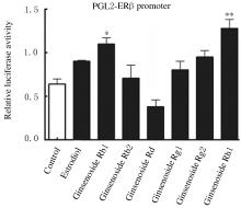
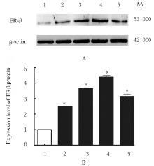
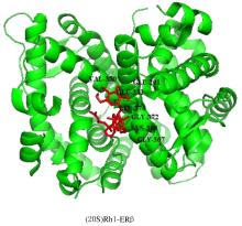
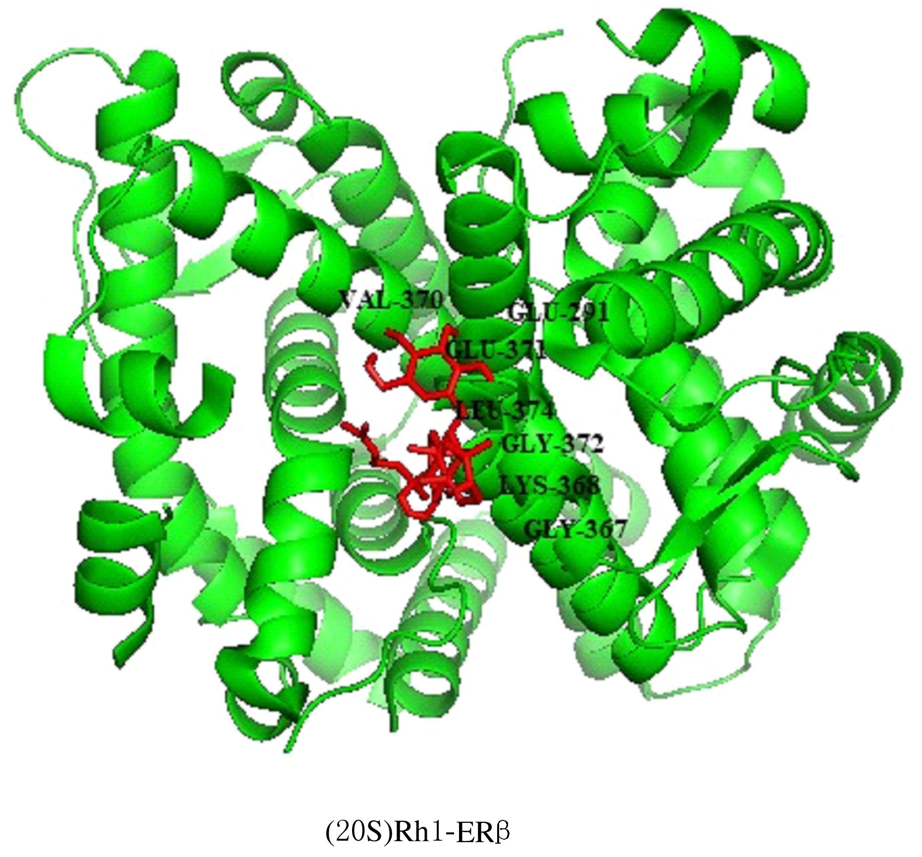
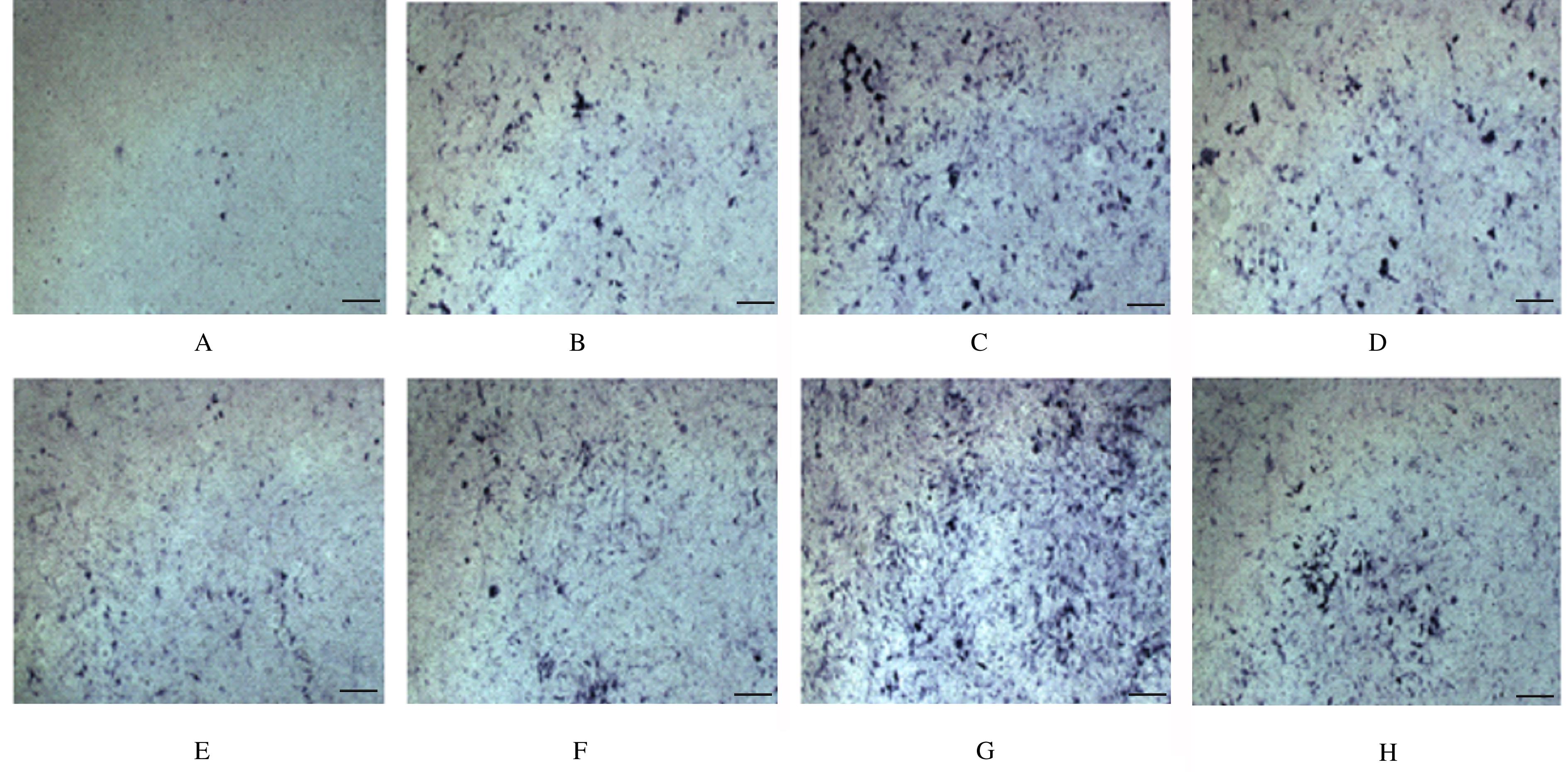
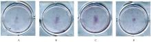
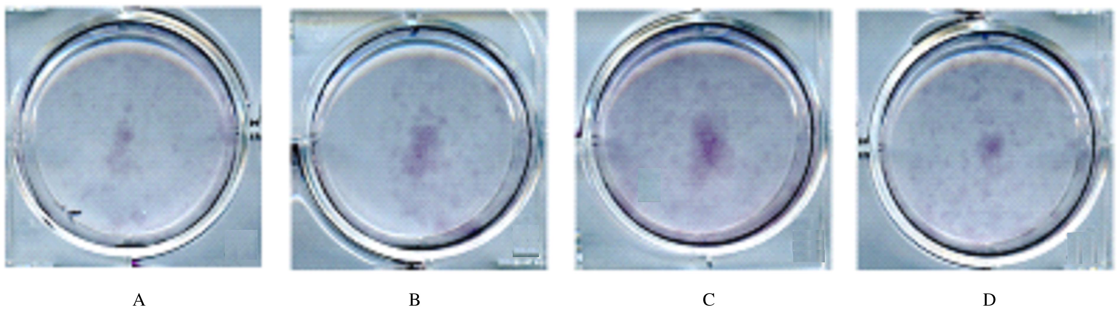
  • Received:2020-12-02 Online:2021-07-28 Published:2021-07-22
  • Contact: Qiu JIANG,Jiang LI E-mail:jiangqiu@163.com;ljiang@gzhmu.edu.cn

摘要: 目的

筛选靶向上调雌激素受体β(ERβ)转录和表达的人参皂苷单体,研究其对MC3T3-E1细胞增殖和分化的影响及其机制。

方法

采用PGL2-ERβ和内参Renilla荧光素酶质粒Prep7-Rluc共同转染HEK293T细胞,细胞分为对照组,雌二醇组(1×10⁻⁶ mmol·L-1),人参皂苷Rb1组、Rb2组、Rd组、Rg1组、Rg2组和Rh1组(1×10⁻⁵ mmol·L-1),通过双荧光素酶报告基因实验检测各组细胞双荧光素酶活性。进一步将MC3T3-E1细胞分为对照组、雌二醇组和不同浓度(1×10⁻⁶、1×10⁻⁵及1×10⁻⁴ mmol·L-1)人参皂苷Rh1组,采用Western blotting法检测各组MC3T3-E1细胞中ERβ蛋白表达水平。通过Auto Dock分子对接实验,模拟人参皂苷Rh1与ERβ蛋白分子的结合情况。MC3T3-E1细胞分为对照组、雌二醇组和不同浓度(5×10⁻⁶、1×10⁻⁵、5×10⁻⁵、1×10⁻⁴、1×10⁻³及5×10⁻³ mmol·L-1)人参皂苷Rh1组,分别作用24、48和72 h后,采用CCK-8法检测各组细胞增殖率。MC3T3-E1细胞分为对照组、雌二醇组(1×10⁻⁶ mmol·L-1)和不同浓度(1×10⁻⁵及1×10⁻⁴ mmol·L-1)人参皂苷Rh1组,配制成骨诱导液,分别诱导MC3T3-E1细胞7、14和21 d,采用碱性磷酸酶(ALP)染色和茜素红染色检测细胞中ALP和钙化结节染色面积,观察人参皂苷Rh1对MC3T3-E1细胞成骨分化的影响。

结果

双荧光素酶报告基因实验,各组HEK293T细胞中转染ERβ-PGL2质粒后,与对照组比较,1×10-5 mmol·L-1人参皂苷Rh1组细胞荧光素酶活性明显升高(P<0.05)。 Western blotting法检测,与对照组比较,不同浓度人参皂苷Rh1组细胞中ERβ蛋白表达水平明显升高(P<0.05),且1×10⁻⁴ mmol·L-1人参皂苷Rh1组细胞中ERβ蛋白表达水平最高。Auto Dock分析,人参皂苷Rh1可以结合在ERβ蛋白的配体结合口袋内。CCK-8实验,培养24、48和72 h后,与对照组比较,1×10-5、5×10⁻⁵、1×10⁻⁴和1×10⁻³ mmol·L-1人参皂苷Rh1组MC3T3-E1细胞增殖率均明显升高(P<0.01),其中72 h时1×10⁻⁴ mmol·L-1人参皂苷Rh1组细胞增殖率最高。成骨诱导分化后,与对照组比较,不同浓度人参皂苷Rh1组细胞中ALP染色面积明显增加,1×10-4 mmol·L-1人参皂苷Rh1组细胞中ALP染色面积最大,而且14 d时ALP染色面积较7 d时明显增加,具有时间和浓度依赖性;与对照组比较,不同浓度人参皂苷Rh1组细胞矿化结节茜素红染色面积明显增加。

结论

人参皂苷Rh1能够明显促进成骨细胞的增殖和分化,其机制可能与其靶向上调细胞中ERβ的转录和表达有关。

关键词: 人参皂苷Rh1, 骨质疏松症, MC3T3-E1细胞, 雌激素受体β

Abstract: Objective

To screen the ginsenoside monomer targetedly up-regulating the transcription and expression of estrogen receptor β (estrogen receptor β, ERβ), and to explore its effect on the proliferation and differentiation of MC3T3-E1 cells and its possible mechanism.

Methods

PGL2-ERβ was co-transfected into the HEK293T cells with Renilla luciferase plasmid Prep7-Rluc, then the HEK293T cells were divided into control group, estradiol group (1×10⁻⁶ mmol·L-1), ginsenoside Rb1, Rb2, Rd, Rg1, Rg2 and Rh1 groups (1×10⁻⁵ mmol·L-1).The dual luciferase activities in various groups were detected by dual luciferase reporter gene assay. The MC3T3-E1 cells were further divided into control group, estradiol group (1×10⁻⁶ mmol·L-1), ginsenoside Rh1 group (1×10⁻⁶ mmol·L-1, 1×10⁻⁵ mmol·L-1 and 1×10⁻⁴ mmol·L-1). The expression levels of ERβ protein in the MC3T3-E1 cells in various groups were detected by Western blotting method. The binding of ginsenoside Rh1 to ERβ protein was simulated by Auto Dock expreriment.The MC3T3-E1 cells were divided into control group, estradiol group (1×10⁻⁶ mmol·L-1), ginsenoside Rh1 group (5×10⁻⁶mmol·L-1, 1×10⁻⁵ mmol·L-1, 5×10⁻⁵ mmol·L-1, 1×10⁻⁴ mmol·L-1, 1×10⁻³ mmol·L-1 and 5×10⁻³ mmol·L-1), after treated for 24,48 and 72 h, the cell proliferation rates in various groups were detected by CCK-8 method. The MC3T3-E1 cells were divided into control group, estradiol group (1×10⁻⁶ mmol·L-1),and ginsenoside Rh1 groups (1×10⁻⁵ mmol·L-1 and 1×10⁻⁴ mmol·L-1). The MC3T3-E1 cells were induced with osteogenic induction solution for 7, 14 and 21 d, respectively; alkaline phosphatase (ALP) staining and alizarin red staining were used to detect the staining areas of ALP and calcified nodules in the MC3T3-E1 cells and observe the effect of ginsenoside Rh1 on the osteogenic differentiation of the MC3T3-E1 cells.

Results

In double luciferase reporter gene experiment, compared with control group, the luciferase activity of cells in ginsenoside Rh1 group (1×10⁻⁵ mmol·L-1) was significantly increased after ERβ-PGL2 plasmid was transfected into the 293T cells (P<0.05).The results of Western blotting method showed that compared with blank control group, the expression levels of ERβ protein in the MC3T3-E1 cells in different concentrations of ginsenoside Rh1 groups were significantly increased (P<0.05), and the expression level of ERβ protein reached the highest at the concentration of 1×10⁻⁴ mmol·L-1.In Auto Dock analysis, ginsenoside Rh1 could bind in the ligand binding pocket of ERβ protein. In CCK-8 experiment, compared with blank control group, the proliferation activities of MC3T3-E1 cells in 1×10-5,5×10-5,1×10⁻⁴ and 1×10⁻³ mmol·L-1 ginsenoside Rh1 groups after cultured for 24,48 and 72 h were significantly increased (P<0.01), and the proliferation rate of cells in 1×10⁻⁴ mmol·L-1 ginsenoside group reached the highest at 72 h. After osteogenic in differentiation, compared with control group, the ALP staining areas in different concentrations of ginsenoside Rh1 groups were significantly increased; when the ginsenoside concentration was 1×10⁻⁴ mmol·L-1, the staining area of ALP was the largest, and the staining area of ALP at 14 d was significantly increased compared with that at 7 d, in a time-and concentration-dependent manner;compared with control group,the alizarin red staining areas of mineralized nodules in different concentrations of ginsenoside Rh1 groups were significantly increased.

Conclusion

Ginsenoside Rh1 can significantly promote the proliferation and differentiation of osteoblasts, and its mechanism may be related to the up-regulation of ERβ transcription and expression.

Key words: ginsenoside Rh1, osteoporosis, MC3T3-E1 cell, estrogen receptor β

中图分类号: 

  • R285.5