吉林大学学报(医学版) ›› 2021, Vol. 47 ›› Issue (4): 978-983.doi: 10.13481/j.1671-587X.20210422

• 基础研究 • 上一篇    下一篇

金银花提取物对LPS诱导的急性前部葡萄膜炎小鼠的抗炎作用及其机制

李康宁,宋志领,贾利龙,楚妙(),熊朝晖   

  1. 河北医科大学第一医院眼科,河北 石家庄 050031
  • 收稿日期:2020-12-16 出版日期:2021-07-28 发布日期:2021-07-22
  • 通讯作者: 楚妙 E-mail:kangkangba2828@163.com
  • 作者简介:李康宁(1976-),女,河北省保定市人,健康管理师,医学硕士,主要从事眼部检查方面的研究。
  • 基金资助:
    河北省卫健委医学科学研究项目(1020140098)

Anti-inflammatory effect of honeysuckle extract on acute anterior uveitis mice induced by LPS and its mechanism

Kangning LI,Zhiling SONG,Lilong JIA,Miao CHU(),Chaohui XIONG   

  1. Department of Ophthalmology,the First Hospital of Hebei Medical University,Shijiazhuang 050031,China
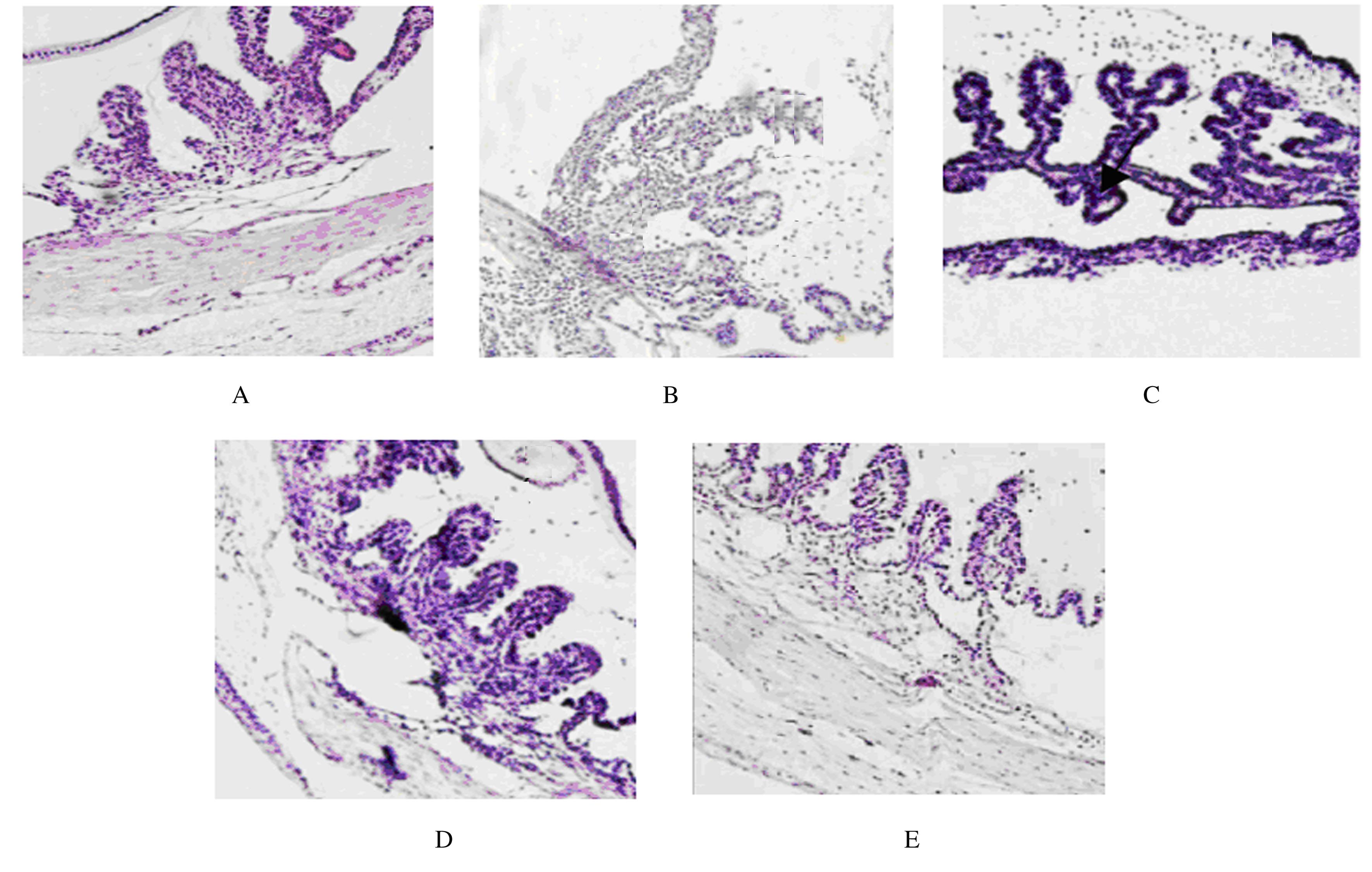
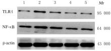
  • Received:2020-12-16 Online:2021-07-28 Published:2021-07-22
  • Contact: Miao CHU E-mail:kangkangba2828@163.com

摘要: 目的

探讨金银花提取物对脂多糖(LPS)诱导的急性前部葡萄膜炎(AAU)小鼠的抗炎作用,并阐明其作用机制。

方法

将50只BALB/c小鼠随机分为对照组,模型组,低、中和高剂量金银花提取物组,每组10只。低、中和高剂量金银花提取物组小鼠灌胃给予250、500 和 750 mg·kg-1金银花提取物,对照组和模型组小鼠均给予等体积生理盐水。连续给药15 d后,除对照组外其余各组小鼠于玻璃体腔内注射125 mg·L-1LPS建立小鼠AAU模型。采用HE染色观察小鼠眼组织病理形态表现,采用实时荧光定量PCR法(RT-qPCR)和Western blotting法检测小鼠眼组织中Toll样受体4(TLR4)及核因子κB(NF-κB)mRNA和蛋白表达水平,采用酶联免疫吸附测定法(ELISA)检测小鼠眼组织中肿瘤坏死因子α(TNF-α)、白细胞介素6(IL-6)和白细胞介素10(IL-10)水平。

结果

对照组小鼠眼组织结构完整,无炎症细胞浸润;模型组小鼠视网膜水肿,葡萄膜组织中出现大量炎症细胞浸润;与模型组比较,低、中和高剂量金银花提取物组小鼠葡萄膜组织中炎症细胞浸润不同程度减轻。与对照组比较,模型组小鼠眼组织中TLR4及NF-κB mRNA和蛋白表达水平明显升高(P<0.05),TNF-α、IL-6和IL-10水平明显升高(P<0.05);与模型组比较,低、中和高剂量金银花提取物组小鼠眼组织中TLR4及NF-κB mRNA和蛋白表达水平明显降低(P<0.05),TNF-α、IL-6和IL-10水平明显降低(P<0.05);与低剂量金银花提取物组比较,中和高剂量金银花提取物组小鼠眼组织中TLR4及NF-κB mRNA和蛋白表达水平明显降低(P<0.05),TNF-α、IL-6和IL-10水平明显降低(P<0.05);与中剂量金银花提取物组比较,高剂量金银花提取物组小鼠眼组织中TLR4及NF-κB mRNA和蛋白表达水平明显降低(P<0.05),TNF-α、IL-6和IL-10水平明显降低(P<0.05)。

结论

金银花提取物可通过TLR4/NF-κB信号通路下调炎症细胞表达,减轻AAU小鼠的眼部组织炎症反应,且呈剂量依赖性。

关键词: 金银花提取物, 急性前部葡萄膜炎, 脂多糖, 小鼠

Abstract: Objective

To investigate the anti-inflammatory effect of honeysuckle extract on lipopolysaccharide (LPS)-induced acute anterior uveitis (AAU) in the mice, and to illuminate the mechanism.

Methods

Fifty BALB/c mice were randomly divided into control group, model group, and low, medium and high doses of honeysuckle extract groups(n=10).The mice in low, medium and high doses of honeysuckle extract groups were given 250, 500 and 750 mg·kg-1 honeysuckle extract by gavage, and the mice in control group and model group were given normal saline. After administration for 15 d,the mice in other groups except control group were injected with 125 mg·L-1 LPS in the vitreous cavity to establish the AAU mouse models. HE staining was used to observe the pathomorphology of the eye tissue of the mice.Real-time fluorescence quantitative PCR(RT-qPCR) and Western blotting methods were used to detect the expression levels of Toll-like receptor 4(TLR4) and nuclear factor-κB(NF-κB) mRNA and proteins in eye tissue of the mice.ELISA was used to determine the levels of tumor necrosis factor-α (TNF-α), interleukin-6 (IL-6) and interleukin-10 (IL-10) in eye tissue of the mice.

Results

The eye tissue of mice in control group was intact and had no inflammatory cell infiltration. Retinal edema occurred and a large number of inflammatory cells infiltrated in tissue of the mice in model group. Compared with model group, the infiltration of inflammatory cells in uvea tissue of the mice in low, medium and high doses of honeysuckle extract groups were decreased to different degrees.Compared with control group, the levels of TLR4 and NF- κB mRNA and proteins in model group were significantly increased (P<0.05), while the levels of TNF-α, IL-6 and IL-10 were significantly increased (P<0.05). Compared with model group,the levels of TLR4 and NF-κB mRNA and proteins in low, medium and high doses of honeysuckle extract groups were significantly decreased (P<0.05), while the levels of TNF-α, IL-6 and IL-10 were significantly decreased (P<0.05). Compared with low dose of honeysuckle extract group,the levels of TLR4 and NF-κB mRNA and proteins in eye tissue of the mice in medium and high doses of honeysuckle extract groups were significantly decreased (P<0.05),while the levels of TNF-α, IL-6 and IL-10 were significantly decreased(P<0.05). Compared with medium dose of honeysuckle extract group,the levels of TLR4 and NF-κB mRNA and proteins in high dose of honeysuckle extract group were significantly decreased (P<0.05), while the levels of TNF-α, IL-6 and IL-10 were significantly decreased(P<0.05).

Conclusion

Honeysuckle extract can down-regulate the expression of inflammatory cells and reduce the inflammatory response of eye tissue in the AUU mice through the TLR4/NF-κB signaling pathway.

Key words: honeysuckle extract, acute anterior uveitis, lipopolysaccharide, mice, anti-inflammatory effects

中图分类号: 

  • R773.9