吉林大学学报(医学版) ›› 2021, Vol. 47 ›› Issue (4): 1038-1042.doi: 10.13481/j.1671-587X.20210431

• 方法学 • 上一篇    下一篇

原代SD大鼠主动脉内皮细胞培养方法的建立及其鉴定

唐元瑜1(),刘海琴2,马华根3   

  1. 1.福建中医药大学中医学院中医基础理论教研室,福建 福州 350122
    2.福建中医药大学 中西医结合学院,福建 福州 350122
    3.北京中医药大学中医学院金匱教研室,北京 100029
  • 收稿日期:2020-11-25 出版日期:2021-07-28 发布日期:2021-07-22
  • 通讯作者: 唐元瑜 E-mail:2422198977@qq.com
  • 作者简介:唐元瑜(1971-),男,四川省攀枝花市人,副教授,医学博士,主要从事血管性疾病基础和临床方面的研究。
  • 基金资助:
    国家自然科学基金项目(81072714);福建省科技厅自然科学基金项目(2017J01545)

Establishment of cultural method of primary SD rat aortic endothelial cells and their identification

Yuanyu TANG1(),Haiqin LIU2,Huagen MA3   

  1. 1.Teaching and Research Office of Basic Theory of Traditional Chinese Medicine,College of Traditional Chinese Medicine,Fujian University of Traditional Chinese Medicine,Fuzhou 350122,China
    2.College of Integrated Traditional Chinese and Western Medicine,Fujian University of Traditional Chinese Medicine,Fuzhou 350122,China
    3.Teaching and Research Office of Jin Gui,College of Traditional Chinese Medicine,Beijing University of Traditional Chinese Medicine,Beijing 100029,China
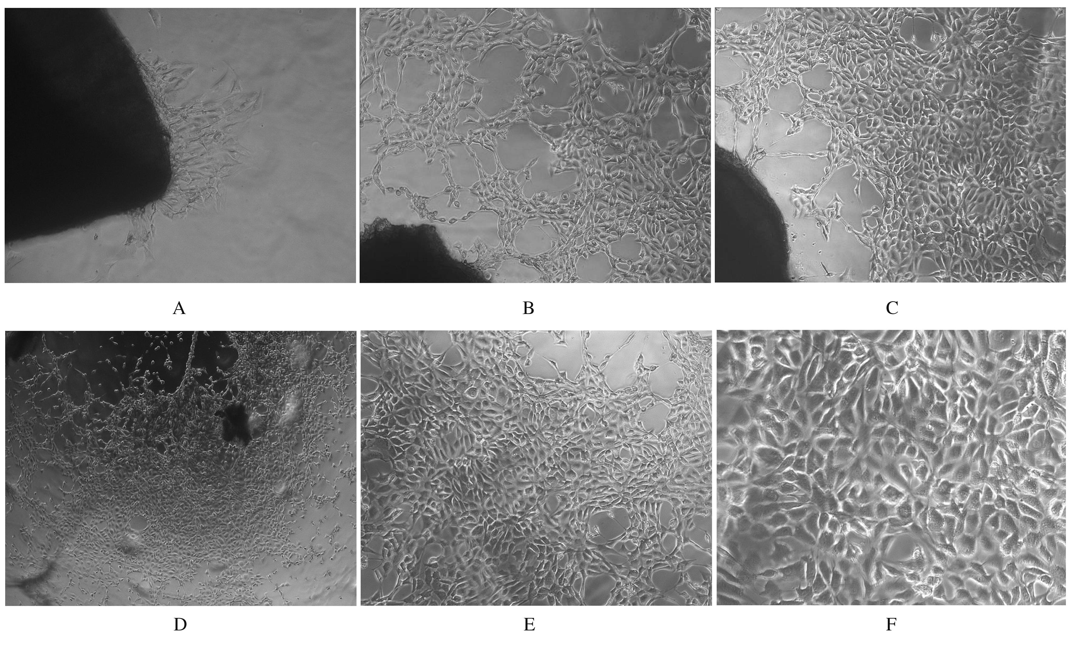
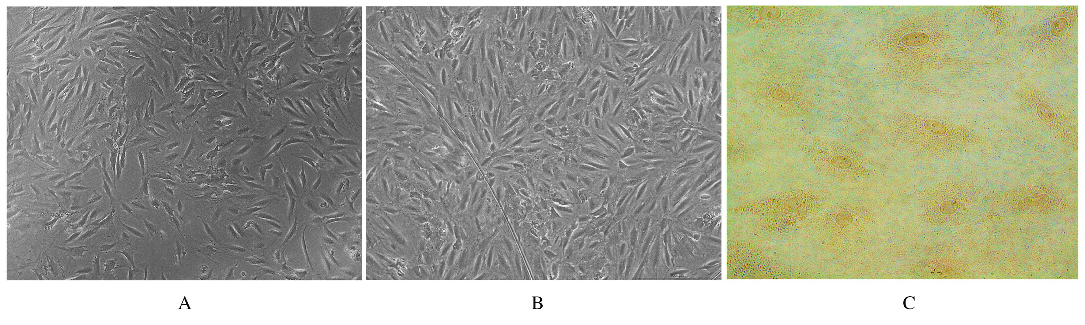
  • Received:2020-11-25 Online:2021-07-28 Published:2021-07-22
  • Contact: Yuanyu TANG E-mail:2422198977@qq.com

摘要: 目的

建立一种简便高效的原代SD大鼠主动脉内皮细胞(AECs)培养方法,为探索AECs的分子生物学特性及开展心脑血管疾病研究提供重要的载体和工具细胞。

方法

取SD大鼠1只,无菌打开胸腹腔,分离出主动脉,剔除血管外结缔组织和脂肪,将主动脉内膜外翻,切成长为1.0~1.5 mm的血管段后接种于培养瓶中,7 d后去除血管段继续培养。通过细胞形态表现观察和第Ⅷ因子(FⅧ)相关抗原免疫细胞化学染色鉴定目的细胞。

结果

接种3 d后,有少量细胞从血管段周围迁移出,形成岛屿状细胞团;10~12 d后,细胞集落逐渐融合成片,铺满瓶底,呈典型的“铺路石样”镶嵌式排列。FⅧ相关抗原免疫细胞化学染色检测,细胞的胞核和胞质淡染成棕红色,表达为阳性,阳性细胞率达98%以上。

结论

主动脉内膜外翻切段贴壁培养法能够成功分离培养出原代大鼠AECs。

关键词: 主动脉, 血管内皮细胞, 内膜外翻切段贴壁培养法, 原代培养, 免疫细胞化学染色, 大鼠,SD

Abstract: Objective

To establish a simple and efficient cultivating method of the primary SD rat aortic endothelial cells(AECs),and to provide an important carrier and tool cells for the exploration of the molecular biologial characteristics of AECs and the study of cardiovascular and cerebrovascular diseases.

Methods

One SD rat was selected, the chest was opened, and the aorta was removed in sterile condition; the connective tissue and fat outside the blood vessel were removed;the aortic tunica intima was everted and the aorta were cut into 1.0-1.5 mm vascular segments,and the vascular segments were placed in a culture bottle for primary culture. Then the vascular segments were removed 7 d later. The cultured cells were identified by morphological observation and immunocytochemical staining of factor Ⅷ(FⅧ) related antigen.

Results

After 3 d of culture, a small number of cells migrated from the vascular segments to form the island cell mass. From the 10th to 12th days, the adherent cells covered most of the culture bottle surface, and presented a typical “paving stone” mosaic arrangement. The results of immunocytochemical staining of FⅧ showed that the nucleus and the cytoplasm were brownish red,and the expression was positive; the positive cell rate was more than 98%.

Conclusion

Adherent culture method of aortic intima valgus can successfully isolate and culture the primary rat AECs.

Key words: aorta, vascular endothelial cells, intima valgus adherent culture method primary culture, immunocytochemical staining, rats,SD

中图分类号: 

  • R329.21