吉林大学学报(医学版) ›› 2021, Vol. 47 ›› Issue (6): 1462-1468.doi: 10.13481/j.1671-587X.20210616

• 基础研究 • 上一篇    下一篇

头孢曲松对蛛网膜下腔出血大鼠星形胶质细胞活化和炎症反应的影响

龚泽华1,2,刘俊杰1,2,徐继伟2,李建民1,2()   

  1. 1.华北理工大学临床医学院实验中心,河北 唐山 063000
    2.华北理工大学附属医院神经外科,河北 唐山 063000
  • 收稿日期:2021-03-25 出版日期:2021-11-28 发布日期:2021-12-14
  • 通讯作者: 李建民 E-mail:2360796486@qq.com
  • 作者简介:龚泽华(1995-),男,浙江省湖州市人,在读硕士研究生,主要从事神经损伤和神经保护方面的研究。
  • 基金资助:
    河北省卫健委医学科学研究项目(20190106);河北省卫健委高级卒中中心建设人才培养项目(361036);河北省唐山市科技局科技支撑项目(18130232a);华北理工大学青年基金项目(Z201736)

Effects of ceftriaxone on activation of astrocytes and inflammation reaction in rats with subarachnoid hemorrhage

Zehua GONG1,2,Junjie LIU1,2,Jiwei XU2,Jianmin LI1,2()   

  1. 1.Experimental Center,College of Clinical Medicine,North China University of Science and Technology,Tangshan 063000,China
    2.Department of Neurosurgery,Affiliated Hospital of North China University of Science and Technology,Tangshan 063000,China
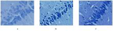
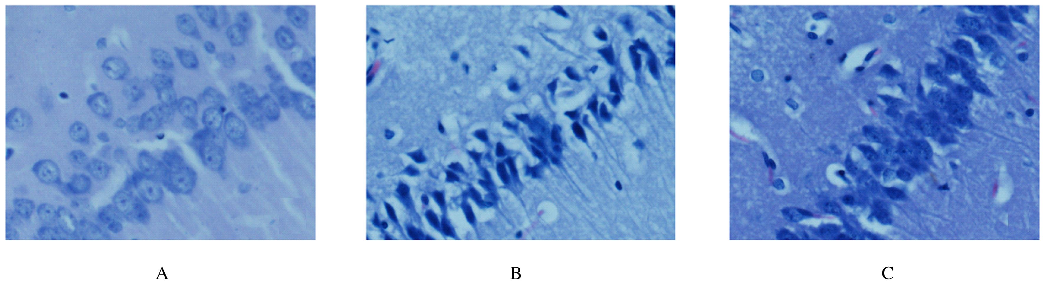
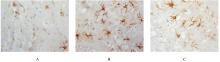
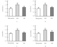
  • Received:2021-03-25 Online:2021-11-28 Published:2021-12-14
  • Contact: Jianmin LI E-mail:2360796486@qq.com

摘要: 目的

探讨头孢曲松(CEF)对蛛网膜下腔出血(SAH)大鼠的神经保护作用,并阐明其可能的作用机制。

方法

将48只清洁级雄性SD大鼠随机分为假手术组、SAH组和CEF组,每组16只。采用改良后的血管内穿刺法制备SAH大鼠模型,CEF组大鼠术前5 d开始腹腔注射给药,药物剂量为200 mg·kg-1。模型建立24 h后,对各组大鼠进行神经功能评分,HE染色法观察各组大鼠脑组织神经细胞形态表现并计算其神经细胞坏死数,免疫组织化学染色检测各组大鼠脑组织中星形胶质细胞的活化数,ELISA法检测各组大鼠脑组织中白细胞介素1β(IL-1β)、白细胞介素33(IL-33)、白细胞介素6(IL-6)和肿瘤坏死因子α(TNF-α)水平。

结果

与假手术组比较,SAH组大鼠组神经功能评分明显降低(P<0.05);与SAH组比较,CEF组大鼠神经功能评分明显升高(P<0.05)。HE染色,假手术组大鼠神经细胞分布排列有序、形态规整,细胞匀称,细胞核居中,染色清楚;SAH组大鼠的神经细胞排列杂乱,形态不规则,呈锥状,胞核收缩、破裂和溶解;CEF组大鼠脑组织神经细胞结构较完整,胞核固缩减轻。与假手术组比较,SAH组大鼠神经细胞坏死数明显增加(P<0.05);与SAH组比较,CEF组大鼠神经细胞坏死数明显减少(P<0.05)。免疫组织化学染色,活化的星形胶质细胞形态多呈“分支状”,细胞体较小,以细胞质染色为主,突起增粗延长且长短不一。与假手术组比较,SAH组大鼠星形胶质细胞活化数明显增多(P<0.05);与SAH组比较,CEF组大鼠星形胶质细胞活化数明显减少(P<0.05)。ELISA法检测,与假手术组比较,SAH组大鼠脑组织中IL-1β、IL-33、IL-6和TNF-α水平明显升高(P<0.05);与SAH组比较,CEF组大鼠脑组织中IL-1β、IL-33、IL-6和TNF-α水平明显降低(P<0.05)。

结论

CEF能够减轻SAH大鼠脑组织神经细胞的坏死,提高大鼠神经功能评分,具有神经保护作用,其机制可能与抑制星形胶质细胞活化和减轻炎症反应有关。

关键词: 蛛网膜下腔出血, 头孢曲松, 炎症反应, 星形胶质细胞

Abstract: Objective

To investigate the neuroprotective effect of ceftriaxone (CEF) in the rats with subarachnoid hemorrhage (SAH) ,and to clarify its possible mechanism.

Methods

A total of 48 male SD rats were randomly divided into sham operation group, SAH group, and CEF group, and there were 16 rats in each group. The SAH rat model was established by improved endovascular puncture. The rats in CEF group were injected intraperitoneally 5 d before the establishment of SAH model, and the drug dose of CEF was 200 mg·kg-1.After the establishment of SAH model for 24 h, the neurological function scores of the rats in various groups were evaluated. The morphology of nerve cells in brain tissue of the rats in various groups was observed by HE staining, and the numbers of necrosis nerve cells were calculated. The numbers of activated astrocytes in brain tissue of the rats in various groups were examined by immunohistochemistry. The levels of interleukin-1β(IL-1β), interleukin-33(IL-33), interleukin-6(IL-6), and tumor necrosis factor-α(TNF-α) in brain tissue of the rats in various groups were detected by ELISA method.

Results

Compared with sham operation group, the neurological function score of the rats in SAH group was significantly decreased (P<0.05). Compared with SAH group, the neurological function score of the rats in CEF group was significantly increased (P<0.05). The HE staining results showed that the distribution of nerve cells in brain tissue of the rats in sham operation group was orderly and regular,the cells were symmetrical, and the nucleus was in the middle and stained clearly; the nerve cells in brain tissue of the rats in SAH group were disordered, irregular and cone-shaped, and the nuclear were shrinkaged, ruptured and dissolved; in CEF group, the structure of nerve cells in brain tissue was intact and the nuclear pyknosis was alleviated. Compared with sham operation group, the number of necrosis nerve cells in brain tissue of the rats in SAH group was significantly increased (P<0.05);compared with SAH group, the number of necrosis nerve cells in brain tissue of the rats in CEF group was significantly decreased (P<0.05). The immunohistochemical staining results showed that most of the activated astrocytes were “branched” in shape, with small cell bodies, mainly cytoplasmic staining, and the processes were thickened and lengthened with different lengths. Compared with sham operation group, the number of activated astrocytes in brain tissue of the rats in SAH group was significantly increased (P<0.05); compared with SAH group, the number of activated astrocytes in brain tissue of the rats in CEF group was significantly decreased (P<0.05).The ELISA assay results showed that the levels of IL-1β, IL-33, IL-6 and TNF-α in brain tissue of the rats in SAH group were significantly higher than those in sham operation group (P<0.05);compared with SAH group, the levels of IL-1β, IL-33, IL-6,and TNF-α in brain tissue of the rats in CEF group were significantly decreased (P<0.05).

Conclusion

CEF can reduce the necrosis of nerve cells in brain tissue of the rats with SAH, improve the neurological function score of the rats, and has a neuroprotective effect; its mechanism may be related to inhibiting the activation of astrocytes and alleviating the inflammatory reaction.

Key words: subarachnoid hemorrhage, ceftriaxone, inflammatory reaction, astrocytes

中图分类号: 

  • R743.35