吉林大学学报(医学版) ›› 2022, Vol. 48 ›› Issue (2): 348-355.doi: 10.13481/j.1671-587X.20220211

• 基础研究 • 上一篇    下一篇

胡桃醌对宫颈癌细胞增殖的抑制作用及其机制

赵行宇1,杨欣1,朱志华1,何涵2,宋梓桐1,张巍1()   

  1. 1.吉林医药学院生化教研室,吉林 吉林 132013
    2.延边大学基础医学院生化教研室,吉林 延吉 133000
  • 收稿日期:2021-06-21 出版日期:2022-03-28 发布日期:2022-05-10
  • 通讯作者: 张巍 E-mail:jlmmczw@163.com
  • 作者简介:赵行宇(1970-),男,安徽省五河县人,副教授,医学硕士,主要从事抗肿瘤药物方面的研究。
  • 基金资助:
    吉林省科技厅科技发展计划项目(20190701062GH);吉林省教育厅“十三五”科学技术研究项目(JJKH20200453KJ)

Inhibitory effect of juglone on proliferation of cervical cancer cells and its mechanism

Xingyu ZHAO1,Xin YANG1,Zhihua ZHU1,Han HE2,Zitong SONG1,Wei ZHANG1()   

  1. 1.Department of Biochemistry,Jilin Medical College,Jilin 13201,China
    2.Department of Biochemistry,School of Basic Medical Sciences,Yanbian University,Ynabian 133000,China
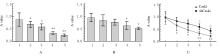
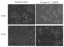
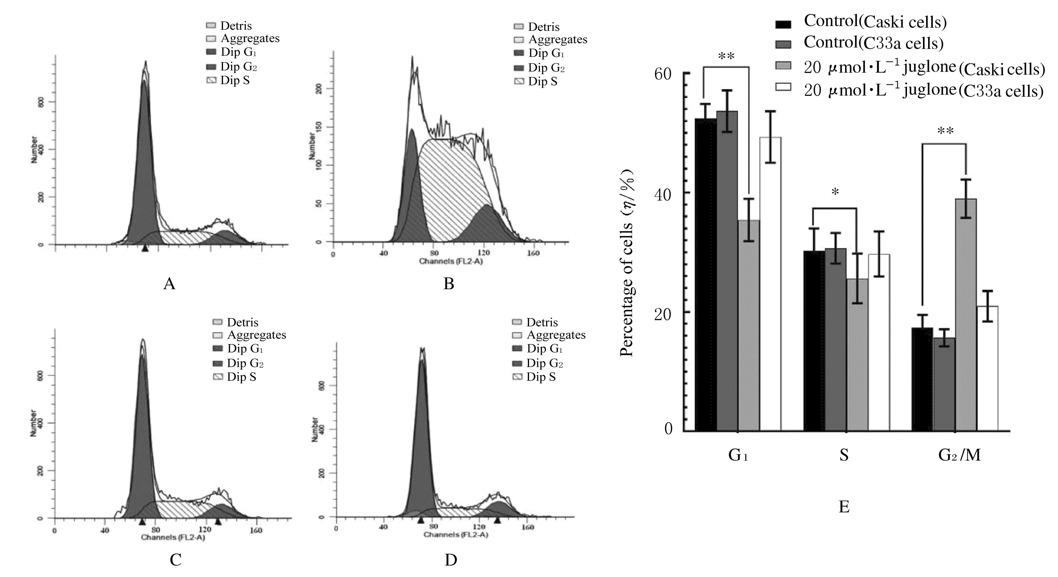
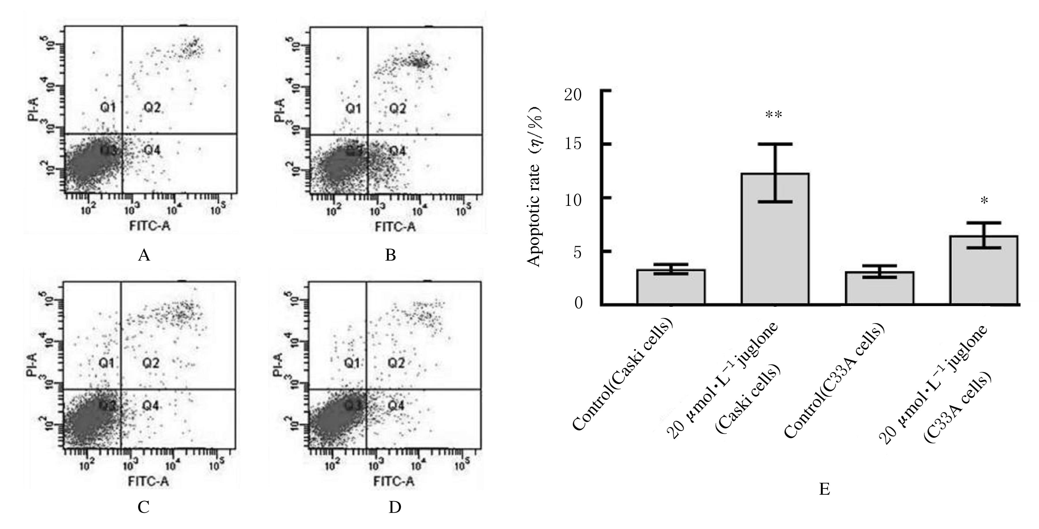
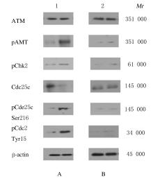
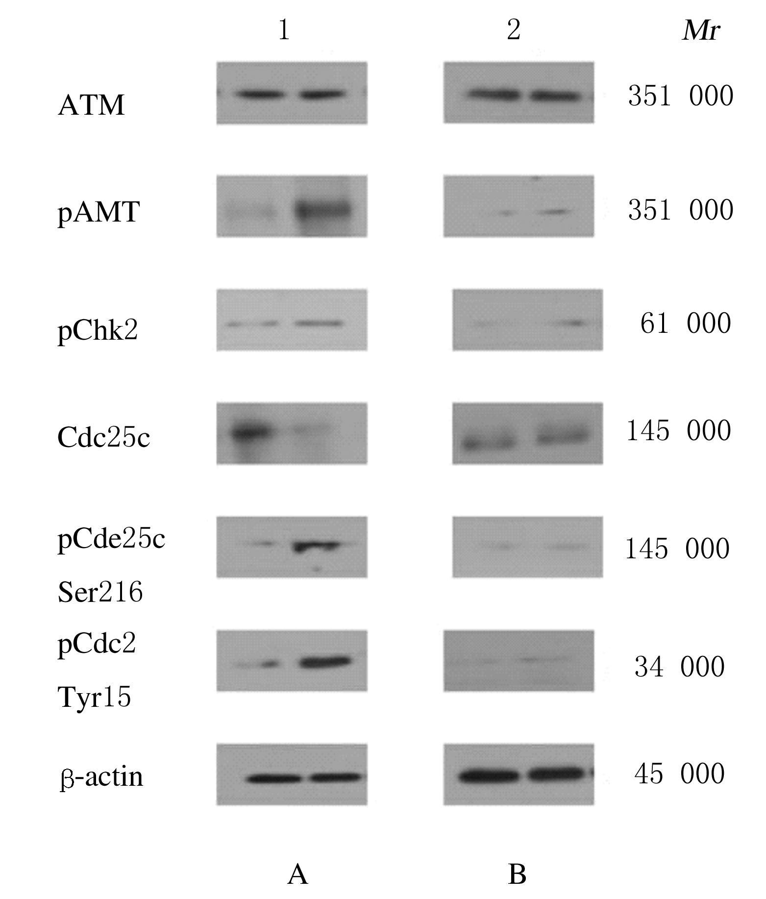
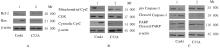
  • Received:2021-06-21 Online:2022-03-28 Published:2022-05-10
  • Contact: Wei ZHANG E-mail:jlmmczw@163.com

摘要: 目的

比较胡桃醌对人乳头瘤病毒(HPV)阳性宫颈癌Caski细胞、HPV阴性C33A细胞和敲减脯氨酰顺反异构酶1(Pin1)基因的Caski细胞(shCaski细胞)增殖的抑制作用,探讨其可能的作用机制。

方法

构建表达Pin1 shRNA慢病毒载体用于感染细胞,筛选得到稳定的shCaski细胞。取对数生长期的Caski、shCaski和C33A细胞,分为正常对照组、10、20、50和100 μmol·L-1胡桃醌组,胡桃醌处理24 h后,采用MTT法检测各组的细胞增殖活性。取对数生长期的Caski细胞和C33A细胞,分为对照组和20 μmol·L-1胡桃醌组,各组细胞处理24 h后,流式细胞术检测各组不同周期细胞百分率和早期凋亡率,Hoechst33258荧光染色观察各组细胞形态表现,Western blotting法检测各组细胞周期和凋亡相关蛋白表达水平。

结果

MTT实验,与正常对照组比较,不同浓度胡桃醌组Caski细胞增殖活性明显降低(P<0.05或P<0.01); 50和100 μmol·L-1胡桃醌组C33A和shCaski细胞增殖活性明显降低(P<0.05)。Hoechst 33258染色,与对照组比较,20 μmol·L-1胡桃醌组Caski细胞和C33A细胞中均可观察到核碎解增加,Caski细胞核碎解数量较C33A细胞多。流式细胞术检测,与对照组比较,20 μmol·L-1胡桃醌组Caski细胞G2期细胞百分率明显升高(P<0.01),20 μmol·L-1胡桃醌组C33A细胞G2期细胞百分率差异无统计学意义(P>0.05);与对照组比较,20 μmol·L-1胡桃醌组Caski细胞早期凋亡率明显升高(P<0.01),20 μmol·L-1胡桃醌组C33A细胞早期凋亡率差异无统计学意义(P>0.05);Western blotting法检测,与C33A细胞比较,Caski细胞中Pin1蛋白表达量明显升高;胡桃醌组Caski细胞和shCaski细胞中Pin1蛋白表达量明显低于对照组;与对照组比较,胡桃醌组C33A细胞中细胞周期相关蛋白表达水平明显改变;Caski细胞中磷酸化ATM(Patm)、磷酸化细胞周期检查点激酶2(pChk2)、216位丝氨酸磷酸化细胞分裂周期蛋白25c(pCdc25c Ser216)和pCdc25c Tyr216蛋白表达量升高,磷酸化细胞分裂周期蛋白25c(pCdc25c)蛋白表达量降低,Cdk1蛋白表达量变化不明显;与对照组比较,胡桃醌处理组Caski细胞中Bcl-2蛋白和线粒体CytC蛋白表达量降低,胞质CytC、Bax、Cleaved Caspase-3和Cleaved PARP蛋白表达量升高。

结论

胡桃醌对HPV阳性宫颈癌细胞增殖抑制及促凋亡作用效果均强于HPV阴性细胞,其机制可能与胡桃醌特异性抑制Pin1基因有关。

关键词: 胡桃醌, 人乳头瘤病毒, 宫颈癌细胞, Pin1基因

Abstract: Objective

To compare the inhibitory effects of juglone on the proliferation of human papilloma virus(HPV)-positive cervical cancer Caski cells, negative C33A cells and Caski cells knocked down the Pin1 gene(shCaski cells),and to explore its possible mechanism.

Methods

A lentiviral vector expressing Pin1 shRNA was constructed and used to infect the cells, and shCaski cells were obtained. The Caski, shCaski and C33A cells in the logarithmic growth phase were selected and divided into normal control group and 10, 20, 50 and 100 μmol·L-1 juglone groups. After juglone treatment for 24 h, the cell proliferation activities in various groups were detected by MTT method. The Caski cells and C33A cells in the logarithmic growth phase were selected and divided into control group and 20 μmol·L-1 juglone group. After 24 h of treatment of the cells in each group, the percentages of cells in different cell cycles and the early apoptotic rates in various groups were detected by flow cytometry. Hoechst33258 fluorescence staining was used to observe the morphology of nucleus; Western blotting method was used to detect the expressions of cell cycle and apoptosis-related proteins.

Results

The MTT experiment showed that the proliferation activities of the Caski cells in different concentrations of juglone groups were significantly decreased (P<0.05or P<0.01) compared with normal control group, while the proliferation activities of C33A and shCaski cells in 50 and 100 μmol·L-1 juglone groups were significantly decreased(P<0.05).The results of Hoechst 33258 staining showed that compared with control group, both the Caski cells and C33A cells in 20 μmol·L-1 juglone group showed increased nuclear fragmentation, and the number of nuclei fragmentation of Caski cells was more than that of C33A cells. The results of flow cytometry showed that compared with control group, the percentage of Caski cells in G2 phase in 20μmol·L-1 juglone group was increased significantly (P<0.01); while there was no significant difference in the percentage of C33A cells in G2 phase in 20 μmol·L-1 juglone group(P>0.05);compared with control group,the early apoptotic rate of Caski cells in 20 μmol·L-1 juglone group was increased significantly (P<0.01); while the early apoptotic rate of C33A cells in 20 μmol·L-1 juglone group was also increased, but there was no significantly difference(P>0.05). The Western blotting results showed that compared with the C33A cells,the expression amount of Pin1 protein in the Caski cells was more higher; and the expression amount of Pin1 protein in Caski and shCaski cells in 20 μmol·L-1 juglone group were decreased. Compared with control group, the expression amounts of cell cycle-related proteins in the C33A cells in 20 μmol·L-1 juglone group did not change significantly; the expression amount of pATM, pChk2, pCdc25c Ser216 and pCdc25c Tyr216 in the Caski cells in 20 μmol·L-1 juglone group were increased, while the expression amount of pCdc25c protein was decreased in 20 μmol·L-1 juglone group;the expression amount of Cdk1 protein didn’t change obviously. Compared with control group, the amounts of Bcl-2 and mitochondrial CytC Caski cells in 20 μmol·L-1 juglone group were decreased, but the expression amounts of cytoplasmic CytC,Bax,Cleaved Caspase-3 and Cleaved PARP proteins in the Caski cells in 20 μmol·L-1 juglone group were increased.

Conclusion

Juglone has stronger inhibitory and pro-apoptotic effects in the HPV-positive cervical cancer cells than negative cells; its mechanism may be related to the specific inhibition of Pin1 gene by juglone.

Key words: Juglone, Human papilloma virus, Cervical cancer cells, Pin1 gene

中图分类号: 

  • R737.33