吉林大学学报(医学版) ›› 2022, Vol. 48 ›› Issue (4): 1003-1009.doi: 10.13481/j.1671-587X.20220421

• 临床研究 •    

IL-17A在非小细胞肺癌组织中的表达及其通过NF-κB信号通路对VEGF表达的调控作用

何程远1,杨红宇2,谭钰晶3,苏杭3,李泓澍1,历春1()   

  1. 1.北华大学基础医学院免疫学教研室,吉林 吉林 132013
    2.吉林省吉林市化工医院病理科,吉林 吉林 132001
    3.北华大学理学院 吉林省中药生物技术创新中心,吉林 吉林 132013
  • 收稿日期:2021-10-19 出版日期:2022-07-28 发布日期:2022-07-26
  • 通讯作者: 历春 E-mail:lichunjl@126.com
  • 作者简介:何程远(1997-),男,吉林省吉林市人,在读硕士研究生,主要从事肿瘤免疫学方面的研究。
  • 基金资助:
    吉林省科技厅自然科学基金自由探索重点项目(YDZJ202101ZYTS089);吉林省卫健委科技项目(2020J017);北华大学研究生创新项目(2021018)

Expression of IL-17A in non-small cell lung cancer tissue and its regulation on VEGF expression via NF-κB signaling pathway

Chengyuan HE1,Hongyu YANG2,Yujing TAN3,Hang SU3,Hongshu LI1,Chun LI1()   

  1. 1.Department of Immunology,College of Basic Medical Sciences,Beihua University,Jilin 132013,China
    2.Department of Pathology,Jilin Chemical Technology Hospital,Jilin Province,Jilin 132001,China
    3.Jilin Province Chinese Medicine Biotechnology Innovation Center,College of Sciences,Beihua University,Jilin 132013,China
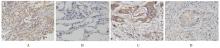
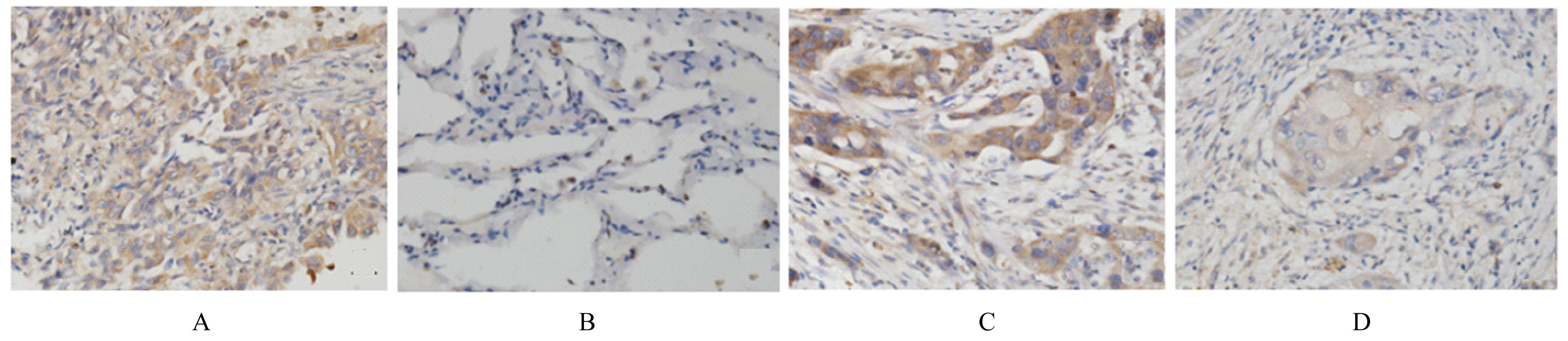
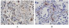
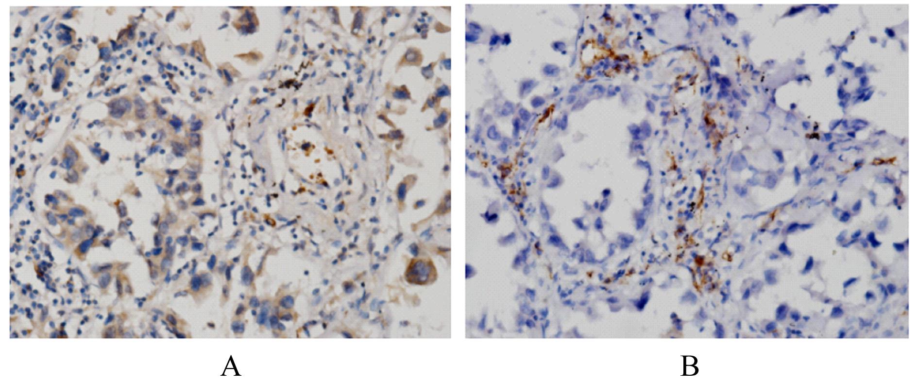
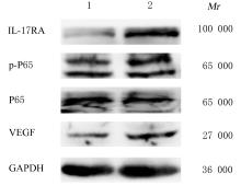
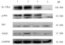
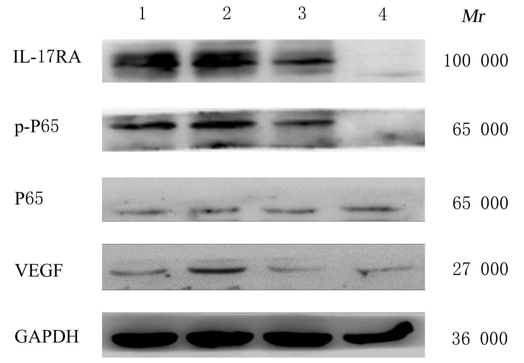
  • Received:2021-10-19 Online:2022-07-28 Published:2022-07-26
  • Contact: Chun LI E-mail:lichunjl@126.com

摘要: 目的

探讨白细胞介素17A(IL-17A) 在非小细胞肺癌 (NSCLC)发生发展中的作用及其通过核转录因子κB(NF-κB)信号通路对血管内皮生长因子 (VEGF)的调控作用,阐明其相关机制。

方法

收集NSCLC术后石蜡标本55例,免疫组织化学染色法检测正常肺组织和NSCLC组织中IL-17A和CD31的表达,分析IL-17A阳性表达率与NSCLC患者临床病理参数的关系,Pearson相关分析法分析IL-17A表达与CD31标记的微血管密度(MVD)的相关性。选取对数生长期的人肺腺癌A549细胞,将A549细胞分为对照组(A549细胞)、外源性重组人IL-17A(rhIL-17A)组、BAY11-7082组(NF-κB信号通路抑制剂)和联合组(rhIL-17A+BAY11-7082),Western blotting法检测各组A549细胞中IL-17受体A(IL-17RA)、P65、磷酸化P65(p-P65)和VEGF蛋白表达水平,MTT法检测各组A549细胞培养上清液作用人脐静脉内皮细胞(HUVECs) 48 h后各组细胞增殖活性。

结果

NSCLC组织中IL-17A阳性表达率明显高于正常肺组织(P<0.05),低分化组NSCLC患者IL-17A阳性表达率明显高于高-中分化组(P<0.05),NSCLC组织中IL-17A表达与MVD呈正相关关系(r=0.329,P<0.05)。与对照组比较,rhIL-17A组细胞中IL-17RA、p-P65和VEGF蛋白表达水平明显升高(P<0.05),BAY11-7082组细胞中IL-17RA、p-P65和VEGF表达水平明显降低 (P<0.01);与rhIL-17A组比较,联合组细胞中IL-17RA、p-P65和VEGF表达水平明显降低(P<0.05)。各组培养上清液作用HUVECs后,与对照组比较,rhIL-17A组细胞增殖活性明显升高 (P<0.05),BAY11-7082组细胞增殖活性明显降低(P<0.05);与rhIL-17A组比较,联合组细胞增殖活性明显降低(P<0.05)。

结论

IL-17A在低分化NSCLC细胞中高表达,其可通过NF-κB信号通路调控VEGF的表达进而促进HUVECs的增殖。

关键词: 白细胞介素17A, 非小细胞肺癌, 核转录因子κB, 血管内皮生长因子, 人脐静脉内皮细胞

Abstract: Objective

To investigate the role of interleukin-17A (IL-17A) in the occurrence and development of non-small cell lung cancer (NSCLC) and its regulation on vascular endothelial growth factor (VEGF) through the nuclear factor-κB (NF-κB) signaling pathway, and to elucidate their related mechanisms.

Methods

A total of fifty-five postoperative paraffin-embedded specimens of the patients with NSCLC were collected. The expressions of IL-17A and CD31 in normal lung tissue and NSCLC tissue were detected by immunohistochemical staining, the correlations between the positive expression rate of IL-17A and the clinicopathological parameters of NSCLC were analyzed, and the correlation between the expressions of IL-17A and CD31-marked microvessel density (MVD) was analyzed by Pearson correlation analysis. The human lung adenocarcinoma A549 cells at logarithmic growth stage were selected and divided into control group (A549 cells), exogenous recombinant human IL-17A (rhIL-17A) group, BAY11-7082 (NF-κB signaling pathway inhibitor) group and combination group (rhIL-17A+BAY11-7082). Western blotting method was performed to detect the expression levels of IL-17 receptor A (IL-17RA), P65,p-P65 and VEGF proteins in the cells in various groups. The MTT method was used to detect the proliferation abilities of human umbilical vein endothelial cells (HUVECs) in various groups after treated with the A549 cell culture supernatant for 48 h.

Results

The positive expression rate of IL-17A in NSCLC tissue was significantly higher than that in normal lung tissue (P<0.05),the positive expression rate of IL-17A in poor-differentiation group was significantly higher than that in well-moderate differentiation group (P<0.05), and the expression of IL-17A was positively correlated with the MVD in NSCLC tissue (r=0.329, P<0.05).Compared with control group, the expression levels of IL-17RA, p-P65 and VEGF proteins in the cells in rhIL-17A group were increased (P<0.05), and the expression levels of IL-17RA, p-P65 and VEGF proteins in the cells in BAY11-7082 group were decreased (P<0.01). Compared with rhIL-17A group, the expression levels of IL-17RA, p-P65 and VEGF proteins in the A549 cells in combination group were significantly decreased (P<0.05). After treated with A549 cell culture supernatant, compared with control group, the proliferation ability of the HUVEC in rhIL-17A group was increased (P<0.05),and the proliferation ability in the cells in BAY11-7082 group was decreased (P<0.05). Compared with the rhIL-17A group, the proliferation ability of the HUVECs in combination group was significantly decreased (P<0.05).

Conclusion

IL-17A is highly expressed in poorly differentiated NSCLC cells, and it can promote the proliferation of HUVECs by regulating the expression of VEGF via NF-κB signaling pathway.

Key words: Interleukin-17A, Non-small cell lung cancer, Nuclear transcription factor-kappa B, Vascular endothelial growth factor, Human umbilical vein endothelial cells

中图分类号: 

  • R734.2