| 1 |
ZHANG Y Q, ZHANG X Y, LU M Y, et al. Ceramide-1-phosphate and its transfer proteins in eukaryotes[J]. Chem Phys Lipids, 2021, 240: 105135.
|
| 2 |
MISHRA S K, GAO Y G, DENG Y B, et al. CPTP: a sphingolipid transfer protein that regulates autophagy and inflammasome activation[J]. Autophagy, 2018, 14(5): 862-879.
|
| 3 |
SIMANSHU D K, KAMLEKAR R K, WIJESINGHE D S, et al. Non-vesicular trafficking by a ceramide-1-phosphate transfer protein regulates eicosanoids[J]. Nature, 2013, 500(7463): 463-467.
|
| 4 |
GOMEZ-LARRAURI A, PRESA N, DOMINGUEZ-HERRERA A, et al. Role of bioactive sphingolipids in physiology and pathology[J]. Essays Biochem, 2020, 64(3): 579-589.
|
| 5 |
MACEYKA M, SPIEGEL S. Sphingolipid metabolites in inflammatory disease[J]. Nature, 2014, 510(7503): 58-67.
|
| 6 |
WANG J, LI C, JIANG Y J, et al. Effect of ceramide-1-phosphate transfer protein on intestinal bacterial translocation in severe acute pancreatitis[J]. Clin Res Hepatol Gastroenterol, 2017, 41(1): 86-92.
|
| 7 |
ZHANG Y Q, JI S Y, ZHANG X Y, et al. Human CPTP promotes growth and metastasis via sphingolipid metabolite ceramide and PI4KA/AKT signaling in pancreatic cancer cells[J]. Int J Biol Sci, 2022, 18(13): 4963-4983.
|
| 8 |
VASAIKAR S V, STRAUB P, WANG J, et al. LinkedOmics: analyzing multi-omics data within and across 32 cancer types[J]. Nucleic Acids Res, 2018, 46(D1): D956-D963.
|
| 9 |
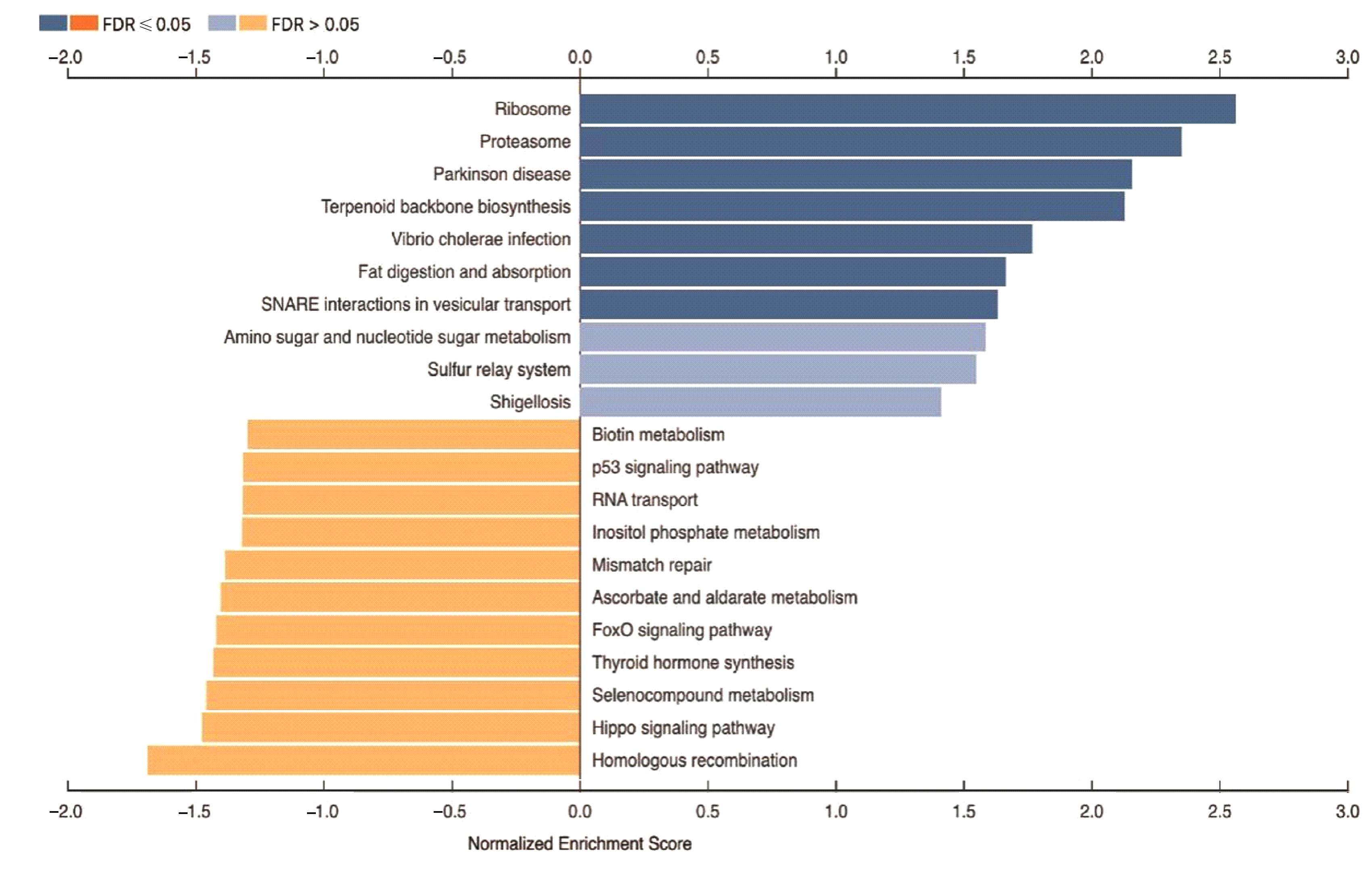
CHUN K H. Discovery of cellular RhoA functions by the integrated application of gene set enrichment analysis[J]. Biomol Ther, 2022, 30(1): 98-116.
|
| 10 |
ZHAO L Z, LIANG J S, ZHONG W, et al. Expression and prognostic analysis of BGN in head and neck squamous cell carcinoma[J]. Gene, 2022, 827: 146461.
|
| 11 |
AUPÉRIN A. Epidemiology of head and neck cancers: an update[J]. Curr Opin Oncol, 2020, 32(3): 178-186.
|
| 12 |
WARNAKULASURIYA S. Living with oral cancer: epidemiology with particular reference to prevalence and life-style changes that influence survival[J]. Oral Oncol, 2010, 46(6): 407-410.
|
| 13 |
BAUMEISTER P, ZHOU J F, CANIS M, et al. Epithelial-to-mesenchymal transition-derived heterogeneity in head and neck squamous cell carcinomas[J]. Cancers, 2021, 13(21): 5355.
|
| 14 |
ZHANG S Z, XIE L, SHANG Z J. Burden of oral cancer on the 10 most populous countries from 1990 to 2019: estimates from the global burden of disease study 2019[J].Int J Environ Res Public Health,2022,19(2): 875.
|
| 15 |
BRAY F, FERLAY J, SOERJOMATARAM I, et al. Global cancer statistics 2018: GLOBOCAN estimates of incidence and mortality worldwide for 36 cancers in 185 countries[J]. CA Cancer J Clin, 2018, 68(6): 394-424.
|
| 16 |
ASTHANA S, LABANI S, KAILASH U, et al. Association of smokeless tobacco use and oral cancer: a systematic global review and meta-analysis[J]. Nicotine Tob Res, 2019, 21(9): 1162-1171.
|
| 17 |
SAXENA R, PRASOODANAN P K V, GUPTA S V, et al. Assessing the effect of smokeless tobacco consumption on oral microbiome in healthy and oral cancer patients[J]. Front Cell Infect Microbiol, 2022, 12: 841465.
|
| 18 |
WEBER M, LUTZ R, OLMOS M, et al. Beyond PD-L1-identification of further potential therapeutic targets in oral cancer[J]. Cancers, 2022, 14(7): 1812.
|
| 19 |
LIU S, SHI C Y, HOU X R, et al. Transcriptional and H3K27ac related genome profiles in oral squamous cell carcinoma cells treated with metformin[J]. J Cancer, 2022, 13(6): 1859-1870.
|
| 20 |
BRENNAN P A, DYLGJERI F, COLETTA R D,et al. Surgical tumour margins and their significance in oral squamous cell carcinoma[J]. J Oral Pathol Med, 2022, 51(4): 311-314.
|
| 21 |
STARZYŃSKA A, SOBOCKI B K, ALTERIO D. Current challenges in head and neck cancer management[J]. Cancers, 2022, 14(2): 358.
|
| 22 |
FAN Z L, YANG L Y, ZHANG N, et al. NEFL promotes invasion and migration of esophageal squamous carcinoma cells via the EGFR/AKT/S6 pathway[J]. Hereditas, 2022, 44(4): 322-334.
|
| 23 |
QIU H Q, ZHANG L, WANG D Z, et al. ZNF488 promotes the invasion and migration of pancreatic carcinoma cells through the akt/mTOR pathway[J]. Comput Math Methods Med, 2022, 2022: 4622877.
|
| 24 |
ZHANG Y H, MOHIBI S, VASILATIS D M, et al. Ferredoxin reductase and p53 are necessary for lipid homeostasis and tumor suppression through the ABCA1-SREBP pathway[J]. Oncogene, 2022, 41(12): 1718-1726.
|
| 25 |
WANG M X, ATTARDI L D. A balancing act: p53 activity from tumor suppression to pathology and therapeutic implications[J]. Annu Rev Pathol, 2022, 17: 205-226.
|
| 26 |
李 震, 刘江凯. p53信号通路调控铁死亡在肝细胞癌中的作用机制[J].临床肝胆病杂志,2023,39(4): 956-960.
|
| 27 |
WANG W, XIAO L N, PAN D, et al. ASF1B enhances migration and invasion of lung cancers cell via regulating the P53-mediated epithelial-mesenchymal transformation (EMT) signaling pathway[J]. Neoplasma, 2022, 69(2): 361-369.
|
| 28 |
MARTINEZ J D, MO Q X, XU Y X, et al. Common genomic aberrations in mouse and human breast cancers with concurrent P53 deficiency and activated PTEN-PI3K-AKT pathway[J]. Int J Biol Sci, 2022, 18(1): 229-241.
|
 )
)