吉林大学学报(医学版) ›› 2022, Vol. 48 ›› Issue (5): 1324-1332.doi: 10.13481/j.1671-587X.20220528

• 临床研究 • 上一篇    

lncRNA GHET1通过Wnt/β-catenin信号通路对子痫前期滋养层细胞生物学行为的影响

黄晓妹,王洪伟(),韩贞艳,韦秋园,黄素静   

  1. 海南医学院第二附属医院产科,海南 海口 570100
  • 收稿日期:2021-10-30 出版日期:2022-09-28 发布日期:2022-11-15
  • 通讯作者: 王洪伟 E-mail:whw780920@163.com
  • 作者简介:黄晓妹(1987-),女,海南省海口市人,主治医师,医学学士,主要从事围产医学和妊娠代谢内分泌疾病方面的研究。
  • 基金资助:
    海南省科技厅基础与应用基础研究计划项目(818QN098)

Effects of lncRNA GHET1 on biological behaviors of trophoblast cells in preeclampsia through Wnt/β-catenin signaling pathway

Xiaomei HUANG,Hongwei WANG(),Zhenyan HAN,Qiuyuan WEI,Sujing HUANG   

  1. Department of Obstetrics,Second Affiliated Hospital,Hainan Medical College,Haikou 570100,China
  • Received:2021-10-30 Online:2022-09-28 Published:2022-11-15
  • Contact: Hongwei WANG E-mail:whw780920@163.com

摘要:

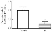
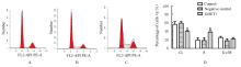
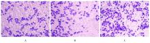
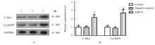
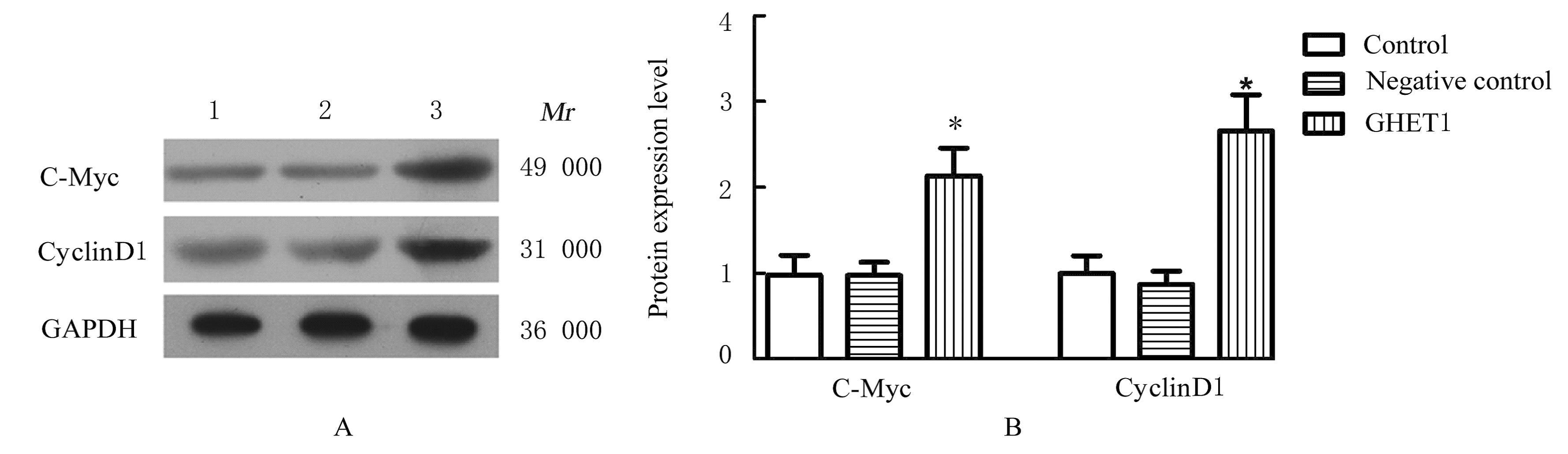
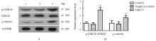
目的 探讨子痫前期(PE)患者胎盘组织中长链非编码RNA胃癌高表达转录本1(lncRNA GHET1)的表达及其对滋养层细胞增殖、细胞周期进展和侵袭能力的影响,并阐明其作用机制。 方法 30名孕产妇分为正常孕产妇组(正常组)15例和PE孕产妇组(PE组)15例,HE染色观察2组研究对象胎盘组织病理形态表现。体外培养滋养层HTR-8细胞,转染过表达lncRNA GHET1及对照序列质粒,并将滋养层细胞分为对照组(常规培养)、GHET1组(转染过表达lncRNA GHET1质粒)和阴性对照组(转染阴性对照序列质粒)。实时荧光定量PCR(RT-qPCR)法检测2组研究对象胎盘组织和各组HTR-8细胞中lncRNA GHET1 mRNA表达水平,CCK-8法检测各组HTR-8细胞增殖率,流式细胞术检测各组不同细胞周期HTR-8细胞百分率,Transwell小室实验检测各组HTR-8细胞侵袭能力,Western blotting法检测各组HTR-8细胞中细胞性骨髓细胞瘤病病毒癌基因(c-Myc)、细胞周期素D1(cyclinD1)、上皮细胞-间充质转化(EMT)相关蛋白E-钙黏蛋白(E-cadherin)和波形蛋白(Vimentin)蛋白表达 水平及Wnt/β-catenin信号通路中磷酸化糖原合成酶激酶3β(p-GSK3β)/糖原合成酶激酶3β(GSK3β)比值和β-连环蛋白(β-catenin)蛋白表达水平。 结果 与正常组比较,PE组患者胎盘组织绒毛发育不良,数量减少,绒毛内及间质血管分布紊乱,血管壁出现纤维素样坏死,钙化区域及绒毛上合体结节增加。与正常组比较,PE组患者胎盘组织中lncRNA GHET1 mRNA表达水平明显降低(P<0.05)。与对照组比较,GHET1组HTR-8细胞中lncRNA GHET1 mRNA表达水平明显升高(P<0.05),阴性对照组HTR-8细胞中lncRNA GHET1 mRNA表达水平差异无统计学意义(P>0.05)。与对照组比较,GHET1组HTR-8细胞增殖率、S期细胞百分率和侵袭细胞数明显升高(P<0.05),阴性对照组上述指标差异无统计学意义(P>0.05)。与对照组比较,GHET1组HTR-8细胞中c-Myc、cyclinD1、Vimentin和β-catenin蛋白表达水平及p-GSK3β/GSK3β比值均明显升高(P<0.05),E-cadherin蛋白表达水平明显降低(P<0.05),阴性对照组上述指标差异无统计学意义(P>0.05)。 结论 过表达lncRNA GHET1可能通过Wnt/β-catenin信号通路促进滋养层细胞增殖、细胞周期进展和细胞侵袭,发挥改善PE进展的作用。

关键词: 长链非编码RNA, 胃癌高表达转录本1, 子痫前期, Wnt/β-catenin信号通路, 细胞增殖, 细胞侵袭

Abstract:

Objective: To investigate the expression of long non-coding RNA gastric carcinoma high expression transcript 1 (lncRNA GHET1) in placenta tissue of the preeclampsia (PE) patients and its effects on the proliferation, cycle progression and invasion ability of trophoblast cells, and to clarify their related mechanisms. Methods Thirty pregnant and lying-in women were divided into normal group(n=15) and PE group (n=15). The pathomorphology of placenta tissue of the subjects in two groups were observed by HE staining.The HTR-8 cells were cultured in vitro, and transfected with over-expression lncRNA GHET1 and control sequence plasmids, and the cells were divided into control group (conventional culture), GHET1 group (trasfected with over-expressed lncRNA GHET1 plasmid) and negative control group (transfected with negative control sequence plasmid).Real-time fluorescence quantitative PCR (RT-qPCR) method was used to detect the expression levels of lncRNA GHET1 mRNA in placenta tissue of the subjects in two groups and HTR-8 cells in various groups. The proliferation rates of HTR-8 cells in various groups were measured by CCK-8 method. The percentages of HTR-8 cells at different cell cycles in various groups were detected by flow cytometry. The invasion abilities of HTR-8 cells in various groups were detected by Transwell chamber method. Western blotting method was used to detect the expression levels of cellular-myelocytomatosis viral oncogene(c-Myc), cyclinD1 and epithelial mesenchymal transformation (EMT)-associated protein E-cadherin,and Vimentin and the expression levels of Wnt/β-catenin signaling pathway-associated proteins and ratio of phosphorylated glycogen synthase kinase 3β (p-GSK3β)/glycogen synthase kinase 3β(GSK3β) and β-catenin proteins in the HTR-8 cells in various groups. Results Compared with normal group, the villi of placenta tissue of the patients in PE group were dysplasia and the amount was decreased, the vascular distribution in villi and interstitium was disorder, the vascular wall showed fibrinoid necrosis, and the calcified area and syncytial nodule on the villi were increased. Compared with normal group, the expression level of lncRNA GHET1 mRNA in placenta tissue of the patients in PE group was significantly decreased (P<0.05). Compared with control group, the lncRNA GHET1 mRNA expression level in the HTR-8 cells in GHET1 group was significantly increased(P<0.05), while there was no significant difference in the indicators mentioned above in negative control group (P>0.05). Compared with control group, the proliferation rate, the percentage of cells at S phase,and the number of invasion cells of HTR-8 cells in GHET1 group were significantly increased (P<0.05), while there were no significant differences in the indicators mentioned above in negative control group(P>0.05). Compared with control group, the expression levels of c-Myc, cyclinD1, Vimentin and β-catenin proteins and ratio of p-GSK3β/GSK3β in the HTR-8 cells in GHET1 group were significantly increased (P<0.05), while the expression level of E-cadherin was significantly decreased (P<0.05); there were no significant differences in the indicators mentioned above in negative control group (P>0.05). Conclusion Over-expression of lncRNA GHET1 in placenta tissue of the PE patients may promote trophoblast cell proliferation, cell cycle progression, and cell invasion through Wnt/β-catenin signaling pathway, and play a role in improving the progression of PE.

Key words: Long non-coding RNA, Gastric cancinoma high expressed transcript 1, Preeclampsia, Wnt/β-catenin signaling pathway, Cell proliferation, Cell invasion

中图分类号: 

  • R714.252