吉林大学学报(医学版) ›› 2024, Vol. 50 ›› Issue (2): 451-456.doi: 10.13481/j.1671-587X.20240218

• 临床研究 • 上一篇    

超声剪切波弹性成像技术检测糖尿病患者不同功能状态下腓肠肌硬度及其临床意义

方雪1,2,康彧2(),沙晓溪2,韩志芬2,张嬿2   

  1. 1.成都中医药大学医学与生命科学学院,四川 成都 611137
    2.成都中医药大学附属医院 超声医学科,四川 成都 610075
  • 收稿日期:2023-04-25 出版日期:2024-03-28 发布日期:2024-04-28
  • 通讯作者: 康彧 E-mail:mofang5474@163.com
  • 作者简介:方 雪(1993-),女,四川省自贡市人,在读硕士研究生,主要从事超声波基础和应用方面的研究。
  • 基金资助:
    国家中医药管理局科技司国家中医临床研究基地科研项目(JDZX2015218)

Detection of gastrocnemius muscle hardness of diabetic patients under different functional states by ultrasonic shear wave elastography and its clinical significance

Xue FANG1,2,Yu KANG2(),Xiaoxi SHA2,Zhifen HAN2,Yan ZHANG2   

  1. 1.School of Medical and Life Sciences,Chengdu University of Traditional Chinese Medicine,Chengdu 611137,China
    2.Department of Ultrasound Medicine,Affiliated Hospital,Chengdu University of Traditional Chinese Medicine,Chengdu 610075,China
  • Received:2023-04-25 Online:2024-03-28 Published:2024-04-28
  • Contact: Yu KANG E-mail:mofang5474@163.com

摘要:

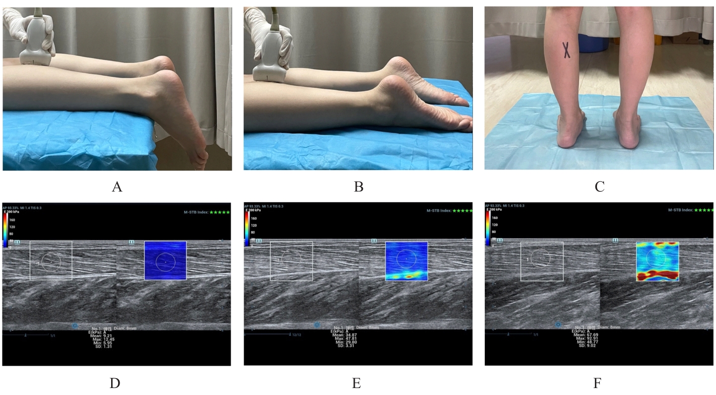
目的 应用超声剪切波弹性成像(SWE)技术检测2型糖尿病患者不同功能状态下腓肠肌的硬度,为临床早期发现骨骼肌损害提供客观化评价依据。 方法 选取70例2型糖尿病患者作为糖尿病组,以70名健康体检者作为对照组。应用SWE技术分别测量2组研究对象腓肠肌在踝关节自然位松弛状态、踝关节跖屈位等张收缩状态和直立位等长收缩状态下的杨氏模量(E)值,并采用体质量指数(BMI)对E值进行标准化(EBMI=E/BMI),比较2组研究对象腓肠肌不同状态下被动硬度和主动硬度的差异,采用Person相关性分析糖尿病患者E值与患者年龄、病程、糖化血红蛋白(HbA1c)水平和糖基化终产物(AGEs)水平的相关性。 结果 2组研究对象腓肠肌在踝关节自然位被动硬度E值和EBMI值比较差异无统计学意义(P>0.05);踝关节跖屈位主动硬度E值比较差异无统计学意义(P>0.05),糖尿病组患者EBMI值组低于对照组(P<0.01);糖尿病组患者直立位主动硬度E值和EBMI值均低于对照组(P<0.01)。糖尿病组患者腓肠肌主动硬度E值与患者病程、HbA1c水平和AGE水平呈负相关关系(r=-0.645,P<0.05;r=-0.741,P<0.05;r=-0.675,P<0.05),与患者年龄无相关性(r=-0.116,P>0.05)。 结论 2型糖尿病患者存在腓肠肌主动硬度减低,被动硬度早期可能不受影响;采用超声SWE技术在直立位等长收缩状态下检测腓肠肌主动硬度有助于发现2型糖尿病患者亚临床阶段肌肉收缩功能减退。

关键词: 糖尿病,2型, 超声检查, 超声剪切波弹性成像, 肌肉硬度

Abstract:

Objective To detect the stiffness of the gastrocnemius muscle in the patients with type 2 diabetes under different functional states by shear wave elastography (SWE) method, and to provide the objective basis for the early clinical detection of skeletal muscle damage. Methods A total of 70 patients with type 2 diabetes were selected as diabetes group, and 70 healthy examinees underwent medical check-ups were regared as control group. SWE was used to detect the Young’s modulus (E) value of the gastrocnemius muscle of the subjects in both groups under three different conditions(neutral ankle position at rest, plantar flexion ankle position during isometric contraction, and upright position during isometric contraction;the E values were normalized by body mass index (BMI) (EBMI=E/BMI); the differences in passive and active stiffness of the gastrocnemius muscle under various conditions of the subjects between two groups were compared; Pearson correlation analysis was used to assess the correlation of the E value in the diabetic patients with patients’ age, disease duration, level of glycated hemoglobin (HbA1c), and level of advanced glycation end-products (AGEs). Results There were no significant differences in the passive stiffness E values and EBMI values of the gastrocnemius muscle of the subjects between two groups under neutral position (P>0.05); there was no significant difference in the active stiffness E values of the gastrocnemius muscle under plantar flexion ankle position(P>0.05), but the EBMI value of the subjects in diabetes group were lower than that in control group (P<0.01). Both E value and EBMI value of active stiffness under upright position of the subjects in diabetes group were lower than those in control group (P<0.01). The E value of active stiffness of the gastrocnemius of the subjects in diabetes group showed a negative correlation with disease duration, HbA1c level, and AGE level (r=-0.645, P<0.05; r=-0.741, P<0.05; r=-0.675, P<0.05), and had no correlation with age (r=-0.116, P>0.05). Conclusion The patients with type 2 diabetes exhibit reduced active stiffness in the gastrocnemius muscle, while passive stiffness may not be affected in the early stage. The use of SWE to measure active stiffness of the gastrocnemius muscle in standing isometric contraction can aid in the detection of subclinical muscle contraction function decline in the patients with type 2 diabetes.

Key words: Diabetes mellitus, type 2, Ultrasonography, Ultrasonic shear wave elastography, Muscle hardness

中图分类号: 

  • R445.1